저작자표시-비영리-변경금지 2.0 대한민국 이용자는 아래의 조건을 따르는 경우에 한하여 자유롭게 l 이 저작물을 복제, 배포, 전송, 전시, 공연 및 방송할 수 있습니다. 다음과 같은 조건을 따라야 합니다: l 귀하는, 이 저작물의 재이용이나 배포의 경우, 이 저작물에 적용된 이용허락조건 을 명확하게 나타내어야 합니다. l 저작권자로부터 별도의 허가를 받으면 이러한 조건들은 적용되지 않습니다. 저작권법에 따른 이용자의 권리는 위의 내용에 의하여 영향을 받지 않습니다. 이것은 이용허락규약(Legal Code)을 이해하기 쉽게 요약한 것입니다. Disclaimer 저작자표시. 귀하는 원저작자를 표시하여야 합니다. 비영리. 귀하는 이 저작물을 영리 목적으로 이용할 수 없습니다. 변경금지. 귀하는 이 저작물을 개작, 변형 또는 가공할 수 없습니다.
CD11b
+
dendritic cells-mediated
immune induction of
inactivated eyedrop vaccine
Eun-Do Kim
Department of Medical Science
The Graduate School, Yonsei University
CD11b
+
dendritic cells-mediated
immune induction of
inactivated eyedrop vaccine
Eun-Do Kim
Department of Medical Science
The Graduate School, Yonsei University
CD11b
+
dendritic cells-mediated
immune induction of
inactivated eyedrop vaccine
Directed by Professor Kyoung Yul Seo
Doctoral Dissertation
submitted to the Department of Medical Science,
the Graduate School of Yonsei University
in partial fulfillment of the requirements
for the degree of Doctor of Philosophy
Eun-Do Kim
This certifies that the Doctoral
Dissertation of Eun-Do Kim
is approved.
The Graduate School
Yonsei University
Acknowledgements
I deeply thanks to my God. He gives me wisdom, intelligence,
cleverness, ideas, understanding and creativeness. He opened the
life of research and has guided me all along my Ph.D. course.
I want to give thanks to Professor Kyoung Yul Seo who has
guided, trusted and supported my all research and learning during
my Ph.D. course. Importantly, I could learn how to investigate as an
independent scientist. It is absolutely worth.
I am grateful to Prof. Jeon-Soo Shin, Prof. Je-Wook Yu, Prof.
Sung Jae Shin and Prof. Sun-Young Chang for much efforts and
patience to review my Ph.D. thesis. I wish to thank Soo Jung Han
who has given many valuable helps and advices for my research
during over a half-decade. I also want to thank special helps from
Dae-Hui Kim and Soonjang Lee. Without their contribution it
would be impossible to finish the work in this winter. I also
appreciate to Dr. Kyuong Sub Choi and Sang Chul Yoon, who gave
me supports, ideas and inspirations.
I want to express gratitude to my parents who always supports
my dreams and vision. Based upon their supports, I could choose to
be a scientist. I want to thanks to Keumdo, Youngdo and Yedo, who
are best brothers and friends to me all the time. Finally, I want to
dedicate this achievement to my beloved fiancée, Sunyoung Kim,
for she waited for me with patience and trust. Her belief that I am
an excellent scientist contributing to a better world is the most
encouraging support.
TABLE OF CONTENTS
GENERAL ABSTRACT ∙∙∙∙∙∙∙∙∙∙∙∙∙∙∙∙∙∙∙∙∙∙∙∙∙∙∙∙∙∙∙∙∙∙∙∙∙∙∙∙∙∙∙∙∙∙∙∙∙∙∙∙∙∙∙∙∙∙∙∙∙∙∙
1
CHAPTER I ∙∙∙∙∙∙∙∙∙∙∙∙∙∙∙∙∙∙∙∙∙∙∙∙∙∙∙∙∙∙∙∙∙∙∙∙∙∙∙∙∙∙∙∙∙∙∙∙∙∙∙∙∙∙∙∙∙∙∙∙∙∙∙∙∙∙∙∙∙∙∙∙∙∙∙∙∙∙∙∙∙∙∙∙
3
ABSTRACT ∙∙∙∙∙∙∙∙∙∙∙∙∙∙∙∙∙∙∙∙∙∙∙∙∙∙∙∙∙∙∙∙∙∙∙∙∙∙∙∙∙∙∙∙∙∙∙∙∙∙∙∙∙∙∙∙∙∙∙∙∙∙∙∙∙∙∙∙∙∙∙∙∙∙∙∙∙∙∙∙∙∙∙∙
4
I. INTRODUCTION ∙∙∙∙∙∙∙∙∙∙∙∙∙∙∙∙∙∙∙∙∙∙∙∙∙∙∙∙∙∙∙∙∙∙∙∙∙∙∙∙∙∙∙∙∙∙∙∙∙∙∙∙∙∙∙∙∙∙∙∙∙∙∙∙∙∙∙∙∙∙
6
II. MATERIALS AND METHODS ∙∙∙∙∙∙∙∙∙∙∙∙∙∙∙∙∙∙∙∙∙∙∙∙∙∙∙∙∙∙∙∙∙∙∙∙∙∙∙∙∙∙∙∙∙ 10
1. Animals ∙∙∙∙∙∙∙∙∙∙∙∙∙∙∙∙∙∙∙∙∙∙∙∙∙∙∙∙∙∙∙∙∙∙∙∙∙∙∙∙∙∙∙∙∙∙∙∙∙∙∙∙∙∙∙∙∙∙∙∙∙∙∙∙∙∙∙∙∙∙∙∙∙∙∙∙∙∙∙ 10
2. Vaccine antigens and adjuvants for mice ∙∙∙∙∙∙∙∙∙∙∙∙∙∙∙∙∙∙∙∙∙∙∙∙∙∙∙∙∙∙∙ 10
3. Immunization for mice ∙∙∙∙∙∙∙∙∙∙∙∙∙∙∙∙∙∙∙∙∙∙∙∙∙∙∙∙∙∙∙∙∙∙∙∙∙∙∙∙∙∙∙∙∙∙∙∙∙∙∙∙∙∙∙∙∙∙∙ 11
4. Viruses for ferrets ∙∙∙∙∙∙∙∙∙∙∙∙∙∙∙∙∙∙∙∙∙∙∙∙∙∙∙∙∙∙∙∙∙∙∙∙∙∙∙∙∙∙∙∙∙∙∙∙∙∙∙∙∙∙∙∙∙∙∙∙∙∙∙∙∙∙∙ 12
5. Vaccination and virus challenge for ferrets ∙∙∙∙∙∙∙∙∙∙∙∙∙∙∙∙∙∙∙∙∙∙∙∙∙∙∙∙ 13
6. Sample collection from mice ∙∙∙∙∙∙∙∙∙∙∙∙∙∙∙∙∙∙∙∙∙∙∙∙∙∙∙∙∙∙∙∙∙∙∙∙∙∙∙∙∙∙∙∙∙∙∙∙∙∙ 13
7. Sample collection from ferrets ∙∙∙∙∙∙∙∙∙∙∙∙∙∙∙∙∙∙∙∙∙∙∙∙∙∙∙∙∙∙∙∙∙∙∙∙∙∙∙∙∙∙∙∙∙∙∙ 14
8. ELISA for detection of Ag-specific Ab ∙∙∙∙∙∙∙∙∙∙∙∙∙∙∙∙∙∙∙∙∙∙∙∙∙∙∙∙∙∙∙∙∙ 14
9. cDNA synthesis and real-time quantitative PCR ∙∙∙∙∙∙∙∙∙∙∙∙∙∙∙∙∙∙∙ 14
10. Protection assay against influenza virus A/California/04/09 ∙ 15
11. Plaque assay ∙∙∙∙∙∙∙∙∙∙∙∙∙∙∙∙∙∙∙∙∙∙∙∙∙∙∙∙∙∙∙∙∙∙∙∙∙∙∙∙∙∙∙∙∙∙∙∙∙∙∙∙∙∙∙∙∙∙∙∙∙∙∙∙∙∙∙∙∙∙∙∙∙∙∙ 15
12. Serological assays in ferrets ∙∙∙∙∙∙∙∙∙∙∙∙∙∙∙∙∙∙∙∙∙∙∙∙∙∙∙∙∙∙∙∙∙∙∙∙∙∙∙∙∙∙∙∙∙∙∙∙∙∙∙ 15
13. Histology for mice ∙∙∙∙∙∙∙∙∙∙∙∙∙∙∙∙∙∙∙∙∙∙∙∙∙∙∙∙∙∙∙∙∙∙∙∙∙∙∙∙∙∙∙∙∙∙∙∙∙∙∙∙∙∙∙∙∙∙∙∙∙∙∙∙∙∙ 16
14. Histology for ferrets ∙∙∙∙∙∙∙∙∙∙∙∙∙∙∙∙∙∙∙∙∙∙∙∙∙∙∙∙∙∙∙∙∙∙∙∙∙∙∙∙∙∙∙∙∙∙∙∙∙∙∙∙∙∙∙∙∙∙∙∙∙∙∙∙ 16
15. Micro-CT ∙∙∙∙∙∙∙∙∙∙∙∙∙∙∙∙∙∙∙∙∙∙∙∙∙∙∙∙∙∙∙∙∙∙∙∙∙∙∙∙∙∙∙∙∙∙∙∙∙∙∙∙∙∙∙∙∙∙∙∙∙∙∙∙∙∙∙∙∙∙∙∙∙∙∙∙∙∙∙∙ 17
16. Electroretinalgram ∙∙∙∙∙∙∙∙∙∙∙∙∙∙∙∙∙∙∙∙∙∙∙∙∙∙∙∙∙∙∙∙∙∙∙∙∙∙∙∙∙∙∙∙∙∙∙∙∙∙∙∙∙∙∙∙∙∙∙∙∙∙∙∙∙∙ 17
17. Data and statistical analyses ∙∙∙∙∙∙∙∙∙∙∙∙∙∙∙∙∙∙∙∙∙∙∙∙∙∙∙∙∙∙∙∙∙∙∙∙∙∙∙∙∙∙∙∙∙∙∙∙∙∙∙ 18
18. Ethics Statement ∙∙∙∙∙∙∙∙∙∙∙∙∙∙∙∙∙∙∙∙∙∙∙∙∙∙∙∙∙∙∙∙∙∙∙∙∙∙∙∙∙∙∙∙∙∙∙∙∙∙∙∙∙∙∙∙∙∙∙∙∙∙∙∙∙∙∙∙∙ 19
III. RESULTS ∙∙∙∙∙∙∙∙∙∙∙∙∙∙∙∙∙∙∙∙∙∙∙∙∙∙∙∙∙∙∙∙∙∙∙∙∙∙∙∙∙∙∙∙∙∙∙∙∙∙∙∙∙∙∙∙∙∙∙∙∙∙∙∙∙∙∙∙∙∙∙∙∙∙∙∙∙∙∙∙∙ 20
1. Significant enhancement of Ag-specific Ab production by
eyedrop influenza vaccine adjuvanted by poly(I:C) ∙∙∙∙∙∙∙∙∙∙∙∙∙∙∙ 20
2. Dose-dependent Ab responses in eyedrop vaccines with
various amounts of Ag and poly(I:C) ∙∙∙∙∙∙∙∙∙∙∙∙∙∙∙∙∙∙∙∙∙∙∙∙∙∙∙∙∙∙∙∙∙∙∙∙∙ 21
3. Comparison of the poly(I:C)-adjuvanted inactivated vaccine
efficacy between eyedrop and intranasal vaccination ∙∙∙∙∙∙∙∙∙∙∙∙ 23
4. Eyedrop split H1N1 influenza vaccine plus poly(I:C)
vaccination can protect mice from lethal influenza virus
challenge ∙∙∙∙∙∙∙∙∙∙∙∙∙∙∙∙∙∙∙∙∙∙∙∙∙∙∙∙∙∙∙∙∙∙∙∙∙∙∙∙∙∙∙∙∙∙∙∙∙∙∙∙∙∙∙∙∙∙∙∙∙∙∙∙∙∙∙∙∙∙∙∙∙∙∙∙∙∙∙∙∙ 24
5. Safety of the administration of eyedrop poly(I:C) plus
inactivated influenza vaccines ∙∙∙∙∙∙∙∙∙∙∙∙∙∙∙∙∙∙∙∙∙∙∙∙∙∙∙∙∙∙∙∙∙∙∙∙∙∙∙∙∙∙∙∙∙∙∙∙ 30
6. Cervarix® administered by eyedrop could induce
Ags-specific immunity in mice ∙∙∙∙∙∙∙∙∙∙∙∙∙∙∙∙∙∙∙∙∙∙∙∙∙∙∙∙∙∙∙∙∙∙∙∙∙∙∙∙∙∙∙∙∙∙∙∙∙∙∙∙∙∙ 34
7. Induction of systemic and mucosal immune response in
ferrets by eyedrop live-attenuated influenza vaccine ∙∙∙∙∙∙∙∙∙∙∙∙∙ 34
8. Eyedrop vaccine protects ferrets from intranasal influenza
virus challenge ∙∙∙∙∙∙∙∙∙∙∙∙∙∙∙∙∙∙∙∙∙∙∙∙∙∙∙∙∙∙∙∙∙∙∙∙∙∙∙∙∙∙∙∙∙∙∙∙∙∙∙∙∙∙∙∙∙∙∙∙∙∙∙∙∙∙∙∙∙∙∙
36
9. Eyedrop vaccine effectively cleared influenza virus in
respiratory organs∙∙∙∙∙∙∙∙∙∙∙∙∙∙∙∙∙∙∙∙∙∙∙∙∙∙∙∙∙∙∙∙∙∙∙∙∙∙∙∙∙∙∙∙∙∙∙∙∙∙∙∙∙∙∙∙∙∙∙∙∙∙∙∙∙∙∙∙
37
10. Influenza eyedrop vaccine is safe in ferret eyes ∙∙∙∙∙∙∙∙∙∙∙∙∙∙∙∙∙∙∙∙∙ 41
IV. DISCUSSION ∙∙∙∙∙∙∙∙∙∙∙∙∙∙∙∙∙∙∙∙∙∙∙∙∙∙∙∙∙∙∙∙∙∙∙∙∙∙∙∙∙∙∙∙∙∙∙∙∙∙∙∙∙∙∙∙∙∙∙∙∙∙∙∙∙∙∙∙∙∙∙∙∙∙∙ 43
V. CONCLUSION ∙∙∙∙∙∙∙∙∙∙∙∙∙∙∙∙∙∙∙∙∙∙∙∙∙∙∙∙∙∙∙∙∙∙∙∙∙∙∙∙∙∙∙∙∙∙∙∙∙∙∙∙∙∙∙∙∙∙∙∙∙∙∙∙∙∙∙∙∙∙∙∙∙∙ 51
REFERENCES ∙∙∙∙∙∙∙∙∙∙∙∙∙∙∙∙∙∙∙∙∙∙∙∙∙∙∙∙∙∙∙∙∙∙∙∙∙∙∙∙∙∙∙∙∙∙∙∙∙∙∙∙∙∙∙∙∙∙∙∙∙∙∙∙∙∙∙∙∙∙∙∙∙∙∙∙∙∙∙ 52
CHAPTER II ∙∙∙∙∙∙∙∙∙∙∙∙∙∙∙∙∙∙∙∙∙∙∙∙∙∙∙∙∙∙∙∙∙∙∙∙∙∙∙∙∙∙∙∙∙∙∙∙∙∙∙∙∙∙∙∙∙∙∙∙∙∙∙∙∙∙∙∙∙∙∙∙∙∙∙∙∙∙
59
ABSTRACT ∙∙∙∙∙∙∙∙∙∙∙∙∙∙∙∙∙∙∙∙∙∙∙∙∙∙∙∙∙∙∙∙∙∙∙∙∙∙∙∙∙∙∙∙∙∙∙∙∙∙∙∙∙∙∙∙∙∙∙∙∙∙∙∙∙∙∙∙∙∙∙∙∙∙∙∙∙∙∙∙
60
I. INTRODUCTION ∙∙∙∙∙∙∙∙∙∙∙∙∙∙∙∙∙∙∙∙∙∙∙∙∙∙∙∙∙∙∙∙∙∙∙∙∙∙∙∙∙∙∙∙∙∙∙∙∙∙∙∙∙∙∙∙∙∙∙∙∙∙∙∙∙∙
61
II. MATERIALS AND METHODS ∙∙∙∙∙∙∙∙∙∙∙∙∙∙∙∙∙∙∙∙∙∙∙∙∙∙∙∙∙∙∙∙∙∙∙∙∙∙∙∙∙
63
1. Mice ∙∙∙∙∙∙∙∙∙∙∙∙∙∙∙∙∙∙∙∙∙∙∙∙∙∙∙∙∙∙∙∙∙∙∙∙∙∙∙∙∙∙∙∙∙∙∙∙∙∙∙∙∙∙∙∙∙∙∙∙∙∙∙∙∙∙∙∙∙∙∙∙∙∙∙∙∙∙∙∙∙∙
63
2. Immunization or administration of fluorescent molecules
63
3. Sample preparation ∙∙∙∙∙∙∙∙∙∙∙∙∙∙∙∙∙∙∙∙∙∙∙∙∙∙∙∙∙∙∙∙∙∙∙∙∙∙∙∙∙∙∙∙∙∙∙∙∙∙∙∙∙∙∙∙∙∙∙
64
4. Cell preparation from draining LN ∙∙∙∙∙∙∙∙∙∙∙∙∙∙∙∙∙∙∙∙∙∙∙∙∙∙∙∙∙∙∙∙∙∙∙
64
5. ELISA for detection of Ag-Specific Ab ∙∙∙∙∙∙∙∙∙∙∙∙∙∙∙∙∙∙∙∙∙∙∙∙∙∙∙
64
6. Flow cytometric analysis ∙∙∙∙∙∙∙∙∙∙∙∙∙∙∙∙∙∙∙∙∙∙∙∙∙∙∙∙∙∙∙∙∙∙∙∙∙∙∙∙∙∙∙∙∙∙∙∙∙∙
65
7. Data and statistical analyses ∙∙∙∙∙∙∙∙∙∙∙∙∙∙∙∙∙∙∙∙∙∙∙∙∙∙∙∙∙∙∙∙∙∙∙∙∙∙∙∙∙∙∙∙∙
65
III. RESULTS ∙∙∙∙∙∙∙∙∙∙∙∙∙∙∙∙∙∙∙∙∙∙∙∙∙∙∙∙∙∙∙∙∙∙∙∙∙∙∙∙∙∙∙∙∙∙∙∙∙∙∙∙∙∙∙∙∙∙∙∙∙∙∙∙∙∙∙∙∙∙∙∙∙∙∙∙∙
66
1. Inoculated eyedrop antigens drains into lymph nodes
through lymphatic drainage ∙∙∙∙∙∙∙∙∙∙∙∙∙∙∙∙∙∙∙∙∙∙∙∙∙∙∙∙∙∙∙∙∙∙∙∙∙∙∙∙∙∙∙∙∙∙
66
2. CD11b
+CD8
+or CD11b
+CD8
-DCs are PE
+cells when
PE-beads are injected by sub-conjunctival injection in
draining lymph nodes ∙∙∙∙∙∙∙∙∙∙∙∙∙∙∙∙∙∙∙∙∙∙∙∙∙∙∙∙∙∙∙∙∙∙∙∙∙∙∙∙∙∙∙∙∙∙∙∙∙∙∙∙∙∙∙
67
3. Sub-conjunctivaly injected antigens can be taken by
resident DCs without the activation of DCs in draining
lymph nodes ∙∙∙∙∙∙∙∙∙∙∙∙∙∙∙∙∙∙∙∙∙∙∙∙∙∙∙∙∙∙∙∙∙∙∙∙∙∙∙∙∙∙∙∙∙∙∙∙∙∙∙∙∙∙∙∙∙∙∙∙∙∙∙∙∙∙∙∙∙∙
68
4. Migratory DCs are dispensable for eyedrop vaccinated
Ag-specific immune induction in dLNs ∙∙∙∙∙∙∙∙∙∙∙∙∙∙∙∙∙∙∙∙∙∙∙∙∙∙∙
71
5. Resident CD11b
+DCs are indispensable to immune
induction for eyedrop vaccine antigens ∙∙∙∙∙∙∙∙∙∙∙∙∙∙∙∙∙∙∙∙∙∙∙∙∙∙∙∙
72
IV. DISCUSSION ∙∙∙∙∙∙∙∙∙∙∙∙∙∙∙∙∙∙∙∙∙∙∙∙∙∙∙∙∙∙∙∙∙∙∙∙∙∙∙∙∙∙∙∙∙∙∙∙∙∙∙∙∙∙∙∙∙∙∙∙∙∙∙∙∙∙∙∙∙∙∙
75
V. CONCLUSION ∙∙∙∙∙∙∙∙∙∙∙∙∙∙∙∙∙∙∙∙∙∙∙∙∙∙∙∙∙∙∙∙∙∙∙∙∙∙∙∙∙∙∙∙∙∙∙∙∙∙∙∙∙∙∙∙∙∙∙∙∙∙∙∙∙∙∙∙∙∙
78
REFERENCES ∙∙∙∙∙∙∙∙∙∙∙∙∙∙∙∙∙∙∙∙∙∙∙∙∙∙∙∙∙∙∙∙∙∙∙∙∙∙∙∙∙∙∙∙∙∙∙∙∙∙∙∙∙∙∙∙∙∙∙∙∙∙∙∙∙∙∙∙∙∙∙∙∙∙∙
79
ABSTRACT (IN KOREAN) ∙∙∙∙∙∙∙∙∙∙∙∙∙∙∙∙∙∙∙∙∙∙∙∙∙∙∙∙∙∙∙∙∙∙∙∙∙∙∙∙∙∙∙∙∙∙∙∙∙∙∙∙∙∙
82
LIST OF FIGURES
CHAPTER I
Figure 1.
CT and poly(I:C) enhances systemic and
mucosal Ab production by eyedrop
vaccination of OVA or HA ∙∙∙∙∙∙∙∙∙∙∙∙∙∙∙∙∙∙∙∙∙∙
22
Figure 2.
Comparison of antibody production by
the amount of hemagglutinin (HA) or
poly(I:C) ∙∙∙∙∙∙∙∙∙∙∙∙∙∙∙∙∙∙∙∙∙∙∙∙∙∙∙∙∙∙∙∙∙∙∙∙∙∙∙∙∙∙∙∙∙∙∙∙∙∙∙
25
Figure 3.
Comparison of Ab production between
intranasal and eyedrop administration
routes ∙∙∙∙∙∙∙∙∙∙∙∙∙∙∙∙∙∙∙∙∙∙∙∙∙∙∙∙∙∙∙∙∙∙∙∙∙∙∙∙∙∙∙∙∙∙∙∙∙∙∙∙∙∙∙∙
26
Figure 4.
Eyedrop inactivated influenza vaccine
plus poly(I:C) administration protects
mice from lethal influenza virus
challenge ∙∙∙∙∙∙∙∙∙∙∙∙∙∙∙∙∙∙∙∙∙∙∙∙∙∙∙∙∙∙∙∙∙∙∙∙∙∙∙∙∙∙∙∙∙∙∙∙∙∙
27
Figure 5.
Comparison of the efficacy of immunity
induction between IM and eyedrop
vaccination ∙∙∙∙∙∙∙∙∙∙∙∙∙∙∙∙∙∙∙∙∙∙∙∙∙∙∙∙∙∙∙∙∙∙∙∙∙∙∙∙∙∙∙∙∙∙∙
29
Figure 6.
Long-term Ag-specific Ab production
induction in eyedrop vaccinated mice ∙∙∙∙∙
31
Figure 7.
No inflammation in the eyes after
administration of HA antigen plus
poly(I:C) in mice ∙∙∙∙∙∙∙∙∙∙∙∙∙∙∙∙∙∙∙∙∙∙∙∙∙∙∙∙∙∙∙∙∙∙∙∙∙∙
32
Figure 8.
No induction of cellular infiltration in
conjunctival tissues after administration
of HA antigen plus poly(I:C) ∙∙∙∙∙∙∙∙∙∙∙∙∙∙∙∙∙∙∙
33
Figure 9.
No detection of contrast medium on the
olfactory bulbs from mice administered
Figure 10. Comparison of Ags-specific Ab
production induction in eyedrop
Cervarix
®vaccinated mice ∙∙∙∙∙∙∙∙∙∙∙∙∙∙∙∙∙∙∙∙∙∙∙
37
Figure 11. Eyedrop vaccination with live attenuated
influenza vaccine (LAIV) elicits
immunological response in ferrets ∙∙∙∙∙∙∙∙∙∙
38
Figure 12. Eyedrop influenza vaccine protects
ferrets from influenza virus challenge ∙∙∙∙∙
39
Figure 13. Successful viral clearance by eyedrop
influenza vaccination ∙∙∙∙∙∙∙∙∙∙∙∙∙∙∙∙∙∙∙∙∙∙∙∙∙∙∙∙∙∙∙
40
Figure 14. Eyedrop inoculation of vaccine-strain
LAIVs does not induce inflammation in
CHAPTER II
Figure 1.
FITC positive cells in lymph nodes and
spleen ∙∙∙∙∙∙∙∙∙∙∙∙∙∙∙∙∙∙∙∙∙∙∙∙∙∙∙∙∙∙∙∙∙∙∙∙∙∙∙∙∙∙∙∙∙∙∙∙∙∙∙∙∙∙∙∙
66
Figure 2.
PE-labeled beads were detected 2 hours
and 24 hours after the inoculation in
draining lymph nodes ∙∙∙∙∙∙∙∙∙∙∙∙∙∙∙∙∙∙∙∙∙∙∙∙∙∙∙∙∙∙∙
67
Figure 3.
Gating strategy on DCs ∙∙∙∙∙∙∙∙∙∙∙∙∙∙∙∙∙∙∙∙∙∙∙∙∙∙∙∙
68
Figure 4.
Gating strategy on resident or migratory
DCs ∙∙∙∙∙∙∙∙∙∙∙∙∙∙∙∙∙∙∙∙∙∙∙∙∙∙∙∙∙∙∙∙∙∙∙∙∙∙∙∙∙∙∙∙∙∙∙∙∙∙∙∙∙∙∙∙∙∙∙
69
Figure 5.
PE-beads were detected in dLNs
administered by ED and SCJ ∙∙∙∙∙∙∙∙∙∙∙∙∙∙∙∙∙∙∙∙
69
Figure 6.
CD11b
+DCs are in highest percentages
of PE-beads positive ∙∙∙∙∙∙∙∙∙∙∙∙∙∙∙∙∙∙∙∙∙∙∙∙∙∙∙∙∙∙∙∙∙
70
Figure 7.
PE-beads positive DCs were detected in
TLR3 KO mice ∙∙∙∙∙∙∙∙∙∙∙∙∙∙∙∙∙∙∙∙∙∙∙∙∙∙∙∙∙∙∙∙∙∙∙∙∙∙∙∙∙
70
Figure 8.
CD11b
+DCs showed highest percentages
of PE
+in TLR3 KO mice ∙∙∙∙∙∙∙∙∙∙∙∙∙∙∙∙∙∙∙∙∙∙∙∙∙
71
Figure 9.
Anti-OVA Ab levels were not decreased
in CCR7 KO mice ∙∙∙∙∙∙∙∙∙∙∙∙∙∙∙∙∙∙∙∙∙∙∙∙∙∙∙∙∙∙∙∙∙∙∙∙
72
Figure 10. Migratory DCs in dLNs were depleted in
OVA+CT eyedrop immunized CCR7 KO
mice ∙∙∙∙∙∙∙∙∙∙∙∙∙∙∙∙∙∙∙∙∙∙∙∙∙∙∙∙∙∙∙∙∙∙∙∙∙∙∙∙∙∙∙∙∙∙∙∙∙∙∙∙∙∙∙∙∙∙
73
Figure 11. The levels of Ag-specific Ab production
was decreased in CD11b KO mice ∙∙∙∙∙∙∙∙∙∙
74
Figure 12. No changes in percentages of resident or
migratory DCs in CD11b KO mice ∙∙∙∙∙∙∙∙∙∙
74
LIST OF TABLES
CHAPTER I
Table 1.
List of influenza viruses used for
vaccination and challenge in this study ∙∙∙
12
Table 2.
List of vaccines tested for the eyedrop
vaccine immune response induction in
LIST OF ABBREVIATIONS
CHAPTER I
CT
cholera toxin
EDV
eyedrop vaccine
CNS
central nervous system
IN
intranasal
S-IgA
secretory-IgA
LAIV
live-attenuated influenza vaccines
OB
olfactory bulb
CALT
conjunctiva-associated lymphoid tissue
TALT
tear-associated lymphoid tissue
poly(I:C)
polyriboinosinic:polyribocytidylic acid
HA
hemagglutinin
ERG
electroretinalgram
OVA
ovalbumin
MPLA
monophosphoryl lipid A
PBS
phosphate-buffered saline
TCID
5050% tissue culture infective doses
IP
intraperitoneal
BALF
bronchoalveolar lavage fluid
ELISA
enzyme-linked immunosorbent assay
dpi
days post infection
PFU
plaque forming units
HI
hemagglutination inhibition
Abs
antibodies
IM
intramuscular
CHAPTER II
EDV
eyedrop vaccine
APCs
antigen presenting cells
dLN
draining lymph nodes
MdLN
mandibular lymph nodes
SPLN
superficial parotid lymph nodes
SCJ
subconjunctival
DCs
dendritic cells
TLR
toll-like receptor
LNs
lymph nodes
CLNs
Cervical lymph nodes
cDCs
classical dendritic cells
OVA
ovalbumin
CT
cholera toxin
PBS
phosphate-buffered saline
MsLNs
mediastinal LNs
PE
phycoerythrin
FACS
fluorescence-activated cell sorting
Ags
antigens
dpi
days post injection
GENERAL ABSTRACT
CD11b+ dendritic cells-mediated immune induction of
inactivated eyedrop vaccine
Eun-Do Kim
Department of Medical Science
The Graduate School, Yonsei University
(Directed by Professor Kyoung Yul Seo)
The eye route has been evaluated as an efficient vaccine delivery routes. However, in order to induce sufficient antibody production with inactivated vaccine, testing of the safety and efficacy of the use of inactivated Ag plus adjuvant is needed. Here, I assessed various types of adjuvants in eyedrop as an anti-influenza serum and mucosal Ab production-enhancer in BALB/c mice. Among the adjuvants, poly (I:C) showed as much enhancement in Ag-specific serum IgG and mucosal IgA antibody production as cholera toxin (CT) after vaccinations with trivalent hemagglutinin-subunits or split H1N1 vaccine Ag in mice. Vaccination with split H1N1 eyedrop vaccine (EDV) Ag plus poly(I:C) showed a similar or slightly lower efficacy in inducing antibody production than intranasal vaccination; the EDV-induced immunity was enough to protect mice from lethal homologous influenza A/California/04/09 (H1N1) virus challenge. Additionally, ocular inoculation with poly(I:C) plus vaccine Ag generated no signs of inflammation within 24 hours: no increases in the mRNA expression levels of inflammatory cytokines nor in the infiltration of mononuclear cells to administration sites. In contrast, CT administration induced increased expression of IL-6 cytokine mRNA and mononuclear cell infiltration in the conjunctiva within 24 hours of vaccination. Moreover, inoculated visualizing materials by eyedrop did not contaminate the surface of the olfactory bulb in
mice; meanwhile, intranasally administered materials defiled the surface of the brain. On the basis of these findings, I propose that the use of inactivated influenza EDV plus poly(I:C) is a safe and effective mucosal vaccine strategy for inducing protective anti-influenza immunity.
Although the efficacy of inactivated EDV was confirmed, the type of antigen presenting cells (APCs) that mediates antigen-specific immune induction has not been reported. Moreover, how the EDV is delivered into the draining lymph nodes (dLN), which are mandibular lymph nodes (MdLN) and superficial parotid lymph nodes (SPLN), is not clarified. In here, I showed that the delivery of proteins or fluorescent beads into the dLN administered by eyedrop or subconjunctival (SCJ) injection is not dependent on the migration of dendritic cells (DCs) or the activation of DCs by TLR stimulation. Instead, the particulates were delivered by flow of lymphatic drainages into the dLN. Among two comprising parts of the dLN, cells in MdLN showed higher levels of percentages of PE-beads+ than SPLN. In MdLN, CD11b+ DCs were in
significantly higher percentages of phycoerythrin (PE)-beads+than other subsets
of DCs do in both resident and migratory DCs. In CD11b knockout mice, the levels of antigen-specific serum IgG or mucosal IgA production were significantly decreased. Thus, it is expected that the strategy targeting resident CD11b+ DCs in MdLN utilizing lymphatic drainage can strengthen the
CHAPTER I
Eyedrop vaccines in mice and ferrets
Induce protective immunity against
ABSTRACT (CHAPTER I)
Eyedrop vaccines in mice and ferrets induce
protective immunity against viral challenges
Eun-Do Kim
Department of Medical Science
The Graduate School, Yonsei University
(Directed by Professor Kyoung Yul Seo)
The eye route has been evaluated as an efficient vaccine delivery routes. However, in order to induce sufficient antibody production with inactivated vaccine, testing of the safety and efficacy of the use of inactivated Ag plus adjuvant is needed. Here, I assessed various types of adjuvants in eyedrop as an anti-influenza serum and mucosal Ab production-enhancer in BALB/c mice. Among the adjuvants, poly(I:C) showed as much enhancement in Ag-specific serum IgG and mucosal IgA antibody production as cholera toxin (CT) after vaccinations with trivalent hemagglutinin-subunits or split H1N1 vaccine Ag in mice. Vaccination with split H1N1 eyedrop vaccine (EDV) Ag plus poly(I:C) showed a similar or slightly lower efficacy in inducing antibody production than intranasal vaccination; the EDV-induced immunity was enough to protect mice from lethal homologous influenza A/California/04/09 (H1N1) virus challenge. Additionally, ocular inoculation with poly(I:C) plus vaccine Ag generated no signs of inflammation within 24 hours: no increases in the mRNA expression levels of inflammatory cytokines nor in the infiltration of mononuclear cells to administration sites. In contrast, CT administration induced increased expression of IL-6 cytokine mRNA and mononuclear cell infiltration in the conjunctiva within 24 hours of vaccination. Moreover, inoculated visualizing materials by eyedrop did not contaminate the surface of the olfactory bulb in
mice; meanwhile, intranasally administered materials defiled the surface of the brain. On the basis of these findings, I propose that the use of inactivated influenza EDV plus poly(I:C) is a safe and effective mucosal vaccine strategy for inducing protective anti-influenza immunity.
Besides, I investigated EDV in pre-clinical development for immunological protection against influenza and for potential side effects involving ocular inflammation and the central nervous system (CNS). Live attenuated influenza EDV, CA07 (H1N1), PZ-4 (H1N2) and Uruguay (H3N2), induced both systemic and mucosal virus-specific antibody responses in ferrets. In addition, EDV resulted in a clinically significant protection against viral challenge, and suppression of viral replication in nasal secretion and lung tissue. Regarding safety, I found that administered EDV flow through the tear duct to reach the base of nasal cavity, and thus do not contact the olfactory bulb. All analyses for potential adverse effects due to EDV, including histological and functional examinations, did not reveal significant side effects. On the basis of these findings, I propose that EDV as effective, while being a safe administration route with minimum local side effects, CNS invasion, or visual function disturbance.
Key words: eyedrop, poly(I:C), influenza, inactivated-vaccine, ferret, live-attenuated influenza vaccines
Eyedrop vaccines in mice and ferrets induce
protective immunity against viral challenges
Eun-Do Kim
Department of Medical Science
The Graduate School, Yonsei University
(Directed by Professor Kyoung Yul Seo)
I. INTRODUCTION
For immunization against influenza, there are two major routes of vaccination: muscular injection and intranasal (IN) administration. Parenteral injection is the most widely and traditionally used method in almost all vaccine regimens; nevertheless, such injections mainly induce serum IgG antibody without inducting secretion of IgA to mucosal surfaces of the respiratory tract, which is the main infection route of the influenza virus. In contrast, intranasal administration induces both systemic IgG and mucosal secretory-IgA (S-IgA) production, initiating mucosal immunity; therefore, intranasal vaccination is more potent than parenteral injection for the prevention of influenza.1,2
Moreover, IN vaccination is advantageous in that is does not require the use of syringes, enabling anyone to readily administer the vaccine without special training.
Recently, some nasal spray live-attenuated influenza vaccines (LAIV), such as FluMist®, were approved by the Food and Drug Administration (FDA) for human use in the United States. However, LAIV can cause some side effects such as sore throat, coryza, and febrile reactions.3 As a result, it is not allowed
for use in pregnant woman and immunodeficient patients, as well as in children under the age of 12 months4 or adults over 50.5Therefore, two major high-risk
groups are excluded from vaccination with the live-virus vaccine. Meanwhile, studies showed that if the inactivated influenza vaccines are intranasally administered, it can induce nerve damage with olfactory bulb (OB)-mediated Ag and adjuvant diffusion into the brain, in the presence of cholera toxin (CT) adjuvant.6,7 Moreover, introduction of inactivated intranasal influenza vaccine
reportedly provoked Bell’s palsy in human.8Thus, many studies have attempted
to devise alternative ways of inducing mucosal immunity to circumvent the side effects of the intranasal influenza vaccines.
Lately, the eye mucosa has come to the forefront as a promising vaccination route. The eye mucosa, which exhibits the common immunological structures of mucosal tissues, including conjunctiva-associated lymphoid tissue (CALT)6,9-11
and tear-associated lymphoid tissue (TALT).12,13 is an inductive site for the
acquirement of systemic and mucosal immunity. The early trials of eyedrop vaccination in avian and bovine models showed that eyedrop vaccination induces protective immunity against Newcastle disease virus and Brucella melitensis, respectively14,15. Additionally, our group was the first to report the
feasibility of the use of eyedrop influenza and Salmonella vaccines in mice.6
Furthermore, eyedrop vaccination does not redirect Ag with CT into the CNS as in intranasal vaccination.6,7
Polyriboinosinic:polyribocytidylic acid (poly(I:C)), a ligand of mammalian toll-like receptor 3, a known receptor for double-stranded RNA, induces interferon alpha/beta production via activation of NF-κB pathways.16 Poly(I:C), therefore,
exerts adjuvant effects by linking the gap between innate and adaptive immunity by enhancing primary antibody responses.17Additionally, the mucosal
adjuvant effect of poly(I:C) against influenza virus has also been shown in intranasally vaccinated inactive-influenza virus hemagglutinin (HA) vaccine plus poly(I:C) in mice.18 Recently, the activity of poly-L-lysine stabilized
poly(I:C) analogue (poly ICLC) as a vaccine adjuvant and innate-immunity activator was assessed in non-human primates and human models, respectively.19,20Thus, clinical trials of the use of poly(I:C) and its safe analogue
may demonstrate the possibility of using potent adjuvant in influenza vaccine immunization.
In the previous study, it was reported that eyedrop vaccination with live-attenuated vaccines, such as influenza virus or Salmonella bacteria, can protect mice from lethal challenge of pathogens.6 However, the vaccines used in the
study were live-attenuated, which may cause unexpected side-effects. Therefore, examining the efficacy of inactivated vaccines immunized by the eye-route is necessary in order to circumvent the use of live-vaccine. However, since it is hard to induce a sufficient immune response by inactivated influenza vaccine-Ag alone, use of adequate adjuvants to induce complete protective immunity is critical to vaccination with inactivated-vaccine Ags.
Meanwhile, the eye mucosa has proven to be an effective Ag delivery route by previous studies in fowls, bovine, goats, and chicken models of immunization.12,21-24 Importantly, a recent investigation in mice determined that
animals immunized with an influenza eyedrop vaccine (EDV) were protected from lethal pathogen infection.6Nevertheless, the clinical significance of these
results is inherently compromised since mice are not natural hosts of influenza. Ferret is one of the most appropriate animal models for the study of influenza EDV for several reasons. First, ferrets are widely used in the study of visual system because their ocular anatomy and physiology are similar to those of humans.25 Second, ferrets have shown to be good model to investigate the
pathogenesis and transmission of influenza since they exhibit a similar level of susceptibility and clinical response to human influenza virus in terms of clinical presentation and respiratory physiology.26,27Lastly, compared to the nonhuman
primates, ferrets are highly preferable in terms of availability, cost of caring, and regulations associated with procurement and maintenance.
Here, I show that poly(I:C) is a potent adjuvant, except for CT, for use in eye-route vaccination for the immunization of killed-influenza virus vaccine Ag. Administration of inactivated influenza vaccine plus poly(I:C) by eyedrops
exerted significantly enhanced production of Ag-specific Ab in both systemic and mucosal immunity, by which mice were protected from lethal influenza virus challenge. Also, there was no signs of inflammation in the eyes after poly(I:C) was administered. In addition, administration of vaccine materials by eyedrops did not contaminate the surface of the brain in contrast to IN, which defiled olfactory bulb regions of the mouse brain.
Moreover, this study aims to evaluate the safety and efficacy of EDV in a ferret model of influenza. Previous investigations detailing immune provocation and acquired protection in mouse models are extended with our analyses in ferrets by providing critical commentary data on clinical presentation and organ histology. Furthermore, I have also been able to address potential safety concerns relating to adverse effects on the CNS and ocular inflammation. On the basis of my findings, I propose that eyedrop vaccination of killed-influenza vaccine along with poly(I:C) or live EDV is an alternative effective and safe preventive mucosal vaccines to induce protective immunity against influenza virus infection in both mice and ferrets.
II. MATERIAL AND METHODS 1. Animals
All studies using mice were performed in strict accordance with the recommendations in the Guide for the Care and Use Committee of Yonsei University Health System. The committee has reviewed and approved the animal study protocol (Approval No: 2011–0137). Specific pathogen-free female BALB/c mice, aged 6-10 wks, were purchased from Charles River Laboratories (Orient Bio, Sungnam, Korea). All mice were maintained in the experimental animal facility under specific pathogen-free conditions at Yonsei College of Medicine (Seoul, Korea) and received sterilized food (Certified Diet MF; Oriental Yeast, Osaka, Japan) and filtered tap water ad libitum. All surgeries were performed after sacrificed by CO2narcosis and every effort was
made to minimize suffering.
For ferret experiments, all experiments related to vaccination and virus infection of animal subjects along with the sample preparation of the ferrets were conducted in strict accordance and adherence to relevant Council of the Republic of Korea and international guidelines regarding animal handling approved by the Animal Use and Care by Laboratory Animal Research Centre (LARC; permit #BLS-ABSL-12-010) of Chungbuk National University (Cheong-ju, Korea), a member of the International Animal Care and Use Committee (IACUC). All experiments using electroretinalgram (ERG) to animal subjects were conducted in strict accordance and adherence to relevant national and international guidelines regarding animal handling approved by the Institutional Animal Care and Use Committee (IACUC) of Yonsei University Health System (Seoul, Korea; permit #2011-0137).
2. Vaccine Ags and adjuvants for mice
The influenza virus subunit vaccines were provided by Dr. Na Gyong Lee (Sejong University, Seoul, Korea). The trivalent HA vaccine comprised the HA subunits from three influenza virus strains: A/New Caledonia/20/99 (H1N1), A/Panama/2007/99 (H3N2) and B/Shandong/7/97. The split H1N1 influenza
virus vaccine was provided by Green Cross Co. (Yongin, Gyeonggi, Korea). The vaccine comprised the split influenza A/California/7/2009 (NYMCX-181) (H1N1) virus. The following commercial vaccines were purchased (all from SANOFI PASTEUR S.A., Lyon, France unless noted) : Avaxim® 160U Adult
Inj., Imovax Polio Inj., Act-HIB®, BCG (Japan BCG Laboratory, Tokyo, Japan),
M-M-R®
II (Merck Sharp & Dohme Corp., PA), Hepavax-Gene®TF inj. (Green
Cross Co., Yongin, Korea), Suduvax®(Green Cross Co., Yongin, Korea), DPT-3
VACCINE inj. (SK Chemical Co., Osan, Korea), and Cervarix®
(GlaxoSmithKline, London, England). Various adjuvants were used including cholera toxin (CT; List Biological Laboratories, Campbell, CA), polyinosinic-polycytidylic acid (poly(I:C), monophosphoryl lipid A (MPLA) from Salmonella minnesota, CpG oligonucleotide (InvivoGen, San Diego, CA), and Imject® Alum (Thermo Scientific, Rockford, IL).
3. Immunization for mice
Prior to experimental manipulation, 6-10-wk-old female mice were anesthetized by intraperitoneal (IP) injection of zoletil (30 mg/kg body weight) and rompun (10 mg/kg body weight). For conjunctival immunization, 100 μg of ovalbumin (OVA) (Sigma-Aldrich, St. Louis, MO) and various adjuvants (0.1 μg to 10 μg poly(I:C) or 10 μg monophosphoryl lipid A (MPLA) or 2 μg CT or 1 mg Imject® Alum) were suspended in 5 μl of phosphate-buffered saline (PBS) and dropped weekly for 3 consecutive wks onto both conjunctival sacs by a micropipette. In some experiments, mice were immunized with 1 μg of trivalent-HA vaccine or 1 μg of H1N1 split vaccine with various adjuvants resuspended in 15 μl or 10 μl of PBS, respectively, and subsequently dropped once more at a 2-wk interval. For HA Ag-dose dependent immunization experiments, 0.5 μg or 1 μg of HA Ags plus 10 μg poly(I:C) were resuspended in 10 μl of PBS, and 2 μg of HA Ags plus 10 μg poly(I:C) were resuspended in 15 μl of PBS. For IN immunization, mice were administered with 1 μg of trivalent-HA vaccine or 1 μg of H1N1 split vaccine with various adjuvants resuspended in 20 μl of PBS and subsequently vaccinated once more at a 2-wk interval. For long-term immunity induction experiments, mice were immunized
with 1 μg of H1N1 split vaccine plus 10 μg of poly(I:C) resuspended in 10 μl of PBS three times at 2-wk intervals.
4. Viruses for ferrets
The live-attenuated viruses (Table. 1) used in this study were adapted in egg at 27℃ (cold adaptation, ca) more than 10 times. The inefficient growth of the ca, live-attenuated viruses compared to wild type corresponding viruses (10-100 fold decreases) was confirmed at 37℃. The human and animal infectious influenza viruses (Table 1) used for challenge in this study were provided by Chungbuk National University (Cheongju, South Korea) and amplified in 10-day-old embryonated chicken eggs. Viruses were serially diluted (10-fold) prior to infection in Mardin-Darby canine kidney cells and the 50% tissue culture infective doses (TCID50) was then calculated by the Reed-Muench method.28
Stock viruses were kept at -82°C, and thawed right before use. Viral growth was determined by observing changes in cellular morphology (cytopathic effects) and hemagglutinin (HA) assay.
Table 1. List of influenza viruses used for vaccination and challenge in this study
Group
(subtype) Virus strain Abbreviation
Challenge dose (TCID50) Similarity H1N1 Vaccination A/California/7/09 x PR8§ CA07 (H1N1) -
-Challenge A/California/04/09 CA04 (H1N1) 105 Homologous H1N2 Vaccination Sw/Korea/PZ4/06
† PZ-4 (H1N2) -
-Challenge A/Sw/Korea/1204/09 Sw09 (H1N2) 106 Heterologous H3N2 Vaccination
A/Uruguay/716/07x PR8§,‡
Uruguay
(H3N2) -
-Challenge A/Hongkong/68 HK68 (H3N2) 106 Heterologous
§A/Puerto Rico/8/34; this virus is used as a backbone strain. †Cross-reactive HI
titer between Sw/Korea/PZ4/06 and Sw/Korea/1204/09 is 2 fold lower than the HI titer of homologous Sw/Korea/PZ4/06 virus (160 Vs 320 HI units). ‡
5. Vaccination and virus challenge for ferrets
15- to 16-wk-old ferrets were purchased from Marshall BioResources (North Rose, NY, USA). All animals were confirmed seronegative for all vaccine strains of influenza A viruses used in this study, including CA07 (H1N1), PZ-4 (H1N2), and Uruguay (H3N2) by serologic assay. For conjunctival immunizations, ferrets were anesthetized by inhalation of isoflurane, and then 10^5 TCID50 of the CA07 (H1N1), PZ-4 (H1N2), and Uruguay (H3N2) LAIVs
in 100 µL PBS were dropped to each eye (200 μL/head) twice with a two wk interval. Two wks after the second immunization, ferrets were intranasally instilled with 1.0 mL volume of 10^5 TCID50/mL for the CA04 (H1N1) and
10^6 TCID50/mL for the Sw09 (H1N2) and HK68 (H3N2) for virus challenge.29
Body temperatures and body weights of infected ferrets were monitored daily. There was no death after the viral infection.
6. Sample collection from mice
At two wks or one year after the final immunization, serum was obtained by retro-orbital bleeding. Tear-wash samples were obtained by lavaging with 10 μl of PBS per eye. Saliva was obtained following IP injection of mice with pilocarpine (500 mg/kg body weight; Sigma-Aldrich). Fecal extract was obtained by adding weighed feces to PBS containing 0.1% sodium azide. The feces samples were mixed by vortexing and subsequently centrifuged, and the supernatants were collected for assay. Vaginal wash samples were collected by lavage with 100 μl of PBS. To obtain bronchoalveolar lavage fluid (BALF), tracheas were cannulated after exsanguination, and the lungs were washed with 1 ml of PBS. After the mice were sacrificed, nasal wash samples were obtained by flushing 100 μl of PBS through the anterior (oral) entrance of the nasal passages using a pipette.
7. Sample collection from ferrets
The ferrets were anesthetized with Zoletil 50® (125 mg zolazepam and 125 mg tiletamine hydrochloride [Vibrac, Carros, France]; 0.2 mg/kg of body weight) and Rompun® (2% xylazine hydrochloride [Bayer Animal Health, Leverkusen, Germany]; 5 mg/kg of body weight) administered intramuscularly 10 min
before sampling. Serum samples from ferrets were collected for HI assays two wks after each vaccination. Nasal secretion samples were collected in 1X PBS with antibiotics two wks after every vaccination for HI assay or at 1, 3, 5, 7, and 9 days post infection (dpi) after viral challenge for titer assays. For the preparation of lung tissue samples, ferrets were euthanatized by CO2inhalation.
The lung tissues were collected at 5 dpi for histopathology or homogenized in 1X PBS containing antibiotics for virus titration. Tissue homogenates of both lobes were clarified by centrifugation at 12,000g, and supernatants were then transferred to fresh tubes. All samples were stored at -82°C until use.
8. ELISA for detection of Ag-specific Ab
Enzyme-linked immunosorbent assay (ELISA) plates (Nunc, Roskilde, Denmark) were coated with OVA (100 μg/ml) or HA vaccine Ag (2 μg/ml) or H1N1 split vaccine (1 μg/ml) in coating buffer and incubated overnight at 4°C. Blocking was done with 1% bovine serum albumin (Sigma-Aldrich) in PBS, and two-fold serially diluted samples were applied to plates. HRP-conjugated goat anti-mouse IgG or IgA Ab (Southern Biotechnology Associates, Birmingham, AL) was added to each well and incubated overnight at 4°C. For color development, tetra-methylbenzidine solution (Thermo Scientific, Rockford, IL) was used. Plates were then measured at 450 nm on an ELISA reader (Molecular Devices, Sunnyvale, CA) after addition of stopping solution (0.5 N HCl). Endpoint titers of Ag-specific Ab were expressed as reciprocal log2 titers of the last dilution that showed > 0.1 absorbance over background levels.
9. cDNA synthesis and real-time quantitative PCR
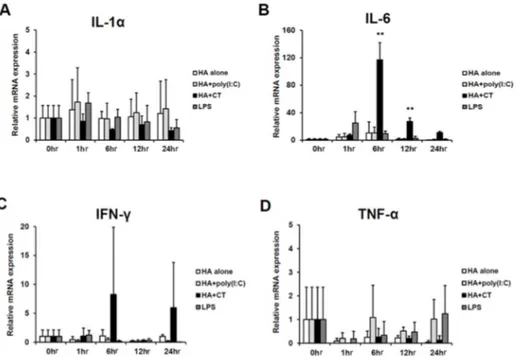
After a wash with nuclease-free water, I homogenized the whole conjunctival or corneal tissue samples at different time points after eye-drop immunization with HA vaccine alone (1 μg), HA plus poly(I:C) (10 μg), or HA plus CT (2 μg). Total RNA was extracted using TRIzol (Invitrogen), and cDNA was synthesized by Superscript II reverse transcriptase with oligo (dT) primer (Invitrogen). cDNA was amplified with HotStart-IT® SYBR® Green qPCR Master Mix (USB, Cleveland, OH) and gene-specific forward and reverse primers on an
ABI 7300 Real- Time PCR system (Applied Biosystems). Results are expressed as mean ± S.D. after normalizing to the expression of β-actin gene using the ΔΔCt method. Primer sequences are available upon request.
10. Protection assay against influenza virus A/California/04/09
At two wks or one year after the final eyedrop or intranasal immunization with 1 μg of H1N1 split vaccine alone or plus 10 μg of poly(I:C), five mice in each group were anesthetized and challenged with 50 μl of mouse-adapted live influenza A/California/04/09 (H1N1) virus suspension (10X LD50; 0.75 TCID50)
via the IN route. Animals were monitored for weight loss and survival every day for 14 days, and there were 3 unexpected deaths in PBS or H1N1 Ag alone administered groups. The specific clinical signs I used to determine when the animals should be euthanized were the loss of 20% of initial bodyweights. Euthanasia was done by CO2 inhalation with a fill rate of about 10% to 30% of
the chamber volume per minute with carbon dioxide, added to the existing air in the chamber.
11. Plaque assay
The measurement of the viral titers in lung tissues in virus-challenged mice were measured as previously described.30 Briefly, the viral titers used in this
study were expressed as the plaque forming units (PFU) calculated by plaque assay. Monolayers of Madin-Darby Canine Kidney cells in 12-well-plates were infected with 10-fold serial dilutions of virus solutions for 45 min at room temperature. After the removal of the solutions, the cells were washed with PBS and overlaid with minimum essential medium, containing 1% low melting agarose (Lonza, ME, USA) and 10 μg/ml of trypsin (Life Technology, NY, USA). After the overlay turned solid, the plates were moved to an incubator with 5% CO2. The plaques formed were fixed by formaldehyde solution and
visualized by staining with crystal violet. 12. Serologic assays in ferrets
Hemagglutination inhibition (HI) assays were performed as previously described to determine the sero-prevalence of vaccinated and challenged
viruses.31 Briefly, serum samples were heat inactivated at 56°C for 30 min and
pretreated with receptor-destroying enzyme (RDE) from Vibrio cholerae (Denka Seiken; Tokyo, Japan) to remove non-specific serum inhibitors. Sera were then analyzed for the presence of virus-specific antibody by HI assays with 0.5% turkey red blood cells (RBC). The HI titer was determined by the reciprocal of the last dilution that contained turkey RBCs with no agglutination. Neutralization tests performed on selected HI-positive sera to confirm results and used to determine MDCK cell infection and expressed as the reciprocal of the highest dilution of serum that gave 50% neutralization of 100 TCID50 of
virus after incubation at 37°C for 72 h.32,33HI assays were performed according
to WHO/World Organization for Animal Health (OIE) recommendations.34
13. Histology for mice
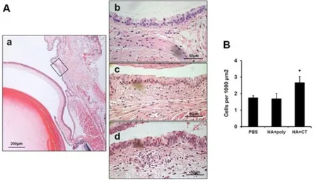
Eye tissues including the conjunctiva and eye balls from controls and 1 μg HA vaccine plus 10 μg poly(I:C) or 2 μg CT treated mice were washed with PBS and fixed in 4% formaldehyde for 24 hr at 4°C. The tissues were dehydrated by gradual soaking in alcohol and xylene and then embedded in paraffin. The paraffin-embedded specimens were cut into 5mm sections and stained with H&E.
14. Histology for ferrets
The lungs or eye tissue of ferrets from each group were harvested at 5 dpi or 24 hour, respectively. Tissues were fixed in 10% neutral-buffered formalin, embedded in paraffin, sectioned (4 µm), and examined in the pathology laboratory of Chungbuk National University Hospital or Yonsei University College of Medicine. Histological assessment was performed by standard hematoxylin and eosin staining and light microscopy at 200X magnification. In blind fashion, either left or right lobes of five lungs were examined with five to seven slides per each lobe. Semi-quantitative analysis of lung inflammation severity in influenza virus-challenged ferrets was performed with some modification as reported elsewhere35for the alveoli.
15. Micro-CT
Imaging was performed as previously described36 with minor modification
using a volumetric CT scanner (NFR-Polaris-G90MVC: NanoFocusRay, Iksan, Korea). Briefly, images were acquired at 65 kVp, 115 μA, and 142-millisecond per frame, and for 700 views. The estimated radiation dose was ~150 mGy using this image acquisition protocol. Images were reconstructed using the volumetric cone-beam reconstruction (FDK) off line mode. The size of reconstruction images was 1,204 x 1,024 pixels, and 512 slices were acquired. The final reconstructed data were converted to the Digital Imaging and Communications in Medicine (DICOM) format to generate 3D-rendered images using 3D-rendering software (Lucion, MeviSYS, Seoul, Korea). For ex vivo brain CT imaging, all BALB/c mice were sacrificed, and the brains of each ocularly or intranasally treated mouse were taken out and CT images thereof were acquired.
16. Electroretinalgram (ERG)
The ferrets were anesthetized with Zoletil 50® (125 mg zolazepam and 125 mg tiletamine hydrochloride [Vibrac, Carros, France]; 0.2 mg/kg of body weight) and Rompun® (2% xylazine hydrochloride [Bayer Animal Health, Leverkusen, Germany]; 5 mg/kg of body weight) administered intramuscularly 10 min before ERG examinations. Pupils were maximally dilated with achieved with 0.5% phenylephrine hydrochloride and 0.5% tropicamide (Mydrin-P®, Santen, Osaka, Japan). Animals were placed in a special holding system to prevent unfavorable movement during full-field ERG recording.
Full-field ERG recording was performed with RETIscan® (Roland Consult, Wiesbaden, Germany) in the same examination room. A Goldring electrode® with diameter of 3 mm (Roland Consult, Wiesbaden, Germany) was placed on the corneal surface as an active electrode using 0.3% hypromellos (GenTeal®, Novartis, Basel, Switzerland), while a second reference electrode was placed on the fornix of the same eye. Concentric subdermal needle electrodes (Roland Consult, Wiesbaden, Germany) were then used as a ground electrode after insertion into the tail of the animal.
Full-field ERG was recorded following the standards of International Society of Clinical Electrophysiology of Vision (ISCEV). Full-field stimulation was produced using Ganzfeld stimulator of the RETIscan unit (Roland Consult, Wiesbaden, Germany), which was positioned just in front of animal’s face. To assess rod responses by ERG, the animals were dark-adapted for 12 hr before anesthesia. A dim white flash with a stimulus strength of 0.01 cd·s/m2was used
for dark-adapted 0.01 ERGs with intervals of 2 sec between flashes. A white 3.0 cd·s/m2 flashes were produced in dark-adapted eye for dark-adapted 3.0 ERGs
and oscillatory potentials recordings. Subsequently, light-adapted ERGs recordings were performed after 15 min of light adaptation. Light-adapted 3.0 ERGs elicited by white flashes at an intensity of 3.0 cd·s/m2 under white
background of 30 cd/m2. Furthermore, 30-Hz flicker responses were recorded
using white light flashes at an intensity of 3.0 cd·s/m2 and a rate of 30 stimuli
per sec (30 Hz). All of responses were amplified at 10,000 times, and were filtered with a band pass between 1 and 300 Hz. Five waveforms for each response were averaged to reduce variability and background noise. ERG abnormality is reflected in alterations in A- and B-wave amplitude. A-wave amplitude measures the trough of the negative deflection as difference from the baseline value. B-wave amplitude measures the difference between the trough of A-wave and the peak of B-wave recording. Recordings of full-field ERG were performed for both eyes prior to the vaccination and 1 day after the treatment, respectively. Vaccination was applied only on right eyes, and left eyes were considered as a control eye.
For statistical analysis of the ERG parameters, the paired t-test was used to assess the difference between the values before and after the vaccination, respectively. The differences were considered to be significant when P-value was less than 0.05.
17. Data and statistical analyses
Data in mice experiments were expressed as the mean ± SD, and statistical analyses were conducted by the ANOVA test (Microsoft Office Excel program). Data in ferret experiments were expressed as the mean ± SD, and statistical
analyses were conducted by the student’s t-test (Microsoft Office Excel program). Number of animals in all groups was 3 or 4. For the data normality test, the Shapiro-Wilk test was used. All data was checked before I use the student’s t-test and concluded that all data came from a normal distribution. 18. Ethics Statement
All experiments involving animal subjects were conducted in strict accordance and adherence to relevant national and international guidelines regarding animal handling as mandated by the Institutional Animal Care and Use Committee (IACUC) of Yonsei University Health System (Seoul, Korea). Approval number is 2011-0137.
III. RESULTS
1. Significant enhancement of Ag-specific Ab production by eyedrop influenza vaccine adjuvanted by poly(I:C)
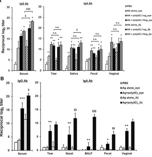
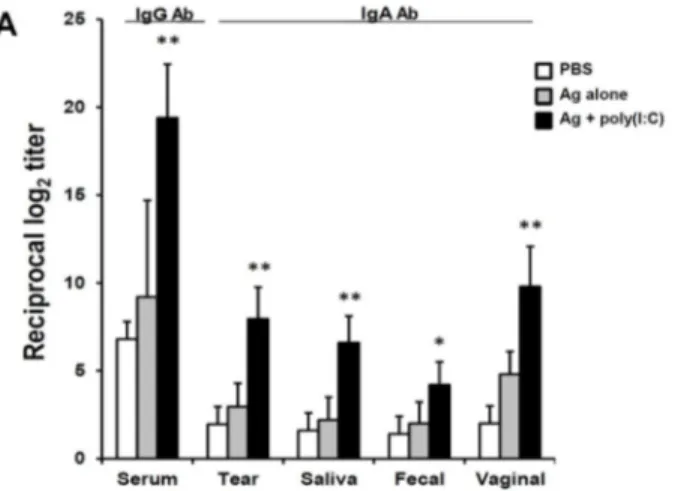
To evaluate the efficacy of various adjuvants in regards to whether they can enhance systemic and mucosal antibody production when used with protein Ag by eye-route vaccination, BALB/c mice were immunized with OVA (100 μg/head) protein plus several conventionally used adjuvants, including CT (2 μg/head), poly(I:C) (10 μg/head), MPLA (10 μg/head), or Imject® Alum (1 mg/head), which is a commercially used alum adjuvant, three times at one-wk intervals. One wk after final immunization, the levels of OVA-specific antibodies (Abs) were measured by ELISA. All mice given OVA plus adjuvant showed significantly higher levels of OVA-specific serum IgG Ab in there sera than those found in mice given PBS or OVA alone (Fig. 1A). Among the adjuvants, CT showed the highest serum IgG Ab production level. However, only mice given OVA plus CT or poly(I:C) showed significantly higher levels of IgA Ab in mucosal compartments (e.g., tear, nasal, fecal, and vaginal washes) than the other adjuvant-treated groups.
To examine whether the Ab production-enhancing effect adjuvanted by CT or poly(I:C) was retained in influenza subunit-vaccine immunized mice, as shown in Fig. 1A, BALB/c mice were immunized with 1 μg of the seasonal influenza vaccine, consisting of trivalent HAs of A/New Caledonia/20/99 (H1N1), A/Panama/2007/97 (H3N2), and B/Shandong/7/97 influenza (B) viruses, with the same adjuvants that were previously used with OVA protein. As shown in Fig. 1B, Ab production levels for serum IgG and mucosal IgA Ab in mice treated with CT or poly(I:C) were significantly higher than those for the other groups. The enhancing effects of MPLA and the Imject® adjuvants were not observed for either types of Abs, compared to mice given PBS or Ag alone. Unexpectedly, the efficacy of Abs-production enhancement of poly(I:C) was similar to that of CT. Moreover, poly(I:C) significantly improved the production of mucosal IgA Ab to as much as CT did in the respiratory passageways, in
nasal cavity wash and BALF samples, which is the main route of influenza infection, indicating that it could be of use as a front-line defense against influenza invasion.37 While CT has been shown to cause CNS damage when it
was administrated intranasally,7,8 there is a report of a lack of a destructive
effect on nasal or brain tissue after administration of poly(I:C).18 Therefore, in
the following experiments, I focused on assessing the adjuvanticity of poly(I:C) via eyedrop administration.
2. Dose-dependent Ab responses in eyedrop vaccines with various amounts of Ag and poly(I:C)
Next, I attempted to elucidate the minimum amount of Ag and poly(I:C) that could effectively induce the enhancement of systemic and mucosal Ab production via eyedrop vaccination. To do so, BALB/c mice were immunized with 10 μg of poly(I:C) and various amounts of HA vaccine, 0.1 μg, 1 μg or 2 μg, via eyedrop vaccination (Fig. 2A). Since the maximum volume of eyedrops per eye is 15 μl and the concentration stocks of HA vaccine and poly(I:C) were 1 μg per 11 μl and 10 μg per 4 μl, respectively, I could not examine the effect of Ag amounts over 2 μg HA vaccine plus 10 μg poly(I:C) in the eyedrop vaccination. As shown in Fig. 2A, 1 μg of HA Ag plus 10 μg poly(I:C) significantly enhanced serum IgG and mucosal IgA Ab production amounts greater than those for mice given doses of 0.1 μg or 2 μg Ag plus 10 μg poly(I:C). Unexpectedly, the enhancement from 2 μg HA eyedrops with 10 μg poly(I:C) was less effective than that from 1 μg HA vaccine Ag eyedrops. Since the total volume of 1 μg or 2 μg HA Ags plus 10 μg poly(I:C) were 10 μl or 15 μl, respectively, it was estimated that the concentration of poly(I:C) was diluted in the doubled volume of 2 μg HA Ag eyedrops and, accordingly, the effect of poly(I:C) was diminished, compared to that for 1 μg Ag plus 10 μg poly(I:C) eyedrops. Thus, for following experiments, I used 1 μg of HA vaccine Ag for eyedrop vaccination.
After the discovery of the effective minimum amount of HA vaccine Ag in the eyedrop vaccination, I checked the dose-dependent efficacy of poly(I:C) in the
Figure 1. CT and poly(I:C) enhances systemic and mucosal Ab production by eyedrop vaccination of OVA or HA. Groups of female BALB/c mice received OVA (100 μg) (A) or HA (1 μg) (B) plus CT (2 μg), poly(I:C) (10 μg), MPLA (10 μg), Imject® Alum (320 μg) resolved in 5 μl of PBS, or 5 μl of PBS alone by drops on both eyes every wk (three times). Ag-specific Ab levels were measured in serum and in various mucosal fluids 1 wk after final vaccination by ELISA. * p < 0.05, ** p < 0.01, *** p < 0.001 versus Ag alone; §§ p < 0.01, §§§ p < 0.001 versus poly(I:C) group; ‘n.s.’, non-significant. Results are representative of three independent experiments, with three mice in each group.
1μg HA EDV in mice. The levels of HA-specific Ab production in all groups of mice in which poly(I:C) was administered together with 1 μg HA-Ag EDV were significantly enhanced, compared to mice treated with 1 μg HA-Ag alone in both serum and the mucosal washes (Fig. 2B). Additionally, there was almost no difference in the adjuvant efficacies of all poly(I:C) administered groups; even administration of 1 μg of poly(I:C) significantly augmented Abs production levels in the 1 μg of HA-Ag eyedrop vaccinated mice to as much as those for mice treated with 30 μg of poly(I:C). Thus, these results indicate that the efficacy of 1 μg poly(I:C) was enough to induce significant Ag-specific immunity when it was vaccinated with HA-Ag via the eye route.
3. Comparison of the poly(I:C)-adjuvanted inactivated vaccine efficacy between eyedrop and intranasal vaccination
Reportedly, vaccination via the IN route is the most effective, among mucosal routes.38 Thus, to examine whether the adjuvant efficacy of poly(I:C) in the
eyedrop vaccination is more effective than that of poly(I:C) administration via other vaccination routes, I compared the efficacy of the immunization of HA vaccine plus poly(I:C) adjuvant via the eye route with that of intranasal vaccination in mice. In Fig. 3A, when the serum IgG Ab levels of the eyedrop and intranasal vaccination groups, which were administered the same amounts of poly(I:C) and HA vaccine, were compared, all of the intranasal vaccination groups showed significantly higher serum IgG Ab responses than their eyedrop vaccination counterparts. However, in the mucosal wash fluids, there was no significant difference in the levels of mucosal IgA Ab-production between the eyedrop and IN vaccination groups, except for saliva fluids. Additionally, the enhancement of IgA Ab production levels in all of the mucosal fluids from mice administered 1 μg poly(I:C) plus HA EDV was significantly similar to that in intra-nasally immunized mice.
Next, to evaluate whether the adjuvanticity of poly(I:C) in eyedrop vaccination is maintained when the influenza virus HA-vaccine Ag is replaced with commercially used inactivated split A/California/04/09 H1N1 (H1N1) influenza
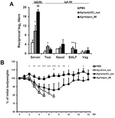
virus vaccine Ag in eyedrops, BALB/c mice were immunized with 1 μg of split H1N1 influenza vaccine plus 10 μg poly(I:C) via eyedrops or intranasally twice at a 2-wk intervals. Two wks after the final immunization, as shown in Fig. 3B, H1N1 vaccine-specific serum IgG Ab production levels in intranasally immunized mice with poly(I:C) plus vaccine Ag were significantly higher than those for eyedrop vaccinated mice. In mucosal fluids, except tear and vaginal wash samples, Ab production levels in the intranasally vaccinated groups were significantly higher than those in the eyedrop groups. However, the levels of Ag-specific Ab production in the poly(I:C) adjuvanted eyedrop vaccinated group were still significantly higher than those in the eyedrop Ag alone treated groups. Therefore, these results indicate that the efficacy of poly(I:C) in eyedrop vaccination is sufficient for use in commercial influenza vaccine Ags, with an efficacy similar with or slightly weaker than that for IN vaccination.
4. Eyedrop split H1N1 influenza vaccine plus poly(I:C) vaccination can protect mice from lethal influenza virus challenge
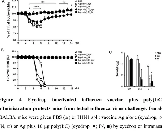
To examine whether the immunity induced by vaccination of inactivated EDV plus poly(I:C) can protect mice from lethal influenza virus challenge, I vaccinated BALB/c mice with two doses of 1 μg of split H1N1 influenza vaccine plus 10 μg of poly(I:C) by eyedrops or IN as a positive control group at a 2-wk intervals. Two wks after the final vaccination, mice were challenged IN with mouse-adapted live influenza A/California/04/09 (H1N1) virus (10X LD50;
0.75 TCID50) and monitored for 2 wks (Fig. 4). Mice in the intranasally Ag +
poly(I:C) treated group maintained their initial bodyweights throughout the monitoring period. Although eyedrop Ag + poly(I:C) treated mice lost about 10 percent of their initial bodyweight (Fig. 4A), all mice began to recover their bodyweights on day 6 and survived against the lethal influenza virus challenge (Fig. 4B). Meanwhile, PBS or Ag alone administered mice both by eyedrop and IN were critically affected by the influenza virus infection (Fig. 4A) and all mice of PBS group and both Ag alone treated groups were sacrificed at humane endpoints (Fig. 4B). Additionally, titers of the challenged influenza virus in lung tissue of mice were measured (Fig. 4C). Proliferating viruses on day 1 began to
reduce on day 4 in both the eyedrop and IN groups, and viruses were significantly cleared on day 7 in both vaccinated groups.
Figure 2. Comparison of antibody production by the amount of hemagglutinin (HA) or poly(I:C). (A). Dose-dependent HA vaccine Ags were administered with 10 μg poly(I:C) resolved in 5 μl of PBS by drops on both eyes two times at a 2-wk interval in female BALB/c mice. HA-specific Ab levels were measured in serum and in various mucosal fluids 2 wks after final vaccination by ELISA. (B) 1 μg of HA vaccine Ag plus dose-dependent