Vertebral compression fractures after spine irradiation
using conventional fractionation in patients with
metastatic colorectal cancer
Woo Joong Rhee, MD, Kyung Hwan Kim, MD, Jee Suk Chang, MD,
Hyun Ju Kim, MD, Seohee Choi, MD, Woong Sub Koom, MD
Department of Radiation Oncology, Yonsei Cancer Center, Yonsei University Health System, Seoul, Korea
Purpose: To evaluate the risk of vertebral compression fracture (VCF) after conventional radiotherapy (RT) for colorectal cancer (CRC) with spine metastasis and to identify risk factors for VCF in metastatic and non-metastatic irradiated spines.
Materials and Methods: We retrospectively reviewed 68 spinal segments in 16 patients who received conventional RT between 2009 and 2012. Fracture was defined as a newly developed VCF or progression of an existing fracture. The target volume included all metastatic spinal segments and one additional non-metastatic vertebra adjacent to the tumor-involved spines.
Results: The median follow-up was 7.8 months. Among all 68 spinal segments, there were six fracture events (8.8%) including three new VCFs and three fracture progressions. Observed VCF rates in vertebral segments with prior irradiation or pre-existing compression fracture were 30.0% and 75.0% respectively, compared with 5.2% and 4.7% for segments without prior irradiation or pre-existing compression fracture, respectively (both p < 0.05). The 1-year fracture-free probability was 87.8% (95% CI, 78.2–97.4). On multivariate analysis, prior irradiation (HR, 7.30; 95% CI, 1.31–40.86) and pre-existing compression fracture (HR, 18.45; 95% CI, 3.42–99.52) were independent risk factors for VCF.
Conclusion: The incidence of VCF following conventional RT to the spine is not particularly high, regardless of metastatic tumor involvement. Spines that received irradiation and/or have pre-existing compression fracture before RT have an increased risk of VCF and require close observation.
Keywords: Spinal neoplasm, Compression fractures, Spinal fractures, Radiotherapy, Colorectal cancer, Risk factors
www.e-roj.org
Introduction
Colorectal cancer (CRC) is the third most commonly diagnosed cancer in males and the second in females, with over 1.2 million estimated new cases worldwide per year [1]. Among more than 2,500 CRC patients in Italy, approximately 10%
had bone metastasis and spinal segments were the most common site of bone metastases, with an incidence of 65% [2]. Radiation therapy (RT) is thought to be an important palliative treatment modality for spine metastasis. Although conventional RT is traditionally the standard of care for palliative treatment of spine metastasis [3-6], spine
Received 20 May 2014, Revised 8 September 2014, Accepted 25 Setember 2014.
Correspondence: Woong Sub Koom, MD, Department of Radiation Oncology, Yonsei Cancer Center, Yonsei University Health System, 50 Yonsei-ro, Seodaemun-gu, Seoul 120-752, Korea. Tel: +82-2-2228-8116, Fax: +82-2-312-9033, E-mail: [email protected]
*This research was presented at the 31th Annual Meeting of Korean Society for Radiation Oncology (KOSRO), October 11, 2013, Seoul, Korea.
CC This is an Open Access article distributed under the terms of the Creative Commons Attribution Non-Commercial License (http://creativecommons.org/
licenses/by-nc/3.0/) which permits unrestricted non-commercial use, distribution, and reproduction in any medium, provided the original work is properly cited.
Radiat Oncol J 2014;32(4):221-230 http://dx.doi.org/10.3857/roj.2014.32.4.221 pISSN 2234-1900 · eISSN 2234-3156
stereotactic body radiation therapy (SBRT) is an emerging treatment option for patients with spine metastasis [7,8]. With the increased use of SBRT, the occurrence of vertebral compression fractures (VCFs) after SBRT has become an important issue. Memorial Sloan-Kettering Cancer Center (MSKCC) reported the first data on VCFs, demonstrating that 27 VCFs developed in a total of 71 spinal segments (39%) after SBRT [9]. The MD Anderson Cancer Center (MDACC) subsequently reported similar findings of 25 VCFs in 123 spinal segments (20%) treated with SBRT [10].
The Spine Oncology Study Group (SOSG) introduced the Spinal Instability Neoplastic Score (SINS) criteria to predict the stability of metastatic vertebral segments [11]. The SINS criteria consist of six components: location, pain, bone lesion, radiographic spinal alignment, vertebral body collapse, and posterolateral involvement of spinal elements. SINS class is determined by the sum of the component scores, and ranges from 0 to 18. A score of 0 to 6 denotes class 1 (stable), 7 to 12 denotes class 2 (potentially unstable), and 13 to 18 denotes class 3 (unstable). Researchers at the University of Toronto evaluated not only the incidence and risk factors of VCF after SBRT, but also the validity of SINS criteria [12]. However, there are no clear data on VCF development after conventional RT, which is still commonly used in real practice.
In this study, we aimed to determine the incidence of VCF after conventional RT for CRC with spine metastasis. In addition, we investigated the risk factors for VCF in both metastatic and non-metastatic irradiated spinal segments. Lastly, we evaluated the role of SINS criteria as an additional predictive factor for VCF in metastatic irradiated spinal segments.
Materials and Methods
1. Patient selection
We retrospectively reviewed 98 spinal segments in 24 patients who were treated with conventional RT between March 2009 and October 2012. All patients were diagnosed with CRC with spine metastasis and had more than 6 months of clinical follow-up. Spinal segments that had a fracture event within the 6-month follow-up period after RT were also included in our study cohort. This study included not only the metastatic vertebra, but also all non-metastatic vertebra involved in the RT volume even if they had pre-existing fractures. Patients with a double primary cancer, a diagnosis of osteoporosis, and those who were receiving bisphosphonate therapy were excluded. Spinal segments that had previously undergone
surgery or a procedure related to vertebral stability, or were treated with SBRT, were also excluded. After patient selection, our final cohort consisted of 68 spinal segments in 16 patients.
2. Treatment and evaluation
All patients were treated with conventional RT. The target volume included all metastatic spinal segments and one additional level of non-metastatic vertebra adjacent to the tumor-involved spines. We evaluated the latest computed tomography (CT) and/or magnetic resonance image (MRI) scans that were taken before planning to determine target volume. The total dose and fractions were determined depending on the patient’s performance status and presence of pre-existing fracture and/or irradiation history. Patients with good performance status were treated with 30 Gy in 10 fractions (n = 52 segments) and patients with poor performance status received either 20 Gy in five fractions (n = 5) or 8 Gy in one fraction (n = 3). For patients with pre-existing fracture or a previously irradiated spine in the RT field, a lower fraction dose was used as follows: 25 Gy in 10 fractions (n = 5), 30 Gy in 25 fractions (n = 2), and 45 Gy in 25 fractions (n = 1).
Pretreatment evaluation of vertebral status was performed by planning CT and diagnostic CT/MRI studies. A VCF was defined as the development of a new VCF or fracture progression in a previously fractured vertebra after irradiation. Fracture progression was defined as more than a 20% reduction in vertebral body height; the same definition was used in MDACC study [10]. In addition, we used CT images to distinguish the type of bone lesion (lytic, blastic, or mixed). MR images were examined to determine paraspinal extension, vertebral body collapse, and posterior element involvement. Both CT and MR images were used to determine the degree and presence of vertebral compression fracture and spine alignment. Development of VCF was identified by follow-up imaging studies (CT or MRI), which were performed at 2- to 4-month intervals. Every vertebral segment with a metastatic lesion was scored according to SINS criteria. We also evaluated the primary tumor pathology, body mass index (BMI), total dose, fractional dose, and use of chemotherapy. Information on general patient characteristics (i.e., age, sex) and presence of pain was collected from medical records.
3. Statistical analysis
All factors were compared by the Fisher exact test. In addition, these factors were also analyzed with logistic regression analysis. Time-to-event data were calculated in months from the start date of RT to the date of the event or last
imaging follow-up if there was no fracture event. Fracture-free-probability (FFP) was analyzed using the Kaplan-Meier estimation method. The log-rank test was used for univariate analysis to compare FFP for each potential risk factor. The multivariate Cox proportional hazards regression model was used to determine the associated effect of potential risk factors that were identified as significant factors on univariate analysis. For all analyses, statistical significance was determined by a p-value <0.05.
Results
The median follow-up duration was 7.8 months (range, 4.9 to 47.1 months). Among 68 spinal segments, there were six fracture events (8.8%) including three new VCFs and three fracture progressions. Patient and treatment characteristics are summarized in Table 1. The median age was 60 years and seven of the 16 patients were male. Among the total 68 spinal segments, 42 vertebral segments (61.8%) showed metastatic involvement, 10 (14.7%) had prior irradiation, and four (5.9%) had pre-existing fracture.
The observed VCF rates in vertebral segments with prior irradiation or pre-existing compression fracture were 30.0% and 75.0%, respectively, compared with 5.2% for those without prior irradiation history and 4.7% for those without pre-existing compression fracture (p < 0.037 and p < 0.002, respectively) (Table 2). The use of chemotherapy during RT and each chemotherapy regimen (data not shown on table) were not related with the development of VCF. After logistic regression analysis, pre-existing compression fracture (p = 0.002) and prior irradiation (p = 0.028) were confirmed as risk factors for VCF. The 1-year FFP was 87.8% (Fig. 1A). The median time to VCF after RT was 5.65 months (range, 4.9 to 8.9 months). In univariate analysis of risk factors for VCF, prior irradiation (p = 0.015), pre-existing compression fracture (p < 0.001), fractional dose less than 3.0 Gy (p = 0.004), and total EQD2 less than 32 Gy (p = 0.006) were identified as significant
factors (Fig. 1B, 1C). Multivariate analysis revealed prior irradiation (HR, 7.30; 95% CI, 1.31–40.86; p = 0.024) and pre-existing compression fracture (HR, 18.45; 95% CI, 3.42–99.52; p = 0.001) as significant risk factors for VCF (Table 3).
Among the 42 metastatic spinal segments we observed five VCFs (11.9%), including two new VCFs and three fracture progressions (Table 4). The rate of VCF in vertebral segments with prior irradiation history was 33.3%, compared with 6.1% for those without prior irradiation history (p = 0.003). After logistic regression analysis, no component was confirmed as
the risk factor for VCF. The 1-year fracture-free probability was 84.4%. The mean and median time to VCF after RT was 6.7 months and 6.2 months, respectively (range, 5.1 to 8.9 months). Univariate analysis confirmed the significance of prior irradiation history (p = 0.031), and pre-existing compression fracture (p < 0.001). Multivariate analysis also demonstrated that prior irradiation (HR, 33.34; 95% CI, 1.77– 630.48; p = 0.019) and pre-existing compression fractures (HR, 23.13; 95% CI, 1.56–342.81; p = 0.022) were significant risk factors for VCF (Table 3).
Among the components of SINS criteria, vertebral body
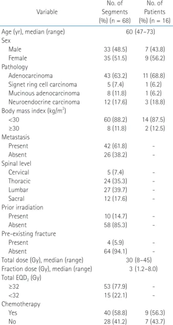
Table 1. Patient characteristics Variable No. of Segments (%) (n = 68) No. of Patients (%) (n = 16) Age (yr), median (range)
Sex Male Female Pathology
Adenocarcinoma Signet ring cell carcinoma Mucinous adenocarcinoma Neuroendocrine carcinoma Body mass index (kg/m2)
<30 ≥30 Metastasis Present Absent Spinal level Cervical Thoracic Lumbar Sacral Prior irradiation Present Absent Pre-existing fracture Present Absent
Total dose (Gy), median (range) Fraction dose (Gy), median (range) Total EQD2 (Gy)
≥32 <32 Chemotherapy Yes No 60 33 (48.5) 35 (51.5) 43 (63.2) 5 (7.4) 8 (11.8) 12 (17.6) 60 (88.2) 8 (11.8) 42 (61.8) 26 (38.2) 5 (7.4) 24 (35.3) 27 (39.7) 12 (17.6) 10 (14.7) 58 (85.3) 4 (5.9) 64 (94.1) 30 3 53 (77.9) 15 (22.1) 40 (58.8) 28 (41.2) (47–73) 7 (43.8) 9 (56.2) 11 (68.8) 1 (6.2) 1 (6.2) 3 (18.8) 14 (87.5) 2 (12.5) -(8–45) (1.2–8.0) -9 (56.3) 7 (43.7) EQD2, equivalent dose in 2 Gy per fraction.
collapse (p = 0.003), SINS classification (p = 0.039) were identified as the significant predictive factors for VCF development in this study (Table 5). However, there was a trend towards more frequent fractures at the junctional spinal level than at other levels. Furthermore, spines with bilateral posterior element involvement showed more fractures than those with no involvement or unilateral posterior element involvement. Finally, SINS class showed statistical significance for an increased development of fractures in metastatic spines
by univariate analysis (p = 0.002), but failed to show statistical significance in multivariate analysis (Table 3, Fig. 1D).
Pain symptoms were reported in almost all of the patients that received palliative RT (14/16). After irradiation of these 14 patients, pain reduction was reported in 12 patients (85.7%); consisted of 7 partial reductions (50.0%) and 5 complete reductions (35.7%). After development of VCF, fracture-induced pain was observed in five vertebral segments with a total of six VCFs and consisted of two cases of pain aggravation and three Table 2. Risk factors for vertebral compression fracture in all spines (n = 68)
Variable No fracture
(n = 62 vertebral segments)
Fracture
(n = 6 vertebral segments) Fractures (%) p-value Age (yr) <60 ≥60 Sex Male Female Pathology Adenocarcinoma Signet ring cell carcinoma Mucinous adenocarcinoma Neuroendocrine carcinoma Body mass index (kg/m2)
<30 ≥30 Metastasis Present Absent Spinal Level Cervical Thoracic Lumbar Sacral Prior irradiation Present Absent Pre-existing fracture Present Absent Total EQD2 (Gy)
≥32 <32
Fraction dose (Gy) <3 ≥3 Chemotherapy Yes No 28 34 31 31 38 5 7 12 54 8 37 25 5 22 25 10 7 55 1 61 50 12 6 56 37 25 5 1 2 4 5 0 1 0 6 0 5 1 0 2 2 2 3 3 3 3 3 3 2 4 3 3 15.2 2.9 6.1 11.4 11.6 0 12.5 0 10.0 0 11.9 3.8 0 8.3 7.4 16.7 30.0 5.2 75.0 4.7 5.7 20.0 25.0 6.7 7.5 10.7 0.101 0.674 0.485 0.601 0.395 0.780 0.037 0.002 0.116 0.143 0.684
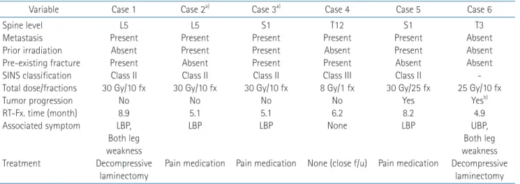
cases of development of new pain. In addition, two cases of spinal cord compression with weakness in both legs occurred and were successfully treated with decompressive surgery (Fig. 2, Table 6). There were 2 cases of tumor progression until the development of VCF among all spinal segments with fracture event, consisted of one metastatic spine and one non-metastatic spine. Vertebral segments with VCF events are summarized in Table 6.
Discussion and Conclusion
The present study identified the incidence and risk factors for VCF after conventional RT in homogenously characterized colorectal cancer patients, and clinical significances as described below. Because conventional RT still has a dominant role in palliative treatment for bone metastases, the results
of our study will provide useful information for physicians. Moreover, although the use of bisphosphonate can delay or decrease development of VCF [2], our cohort contained only CRC patients who rarely use bisphosphonate. Furthermore, it was confirmed that bisphosphonate was not used in all patients. So we could identify the effect of RT on VCF without this confounding factor. The incidence of VCF in our study was 8.8%, suggesting that VCF after conventional RT is not particularly common. Additionally, we identified two risk factors for VCF, prior irradiation and pre-existing compression fracture. Therefore, timely management and close follow-up are required when planning to treat vertebral segments with these risk factors.
Previous studies that focused on VCF after SBRT analyzed several risk factors including SINS criteria and revealed that lytic spinal lesion, spinal misalignment, pre-existing VCF, Fig. 1. Fracture-free probability (FFP) according to various risk factors. (A) Total irradiated spine. (B) With or without prior irradiation
to total spine. (C) With or without pre-existing fracture in total spine. (D) FFP according to the Spinal Instability Neoplastic Score (SINS) class. A No prior irradiation (n = 58) Prior irradiation (n = 10) 0 4 8 12 100 80 60 40 20 16 FFP Time (months) 0 1-yr FFP 87.8% 0 4 8 12 100 80 60 40 20 16 FFP Time (months) 0 p = 0.015 B C 0 4 8 12 100 80 60 40 20 16 FFP Time (months) 0 0 4 8 12 100 80 60 40 20 16 FFP Time (months) 0 p = 0.002 D p < 0.001 No pre-existing fx (n = 64) Pre-existing fx(n = 4) SINS class I (n = 11) SINS class II (n = 29) SINS class III (n = 1)
and vertebral body involvement increased the risk of VCF [9,10,12,13]. In this study, we identified prior irradiation and pre-existing compression fracture as risk factors for VCF, thus pre-existing compression fracture was a consistent risk factor among studies. However, prior irradiation was not identified as a risk factor in the studies of MDACC or the University of Toronto, although both studies evaluated the clinical significance of prior irradiation in VCF development. As the uncontrolled bone metastasis after RT can be the risk factor for VCF, the presence of prior irradiation history have the meaning of both severe radiation induced change and tumor progression even if prior RT. However, exact underlying mechanism of VCF is not well defined, and further research into this association is obviously necessary.
Recently, the SOSG introduced the SINS criteria to predict instability of the metastatic spine. Evaluation of their reliability and validity indicated that the sensitivity and specificity of these criteria in an unstable and potentially unstable group were 95.7% and 79.5%, respectively [14]. Although several groups have studied the relationship between SINS criteria and development of VCF after SBRT, whether SINS criteria can help predict VCF after conventional RT has not been validated. At our best knowledge, there is the only 1 published study that revealed the relationship between SINS criteria conventional RT [15]. They showed that a higher spinal instability score
increased the risk of RT failure and retreatment, independent of performance status, primary tumor, and symptoms. Our study revealed that vertebral body collapse score could predict VCF after conventional RT, consistent with data from MSKCC. In addition, overall SINS class was significantly related with VCF (p = 0.002) in univariate analysis; however, the small number of vertebral body collapsed or class III spinal segments limited the statistical analysis. Further researches with large cases should be performed to confirm our results about the relation between conventional RT and SINS criteria.
In contrast to SBRT, by defining target volume as the involved vertebral segment only, conventional fraction RT traditionally includes 1 to 2 levels of adjacent non-metastatic spines as well as all metastatic spines. Thus, it is important to know the risk and predictive factors for VCF in non-metastatic spines, and to date these factors have not been clearly defined. In our study, development of VCF after irradiation of non-metastatic spine was observed only in one case (4%) among a total of 26 vertebral segments (case 6 of Table 6). Furthermore, not only the VCF development, but also tumor progression was observed in this case, suggesting that tumor progression is more reasonable cause for the development of VCF than radiation effect. Thus, there was no VCF on non-metastatic vertebral segment without tumor involvement in our study cohort, suggesting the inclusion of adjacent metastasis-free Table 3. Significant risk factors for vertebral compression fracture in univariate and multivariate analysis
Variable Univariate Multivariate
1-yr FFP p-value HR 95% CI p-value
All spines Prior irradiation Present Absent Pre-existing fracture Present Absent 92.5 66.7 93.7 25.0 0.015 <0.001 7.304 18.445 1.306–40.856 3.419–99.520 0.024 0.001 Metastatic spines Prior irradiation Present Absent Pre-existing fracture Present Absent SINS classification Class I Class II Class III 90.9 64.8 92.9 25.0 100 81.6 0.0 0.031 <0.001 0.002 33.337 23.129 NA 1.767–630.476 1.560–342.807 NA 0.019 0.022 0.340
vertebra seems to be safe. On the other hand, after irradiation of metastatic spine, VCF was observed in five cases (12%) among a total of 42 vertebral segments, suggesting that irradiation of the metastatic spine is somewhat riskier. One possible cause of VCF is weakening of the bone structure induced by radiation. Al-Omair et al. [16] investigated the pathophysiology of VCF development after SBRT by spine tissue biopsy and revealed that radiation-induced necrosis and fibrosis contributed to VCF development after SBRT with a high
fractional dose. However, the fractional dose in conventional RT is much lower than that of SBRT, suggesting the possibility of another mechanism related to VCF after conventional RT, other than tumor regression. However the exact mechanism is currently unclear and our study did not identify any risk factor for VCF in non-metastatic spines.
We reviewed all cases of VCF in this study and found six fractured vertebral segments. Among these, pain aggravation was observed in five cases, and spinal cord compression Table 4. Risk factors for vertebral compression fracture in metastatic spine (n = 42)
Variable No fracture
(n = 37 vertebral segments)
Fracture
(n = 5 vertebral segments) Fractures (%) p-value Age (yr) <60 ≥60 Sex Male Female Pathology Adenocarcinoma Signet ring cell carcinoma Mucinous adenocarcinoma Neuroendocrine carcinoma Body mass index (kg/m2)
<30 ≥30 Para-spinal extension Present Absent Spinal Level Cervical Thoracic Lumbar Sacral Prior irradiation Present Absent Pre-existing fracture Present Absent Total EQD2 (Gy)
≥32 <32 Fraction dose <3 ≥3 Chemotherapy Yes No 20 17 23 14 15 5 7 10 31 6 4 33 2 9 17 9 6 31 1 36 30 7 2 35 19 18 5 0 2 3 4 0 1 0 5 0 2 3 0 1 2 2 3 2 3 2 3 2 1 4 2 3 20.0 0 8.0 17.6 21.1 0 12.5 0 13.9 0 33.3 8.3 0 10.0 10.5 18.2 33.3 6.1 75.0 5.3 9.1 22.2 33.3 10.3 9.5 14.3 0.070 0.632 0.327 0.585 0.875 0.057 0.003 0.141 0.567 0.323 1
presenting with weakness in both legs was observed in two cases (case 1, 6), especially for case 6, there was newly developed tumor involvement in non-metastatic spine. Decompressive laminectomy and tumor removal were successfully performed. A prophylactic stabilizing procedure (e.g., vertebroplasty) should be considered when the targeted vertebral segment has risk factors for VCF, because VCF can cause pain and neurologic symptoms. In addition, regular interval imaging and clinical follow-up is needed to inspect for the development of VCF.
Special consideration is needed when interpreting the results of this study because of the inherent limitations of a retrospective study. In spite of our best efforts to perform regular follow-up imaging studies with uniform modalities there were some inconsistencies in imaging modality and
follow-up interval among patients. Therefore, the incidence of VCF might be underestimated because of incomplete assessment. As a result of the exclusion criteria, only 68 vertebral segments of 16 patients were included in our study, limiting our statistical power to analyze prognostic factors. However, most patients in our study cohort might be relatively long-term survivors, suggesting that our results address a clinically relevant problem after palliative RT. As our study did not focus on event per patient but rather event per vertebral segment, the reliability of data on the patients’ characteristics could be diminished in cases of multiple fractures in one patient. However, we found six fractures in five patients in this study, indicating only a single case of multiple fractures, thus the reliability of this study is not significantly compromised. Conversely, our study revealed that the presence of a risk Table 5. SINS criteria in metastatic spine (n = 42)
Variable No fracture (n = 37 vertebral segments) Fracture (n = 5 vertebral segments) Fractures (%) p-value Location Rigid (S2–S5) Semi-rigid (T3–T10) Mobile spine (C3–C6, L2–L4) Junctional (occiput–C2, C7–T2, T11–L1, L5–S1) Pain Pain free
Occasional and non-mechanical Mechanical
Bone lesion type Blastic Mixed Lytic Alignment Normal Kyphosis/scoliosis Subluxation/translation Vertebral body collapse ≥50%
<50%
No collapse, but tumor involvement in >50% of body None of the above
Posterior element Not involved Unilateral Bilateral SINS classification Class I Class II Class III 6 4 13 14 2 5 30 0 16 21 34 3 0 1 0 9 27 25 4 8 11 25 0 0 0 0 5 0 1 4 0 1 4 5 0 0 0 2 2 1 1 1 3 0 4 1 0 0 0 26.3 0 16.7 11.8 0 5.9 16.0 12.8 0 0 0 100 18.2 3.6 3.8 20.0 27.3 0 13.8 100 0.081 1 0.392 1 0.003 0.095 0.039
factor in a certain vertebral segment does not increase the fracture risk at other spinal levels.
Overall, the incidence of VCF after conventional RT is not very high. However, close observation is warranted in patients with prior irradiation to the spine and/or pre-existing compression fracture in the spine. In addition, SINS
criteria can be used as an option for predicting fracture risk before performing conventional RT. Further studies are required to confirm these findings and examine the validity of extrapolating our results to bone metastasis in the setting of other tumors.
Fig. 2. (A) A newly developed
ver-tebral compression fracture (VCF) in the T3 vertebral segment at 4.9 months after irradiation with 25 Gy in 10 fractions. There were no meta static lesions, prior irradi-ation, or pre-existing fractures before radiation therapy. Upper back pain and weakness in both legs were observed after VCF and were successfully treated by de-com pressive laminectomy (case 6 in Table 6). (B) VCF progression was observed in the L5 vertebral segment at 8.9 months after irradiation with 30 Gy in 10 fractions. This ver tebral segment had a metastatic lesion and pre-existing fracture. Lower back pain and weakness in both legs were observed after VCF and were successfully treated by de compressive laminectomy (case 1 in Table 6).
A
B
Table 6. Summary of vertebral segments with vertebral compression fracture events
Variable Case 1 Case 2a) Case 3a) Case 4 Case 5 Case 6
Spine level Metastasis Prior irradiation Pre-existing fracture SINS classification Total dose/fractions Tumor progression RT-Fx. time (month) Associated symptom Treatment L5 Present Absent Present Class II 30 Gy/10 fx No 8.9 LBP, Both leg weakness Decompressive laminectomy L5 Present Present Absent Class II 30 Gy/10 fx No 5.1 LBP Pain medication S1 Present Present Present Class II 30 Gy/10 fx No 5.1 LBP Pain medication T12 Present Absent Present Class III 8 Gy/1 fx No 6.2 None
None (close f/u)
S1 Present Present Absent Class II 30 Gy/25 fx Yes 8.2 LBP Pain medication T3 Absent Absent Absent -25 Gy/10 fx Yesb) 4.9 UBP, Both leg weakness Decompressive laminectomy T, thoracic spine; L, lumbar spine; S, sacrum; SINS, Spine Instability Neoplastic Score; fx, fractions; RT-Fx. time, monthly time interval be-tween start date of radiation therapy and fracture event; LBP, lower back pain; UBP, upper back pain; f/u, follow-up.
Conflicts of Interest
No potential conflict of interest relevant to this article was reported.
References
1. Jemal A, Bray F, Center MM, Ferlay J, Ward E, Forman D. Global cancer statistics. CA Cancer J Clin 2011;61:69-90.
2. Santini D, Tampellini M, Vincenzi B, et al. Natural history of bone metastasis in colorectal cancer: final results of a large Italian bone metastases study. Ann Oncol 2012;23:2072-7. 3. Chow E, Zeng L, Salvo N, Dennis K, Tsao M, Lutz S. Update on
the systematic review of palliative radiotherapy trials for bone metastases. Clin Oncol (R Coll Radiol) 2012;24:112-24. 4. Chow E, Harris K, Fan G, Tsao M, Sze WM. Palliative
radiotherapy trials for bone metastases: a systematic review. J Clin Oncol 2007;25:1423-36.
5. Wu JS, Wong R, Johnston M, Bezjak A, Whelan T; Cancer Care Ontario Practice Guidelines Initiative Supportive Care Group. Meta-analysis of dose-fractionation radiotherapy trials for the palliation of painful bone metastases. Int J Radiat Oncol Biol Phys 2003;55:594-605.
6. McQuay HJ, Collins SL, Carroll D, Moore RA. Radiotherapy for the palliation of painful bone metastases. Cochrane Database Syst Rev 2000;(2):CD001793.
7. Sheehan JP, Shaffrey CI, Schlesinger D, Williams BJ, Arlet V, Larner J. Radiosurgery in the treatment of spinal metastases: tumor control, survival, and quality of life after helical tomo-therapy. Neurosurgery 2009;65:1052-62.
8. Kim MS, Keum KC, Cha JH, et al. Stereotactic body radio-therapy with helical tomoradio-therapy for pain palliation in spine
metastasis. Technol Cancer Res Treat 2013;12:363-70. 9. Rose PS, Laufer I, Boland PJ, et al. Risk of fracture after single
fraction image-guided intensity-modulated radiation therapy to spinal metastases. J Clin Oncol 2009;27:5075-9.
10. Boehling NS, Grosshans DR, Allen PK, et al. Vertebral compression fracture risk after stereotactic body radiotherapy for spinal metastases. J Neurosurg Spine 2012;16:379-86. 11. Fisher CG, DiPaola CP, Ryken TC, et al. A novel classification
system for spinal instability in neoplastic disease: an evidence-based approach and expert consensus from the Spine Oncology Study Group. Spine (Phila Pa 1976) 2010;35:E1221-9.
12. Cunha MV, Al-Omair A, Atenafu EG, et al. Vertebral com-pression fracture (VCF) after spine stereotactic body radiation therapy (SBRT): analysis of predictive factors. Int J Radiat Oncol Biol Phys 2012;84:e343-9.
13. Sahgal A, Atenafu EG, Chao S, et al. Vertebral compression fracture after spine stereotactic body radiotherapy: a multi-institutional analysis with a focus on radiation dose and the spinal instability neoplastic score. J Clin Oncol 2013;31:3426-31.
14. Fourney DR, Frangou EM, Ryken TC, et al. Spinal instability neoplastic score: an analysis of reliability and validity from the spine oncology study group. J Clin Oncol 2011;29:3072-7. 15. Huisman M, van der Velden JM, van Vulpen M, et al. Spinal
instability as defined by the spinal instability neoplastic score is associated with radiotherapy failure in metastatic spinal disease. Spine J 2014 Apr 4 [Epub]. http://dx.doi.org/10.1016/ j.spinee.2014.03.043.
16. Al-Omair A, Smith R, Kiehl TR, et al. Radiation-induced vertebral compression fracture following spine stereotactic radiosurgery: clinicopathological correlation. J Neurosurg Spine 2013;18:430-5.