Notes Bull. Korean Chem. Soc. 2012, Vol. 33, No. 2 685 http://dx.doi.org/10.5012/bkcs.2012.33.2.685
NMR Study of Hydrogen Exchange in the DNA Decamer Duplexes Containing
the p53 Response Element
Ae-Ree Lee,a Sung Jae Cho,†,a Hee-Eun Kim, Yeon-Mi Lee, Joon-Hwa Lee,* and Byong-Seok Choi†,* Department of Chemistry and Research Institute of Natural Science, Gyeongsang National University,
Jinju, Gyeongnam 660-701, Korea. *E-mail: joonhwa@gnu.ac.kr
†Department of Chemistry, KAIST, Daejeon 305-701, Korea. *E-mail: byongseok.choi@kaist.ac.kr Received October 5, 2011, Accepted November 24, 2011
Key Words : NMR, Hydrogen exchange, p53 response element, Cancer
P53 is the major tumor suppressor and is a transcription factor involved in various cellular metabolisms, including DNA repair, regulation of cell cycle, apoptotic cell death.1-5 The p53 contains an N-terminal transactivation domain (amino acids 1-42), a proline-rich SH3 target-region (amino acids 60-97), a DNA-binding domain (amino acids 102-292), a tetramerization domain (amino acids 323-356) and a C-terminal regulatory domain (360-393).6
The sequence-specific DNA binding domain (DBD) of p53 plays a critical role for the initiation of biological functions of p53 system.1-5 Deficiency of the p53 function is mainly due to mutations which interfere with the DNA-binding ability of the protein. P53 can recognize specific DNA sequences, which is called as the p53 response element (p53RE).7 The p53RE have two half-site palindromes and each half-site palindrome has 10 base-pair (bp) with a consensus sequence of 5'-RRRCWWGYYY-3', where W can be A or T, and R and Y is purine and pyrimidine bases, respectively. The p53-DBD binds to various p53RE variants with somewhat different binding modes. The p53-DBD had different binding affinity to a variety of p53RE substrates. However, the structural features of the p53-DNA complexes could not explain this sequence selectivity of the p53-DBD. The dynamic property of the p53RE sequences with their structural feature can provide the useful information to
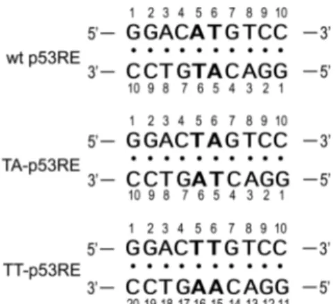
understand the molecular interaction between the p53 and its p53RE. NMR hydrogen exchange data provide information on the thermodynamics and kinetics for base-pair opening and represent a probe of the dynamic motion of the base pairs. Hydrogen exchange data can also be used to probe how DNA duplexes are stabilized by the intermolecular interaction with proteins. Thus, to understand the origin of the sequence selectivity of the p53, the imino proton ex-change rate constants were measured for the DNA decamer duplex containing the consensus p53RE sequence (referred as to wt p53RE, Fig. 1). To further understand the corre-lation between the base pair dynamics and the sequence selectivity of the p53, the exchange rate constants (kex) of the imino protons for the wt p53RE duplex were compared with those of the other p53RE variants (Fig. 1).
Experimental Section
All DNA oligonucleotides were purchased from M-bio-tech Co. (Seoul, Korea). The oligonucleotides were purified by reverse-phase HPLC and desalted by Sephadex G-25 column. DNA duplexes were prepared by dissolving in an NMR buffer (90% H2O/10% D2O solution containing 10 mM sodium phosphate (pH 8.0) and 100 mM NaCl). NMR experiments were carried out on a Varian Inova 600-MHz NMR spectrophotometer (KAIST, Daejeon) equipped with x,y,z-axis pulsed-field gradient triple resonance probe. One-dimensional (1D) NMR data were processed and analyzed with the program FELIX (Accelrys) or VNMR J and 2D data were processed with the program NMRPipe8 and analyzed with the program Sparky.9 The kex of the imino protons were determined as previously described.10 The kex
values of the imino protons were measured by water magnetization transfer experiments.10 The kex for the imino
protons were determined by fitting the data to Eq. (1):10 (1) where R1a and R1w were the independently measured and are
the apparent longitudinal relaxation rates of the imino proton and water, respectively, and I0 and I(t) are the peak
intensities of the imino proton in the water magnetization I t( ) I0 --- =1−2 kex R1w–R1a ( ) --- e( –R1at–e–R1wt) aEqual contribution
Figure 1. DNA sequence contexts of the wt, TA-, and TT-p53RE DNA decamer duplexes.
686 Bull. Korean Chem. Soc. 2012, Vol. 33, No. 2 Notes
transfer experiments at times zero and t, respectively.10,11 In the case that some imino resonances are partially overlapped with each other, the kex data were determined by fitting the
relative peak heights, instead of the peak intensities, to Eq. (1). Results and Discussion
2D NOESY spectra of the wt, TA-, and TT-p53RE duplexes in 90% H2O/10% D2O buffer solution containing
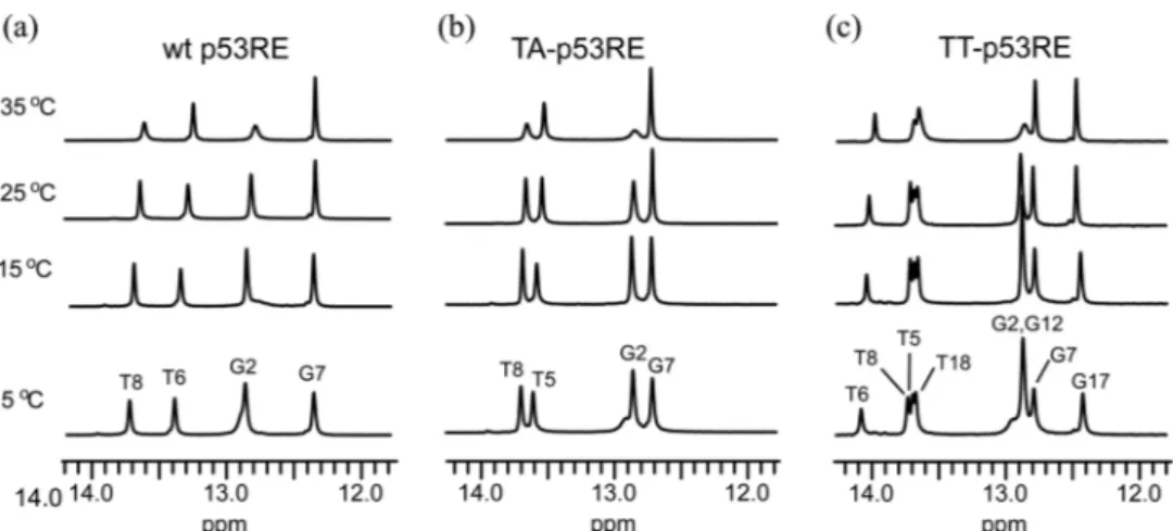
10 mM sodium phosphate (pH 8.0) and 100 mM NaCl were acquired at 5oC with 150 and 300 ms mixing times. The exchangeable imino protons were assigned by the strong G-imino to C-amino or T-G-imino to A-H2 NOE cross peaks in the NOESY spectra. In temperature-dependent imino proton spectra (Fig. 2), all imino proton resonances except those of the terminal G·C base pairs could be observed at 25oC. The G2 (and G12) imino proton resonances in the three duplexes were broadened at 35oC, indicating instabilities of the G2·C9 base pairs in the wt and TA-p53RE duplexes and the corresponding G2·C19 and G12·C9 base pairs in the
TT-p53RE duplexes (Fig. 2).
The kex of the imino protons for the wt p53RE duplex in
90% H2O/10% D2O solution containing 10 mM TRIS-d11
(pH 8.5) and 100 mM NaCl were determined by water magnetization transfer method at 25 oC. The relative peak heights of the water magnetization transfer for the imino proton resonances of the wt p53RE duplex at 25 oC are plotted as a function of delay time in Figure 4. Figure 4(e) shows the kex data of the imino protons of the wt p53RE
duplex determined by fitting to Eq. (1). The central G7 imino proton is the slowest exchanging proton (kex of 0.7 ±
0.1 s−1), indicating that the G7·C4 base pair is the most stable base pair in the wt p53RE duplex. The central T6 imino proton has the kex value of 4.6 ± 0.1 s−1, whereas the kex value
for the T8 imino protons is 25.7 ± 0.1 s−1, indicating that the central two A5·T6 base pairs are relatively more stable com-pared to other A·T base pairs. The G2 imino proton next to the terminal base pair has the largest exchange rate constant of any non-terminal base pairs (kex of 57.4 ± 0.2 s−1).
To further understand the dynamic property of the p53RE Figure 2. Temperature dependence of the imino proton resonances in the 1H-NMR spectra for the (a) wt, (b) TA-, and (c) TT-p53RE DNA duplexes in 90% H2O/10% D2O solution containing 10 mM sodium phosphate (pH 8.0) and 100 mM NaCl. Experimental temperatures are shown on the left of spectra.
Figure 3. 1D imino proton spectra of the water magnetization transfer experiments for the (a) wt, (b) TA-, and (c) TT-p53RE duplexes in 90% H2O/10% D2O solution containing 10 mM TRIS-d11 (pH 8.5) and 100 mM NaCl at 25 oC. The delay times between the selective water inversion and acquisition pulse are indicated on the left of spectra.
Notes Bull. Korean Chem. Soc. 2012, Vol. 33, No. 2 687
sequence, the kex measurements were also performed at 25 oC in the two p53RE variant duplexes. In the TA-p53RE
duplex, where A5T6 sequence at the central region is changed to T5A6 (see Fig. 1), the peak intensity of the T5 imino proton shows larger dependence on the delay time after selective water inversion compared to the T6 of the wt p53RE duplex (Fig. 4(b)). This leads to a 1.5-fold larger kex
value of the T5 imino proton than the T6 imino proton in the wt p53RE duplex (Fig. 4(e)), demonstrating that two T·A base pairs in the central TA sequence are less stable than the corresponding two A·T base pairs in the central AT sequence. This observation is consistent with the previous base-pair opening kinetics study on the DNA decamer duplexes, in which the apparent dissociation constants for the two A·T base-pairs in the central TA step is 1.5-fold larger than those of the corresponding base-pairs in the central TA step.12 However, this difference in the A·T base-pair stability bet-ween the AT and TA sequences does not affect the kex for the
G7 and T8 imino protons of the neighboring base-pairs (Fig. 4(c) and 4(d)). In addition, there is no significant difference in the kex values between two duplexes for the G2·C9 base
pair (Fig. 4(a)). These results indicate that the change from AT to TA sequence at the central region in the p53 response element leads to destabilization of these two base pairs but no destabilization of the neighboring base pairs.
Hydrogen exchange experiments were also performed at 25oC for the TT-p53RE duplex, where the central palind-rome AT sequence is changed to TT/AA sequence at the 5 and 6 positions (see Fig. 1). Like TA-p53RE duplex, the peak intensity of the T5 imino proton shows larger dependence on the delay time compared to the T6 of the wt p53RE duplex (Fig. 4(b)), leading to a 2-fold larger kex value
of the T5 imino proton than the T6 imino proton in the wt p53RE duplex (Fig. 4(e)). Surprisingly, contrast to the T5,
the peak intensity of the T6 imino proton showed a smaller dependence on the delay time (Fig. 4(b)), meaning that the kex value of the T6 imino proton is 4-fold smaller than that of
the T5 imino proton and 2-fold smaller than that of the corresponding T6 imino proton in the wt p53RE duplex (Fig. 4(e)). Similar to the TA-p53RE duplex, this change has no significant effect on the kex values of the neighboring
C4·G17 and G7·C14 base pairs (Fig. 4(a)). Interestingly, the T8 imino proton in the TT-p53RE duplex has 2-fold smaller kex value than that of the wt p53RE duplex, whereas the T18
imino proton shows no significant change in its kex value
(Fig. 4(d) and 4(e)). These results demonstrate that the base pair inversion at the 5 position also leads to affect the stabilities of this base pair as well as the neighboring T6·A15 base pair. The AT base step exhibits the roll of ~−5o to minimize structural hindrance between two adenine base.13 This unusual structural feature at the AT base step can explain why these base-pairs in the TT-p53RE duplex are more stable than the corresponding base-pairs in the wt-p53RE duplex.
In summary, we determined the kex values of the imino
protons in the wild-type p53 response element as well as the variant sequences using NMR spectroscopy. The change of the AT to TA or TT sequences of the p53 response element leads to unusual features in the stabilities of these two base pairs as well as and the neighboring base pairs. This unusual feature of the base pair stabilities in the p53 response ele-ment sequence might strongly interfere with the molecular interaction with p53 proteins. Thus, this hydrogen exchange study can explain why the conversed AT sequence at the central region of the p53 response element is very sensitive to substitution.
Acknowledgments. This Work was supported by the Figure 4. Relative peak height [I(t)/I0] in the water magnetization transfer spectra for the (a) G2/G12, (b) T5/T6, (c) G7/G17, and (d) T8/ T18 imino protons of the wt, TA-, and TT-p53RE duplexes as a function of delay time. Solid lines indicate the best fitting of these data using Eq. (1). (e) The exchange rate constants of the imino protons for the wt, TA-, and TT-p53RE duplexes at 25oC and the error bars represents fitting errors.
688 Bull. Korean Chem. Soc. 2012, Vol. 33, No. 2 Notes
National Research Foundation of Korea Grants (2010-0014199 and NRF-C1ABA001-2010-0020480) funded by the Korean Government (MEST).
References
1. Soussi, T.; Legros, Y.; Lubin, R.; Ory, K.; Schlichtholz, B. Int. J. Cancer 1994, 57, 1.
2. Vogelstein, B.; Lane, D.; Levine, A. J. Nature 2000, 408, 307. 3. Aylon, Y.; Oren, M. Cell 2007, 130, 597.
4. Das, S.; Raj, L.; Zhao, B.; Kimura, Y.; Bernstein, A.; Aaronson, S. A.; Lee, S. W. Cell 2007, 130, 624.
5. Tanaka, T.; Ohkubo, S.; Tatsuno, I.; Prives, C. Cell 2007, 130, 638.
6. Romer, L.; Klein, C.; Dehner, A.; Kessler, H.; Buchner, J. Angew.
Chem. Int. Ed. 2006, 45, 6440.
7. Wei, C. L.; Wu, Q.; Vega, V. B.; Chiu, K. P.; Ng, P.; Zhang, T.; Shahab, A.; Yong, H. C.; Fu, Y.; Weng, Z.; Liu, J.; Zhao, X. D.; Chew, J. L.; Lee, Y. L.; Kuznetsov, V. A.; Sung, W. K.; Miller, L. D.; Lim, B.; Liu, E. T.; Yu, Q.; Ng, H. H.; Ruan, Y. Cell 2006, 124, 207.
8. Delaglio, F.; Grzesiek, S.; Vuister, G. W.; Zhu, G.; Pfeifer, J.; Bax, A. J. Biomol. NMR 1995, 6, 277.
9. Goddard, T. D.; Kneller, D. G. 2003, SPARKY 3. University of California, San Francisco, CA.
10. Lee, J.-H.; Pardi, A. Nucleic Acids Res. 2007, 35, 2965.
11. Choi, K.-H.; Lee, E.-H.; Kim, H.-E.; Lee, Y.-M.; Lee, A.-R.; Park, J.-W.; Choi, M. Y.; Lee, J.-H. Bull. Korean Chem. Soc. 2011, 32, 1754.
12. Park, J.-Y.; Lee, J.-H.; Choi, B.-S. FEBS Lett. 1998, 426, 325. 13. Dickerson, R. E. Methos Enzymol. 1992, 211, 67.