Role of inducible heat shock protein
70 in radiation-induced cell death
Su-Jae Lee,
1Sun-Ah Choi,
1Kang-Hyun Lee,
2Hee-Yong Chung,
3Tae-Hwan Kim,
1Chul-Koo
Cho,
1and Yun-Sil Lee
11Laboratory of Radiation Effect, Korea Cancer Center Hospital, Seoul 139–706, Korea 2Department of Urology, Korea Cancer Center Hospital, Seoul 139–706, Korea
3Department of Microbiology, College of Medicine Hanyang University, Seoul 133–791, Korea
Abstract We previously demonstrated the protective effect of inducible heat shock protein 70 (Hsp70) against gamma radiation. Herein, we extend our studies on the possible role of Hsp70 to ionizing radiation-induced cell cycle regulation. The growth rate of induciblehsp70-transfected cells was 2–3 hours slower than that of control cells. Flow cytometric analysis of cells at G1 phase synchronized by serum starvation also showed the growth delay in the Hsp70-overex-pressing cells. In addition, reduced cyclin D1 and Cdc2 levels and increased dephosphorylated phosphoretinoblastoma (pRb) were observed in induciblehsp70-transfected cells, which were probably responsible for the reduction of cell growth. To find out if inducible Hsp70-mediated growth delay affected radiation-induced cell cycle regulation, flow cytometric and molecular analyses of cell cycle regulatory proteins and their kinase were performed. The radiation-induced G2/M arrest was found to be inhibited by Hsp70 overexpression and reduced p21Wafinduction and its kinase
activity by radiation in the Hsp70-transfected cells. In addition, radiation-induced cyclin A or B1 expressions together with their kinase activities were also inhibited by inducible Hsp70, which represented reduced mitotic cell death. Indeed, hsp70 transfectants showed less induction of induced apoptosis. When treated with nocodazole, radiation-induced mitotic arrest was inhibited by inducible Hsp70. These results strongly suggested that inducible Hsp70 modified growth delay (increased G1 phase) and reduced G2/M phase arrest, subsequently resulting in inhibition of radiation-induced cell death.
INTRODUCTION
Heat shock proteins (Hsps) act as molecular chaperones, contributing to the folding of nascent polypeptides and also to protein transport and degradation. They protect cells and organisms from oxidative damage both in vivo and in vitro (Jacquier-Sarlin et al 1994; Plumier et al 1995) and often prevent cell death (Seo et al 1996). The cyto-protective effect of Hsps has been attributed to one of the major Hsps, Hsp70 (Jaattela and Wissing 1993), a member of the 70-kDa Hsp family, which is proposed to serve as a molecular chaperone (Gething and Sambrook 1992). In-deed, overexpression of Hsp70 protects cells, tissues, and organs from harmful assaults. However, under some con-ditions, the protective action of Hsp70 appears to be un-related to its chaperoning activity. For example, tumor Received 26 October 2000; Revised 13 March 2001; Accepted 14 March 2001.
Correspondence to: Yun-Sil Lee, Tel: 82-2-970-1325; Fax: 82-2-977-0381; E-mail: yslee@mail.kcch.re.kr.
necrosis factor causes cell death by the activation of a signal transduction pathway, leading to apoptosis (Ichijo et al 1997), and this apoptotic process can be prevented by overproduction of Hsp72 (Jaattela et al 1992), seem-ingly appearing to interfere with the apoptotic program such as Jun N-terminal kinase (Gabai et al 1997), extra-cellular signal-regulated protein kinase, and p38 mito-gen-activated protein kinase (Hung et al 1998).
On exposure to ionizing radiation, eukaryotic cells ac-tivate a variety of intracellular signaling pathways, there-by delaying cell cycle progression at ‘‘checkpoints’’ in the G1, S, or G2 phases and activating DNA repair. Cell cycle progression is controlled by specific Cdk-cyclin kinase ac-tivity that phosphorylates and activates proteins essential for execution of events that lead to cell cycle progression. Of the families of Cdk–cyclin kinase inhibitors, the p21Waf
family includes the structurally related proteins, the p21Waf, which are capable of inhibiting a variety of Cdk–
cyclin kinases. In addition, Cdc2, a highly conserved mi-tosis-promoting Cdk, is thought to mediate in G2
pri-274 Lee et al
marily by phosphorylation and appears to operate in a variety of experimental systems. Inhibitory phosphory-lation of Cdc2 is associated with the restriction of the kinase and its mitotic heterodimerization partner cyclin B1 (McDonald et al 1996). There is little direct informa-tion on the role of inducible Hsp70 in the cell cycle con-trol; however, it is present both in the nucleus and cyto-plasm in quiescent cells and begins to enter to the nucleus when the cells are stimulated to enter from G1 to S phase (Milarski and Morimoto 1986). Since our previous study suggested that inducible Hsp70 overexpression endowed cells with radioresistance (Park et al 2000), further study on the relationship between inducible Hsp70-mediated radioresistance and cell cycle regulation was carried out. Herein, we report that inducible Hsp70 decreased cell growth (increased G1 phase) and shortened radiation– induced G2/M phase arrest.
MATERIALS AND METHODS Cell cultures and treatment
The mouse cells of NIH3T3 were propagated in Dulbec-co’s modified Eagle’s medium (DMEM) supplemented with 10% fetal calf serum (FCS), 1% antibiotic/antimy-cotic solution, and 2 mM L-glutamine (all from GIBCO
BRL, Gaithersburg, MD, USA). The cell density was kept subconfluent, and the cells were passaged twice a week.
Vector construction
The detailed procedure to construct MFG.B7.puro vector, which was used for constructing MFG.Hsp70puro, has been published elsewhere (Kwak et al 1998; Park et al 2000). To establish cell line, 1 mg of MFG.Hsp70puro or the MFGpuro plasmids was introduced into cells by li-pofection (Lipofectamine, GIBCO BRL) in serum-free me-dia. Cells were propagated in DMEM supplemented with 10% FCS, 1% antibiotic/antimycotic solution, 2 mM L
-glutamine, and 2mg/mL of puromycin (all from GIBCO BRL). Twenty-four hours after transfection, media were changed and the cells were maintained in medium con-taining 10% serum and 2mg/mL of puromycin. Control cells were transfected with MFGpuro alone. hsp70-trans-fected cells were frequently tested for the expression of Hsp70 by Western blot analysis and found to express high levels of inducible Hsp70 protein.
Irradiation
Cells were exposed to gamma rays with137Cs gamma ray
source (Atomic Energy of Canada Ltd, Canada) with a dose rate of 3.81 Gy/min.
Cell counting
Cellular proliferation was monitored by counting cells us-ing vital dye trypan blue (Sigma, St-Quentin, France), and the viable cells were counted using a photomicro-scope.
[3H]-thymidime incorporation assay
Cells were plated at a confluence of 70–80% in 10-cm Petri dishes. After irradiation, the cells were trypsinized, plat-ed in 96-well plates (53 103cells/well), and incubated
for various periods. After adding 1mCi of [3H]-thymidine
to the incubation medium, the cells were further incu-bated for the last 4 hours before harvest, and the radio-activity of the cells was determined with a scintillation counter (PACKARD, TRI-CARB 4530, Meriden, CT, USA).
G1-phase synchronization
Cells were plated 1 day before a 48-hour serum starva-tion. The medium was then changed to serum-containing medium, and cells were thereafter harvested at various time points to study cell cycle distribution.
Cell cycle analysis
For cell cycle analysis, cells were fixed in 80% ethanol at 48C for at least 18 hours. The fixed cells were then washed once with phosphate-buffered saline (PBS)–ethylenedi-amine-tetraacetic acid (EDTA) (1 mM EDTA in PBS) and resuspended in 1 mL of PBS. After the addition of 10mL of propidium iodide solution (5 mg/mL) and 10 mL of ribonuclease (10mg/mL), the samples were incubated for 30 minutes at 378C and analyzed with a FACScan flow cytometer (Becton Dickinson, Franklin Lakes, NJ, USA).
Detection of apoptosis
For detection of apoptotic cells, cells were plated on glass slides and irradiated with 4 Gy. After 12 or 24 hours, the cells were fixed for 20 minutes in 3.7% formaldehyde, washed with PBS, and incubated with Hoechst 33258 so-lution in PBS (4mg/mL) for 30 minutes at room temper-ature in the dark. Specimens were analyzed with a fluo-rescence microscope (Olympus, Japan) mounted on a Ni-kon Diaphot-TDM, and the percentage of apoptotic cells was determined.
Detection of mitotic index
For detection of mitotic index, cells were plated on glass slides and treated with nocodazole (400 ng/mL) com-bined with 4 Gy of radiation. After 6, 12, 24, or 48 hours,
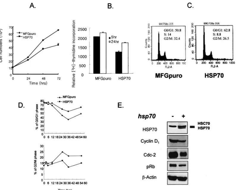
Fig 1. Comparison of cell growth. (A) Vector control and induciblehsp70-transfected cells were incubated for indicated times (0–72 hours). At each time point, viable cells were counted by trypan blue dye exclusion method. Error bar indicates mean6SD from 3 independent experiments. (B) Vector control and induciblehsp70-transfected cells were incubated for indicated times (5 and 24 hours). At each time point, incorporation of [3H]-thymidine was carried out for an additional 4 hours, and the radioactivity of the cells was determined with a scintillation
counter. Error bar indicates mean6SD from 3 independent experiments. (C) Cell cycle distributions of exponentially growing vector control and induciblehsp70-transfected cells were determined using flow cytometry after propidium iodide staining. (D) Cells were preincubated for 20 hours with 300mM mimosine and harvested after incubation for the period indicated. Cell cycle distribution following release from mimosine treatment was analyzed after staining with propidium iodide by a flow cytometer. (E) Protein extracts from growing vector control and inducible hsp70-transfected cells were prepared, separated by SDS-PAGE, and analyzed by Western blot.
the cells were fixed in 70% ethanol. The rest of the pro-cedures to obtain the percentage of mitotic cells are the same as described in the ‘‘Detection of apoptosis’’ section. Mitotic index was calculated by the percentage of mitotic arrested cells.
Polyacrylamide gel electrophoresis and Western blot
For polyacrylamide gel electrophoresis (PAGE) and West-ern blot, cells were solubilized with lysis buffer (120 mM sodium chloride, 40 mM Tris [pH 8.0], 0.1% NP40). Sam-ples were denatured in sample buffer containing 25 mM
Tris hydrochloride (pH 6.8), 2% sodium dodecyl sulfate (SDS), 10% glycerol, 10% 2-mercaptoethanol, and 0.002% bromphenol blue and boiled for 5 minutes. Then equal amounts of proteins (40mg/well) were analyzed on 10% SDS-PAGE. After electrophoresis, proteins were trans-ferred onto a nitrocellulose membrane and processed for immunoblotting. Blots were incubated with a 1:1000 di-lution of antibodies to cell cycle–related proteins; the mouse monoclonal anti-p53 antibody (Calbiochem, On-cogene Research Products, Cambridge, MA, USA); anti– cyclin B1 and anti-Cdc2 antibody (Santa Cruz Biotech-nology Inc, Santa Cruz, CA, USA); the rabbit polyclonal
276 Lee et al
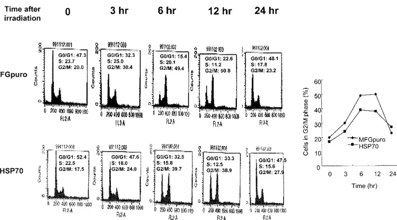
Fig 2. Cell cycle arrest. Cells were harvested following incubation after 4 Gy of radiation for the period indicated, and cell cycle distribution was analyzed after staining with propidium iodide by a flow cytometer.
anti-Cdk2, -Cdk4, –cyclin D1, and -p21Waf antibodies
(Santa Cruz Biotechnology); and –cyclin E antibody (Up-state Biotechnology Inc, Lake Placid, NY, USA). Blots were further incubated with horseradish peroxidase–con-jugated secondary antibody diluted at 1:5000, and specific bands were visualized by chemiluminescence (ECL, Amersham International). Autoradiographs were record-ed onto X-Omat AR films (Eastman Kodak Co).
Immunoprecipitation
Cell lysates were incubated with anti-CDK2, anti–cyclin B1 polyclonal antibody (Santa Cruz Biotechnology), or normal rabbit serum. The immunocomplex was collected on protein A–Sepharose (Sigma) and analyzed by SDS-PAGE using the ECL detection kit.
Immunoprecipitation and immune complex kinase assay
Cell lysates were incubated with a primary antibody, and immunocomplexes were collected on protein A–Sephar-ose beads and resuspended in kinase assay mixture con-taining [g-32P] adenosine triphosphate (ICN) and
GST-pRB (Santa Cruz Biotechnology) or histone H1 (Life Tech-nologies Inc) as substrates. The proteins were separated on SDS-polyacrylamide gels, and bands were detected by autoradiography.
Data analysis and statistics
The points shown in the figures are means of 3 indepen-dent determinations, and Stuindepen-dent’s t-test was used for sta-tistical analysis.
RESULTS Cell growth
When growth curves of control vector and inducible
hsp70-transfected cells were examined by the trypan blue
dye exclusion method, the growth rate of the transfected cells was 2 to 3 hours slower than that of vector control cells (Fig 1A). Relative rate of [3H]-thymidine
incorpora-tion also revealed the growth arrest (Fig 1B), and flow cytometric analysis of DNA content also indicated ele-vated G1 (23.6%) and less S phase (37.1%) in the hsp70-transfected cells (Fig 1C). To examine whether the delay of cell growth by Hsp70 overexpression was due to an alteration of cell cycle, the control vector or hsp70-trans-fected cells were synchronized by serum starvation, then refed with fresh medium plus 10% fetal bovine serum, and cell cycle progression was monitored by measuring cellular DNA contents by flow cytometry. G1 arrest was induced with serum starvation both in the control and
hsp70-transfected cells; however, the cell cycle of the
transfected cells proceeded more slowly than the control cells in serum starvation (Fig 1D). To examine further
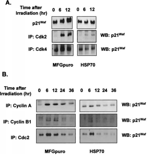
Fig 3. Expression of p21Waf. At the
in-dicated interval after 4 Gy of radiation, protein extracts (60mg) from growing vector control and inducible hsp70-transfected cells were prepared, sepa-rated by SDS-PAGE, and analyzed by Western blotting for p21Waf, or cell
ly-sates (500mg) were immunoprecipitat-ed (IP) with anti-Cdk2 or anti-Cdk4 an-tibodies (A) and anti–cyclin A, cyclin B, or Cdc2 antibodies (B), separated on SDS-polyacrylamide gels, transferred to nitrocellulose, and incubated with anti-p21WAF1 antibody followed by
en-hanced chemiluminescence detection.
whether the cell growth delay by inducible Hsp70 was due to altered expression of cell cycle–related proteins, Western blot analysis was performed. It was found that overexpression of inducible Hsp70 reduced the basal lev-els of Cdc2 and cyclin D1 proteins and increased de-phosphorylation of retinoblastoma (Rb). There was no significant change in the levels of Cdk2, Cdk4, cyclin A, cyclin B1, cyclin E, p21Waf, p27kip, proliferating cell nuclear
antigen, and p53 proteins (data not shown). Since cyclin D1–Cdk4 and cyclin A– or cyclin B1–Cdc2 complexes are known to be involved in the progression from G1 to S, S to G2, and G2 to M phase, respectively, and also pRb dephosphorylation is known to induce the growth arrest in G1 and S transition, it is highly likely that reduced expressions of Cdc2 and cyclin D1 and dephosphorylated pRb are responsible for the cell growth delay in the in-ducible hsp70-transfected cells.
G2/M block
Using flow cytometry to determine if altered cell growth by inducible hsp70 can affect radiation-induced cell cycle
arrest, G2 block in both cell lines was examined by mea-suring accumulation of cells in G2/M at approximately 3, 6, 12, and 24 hours after 4 Gy of radiation. The cell cycle arrest in the Hsp70-overexpressing cells was clearly decreased; G2/M arrest decreased by 20% and 24% at 6 and 12 hours after irradiation, respectively. These data clearly demonstrated that inducible Hsp70 released ra-diation-induced G2/M phase arrest (Fig 2).
p21Wafinduction
When cells were irradiated with 4 or 8 Gy, the increase of total level of p21Wafprotein in the inducible
hsp70-over-expressing cells was much less than in the control cells. We, therefore, examined possible association of p21Waf
with Cdk2 or Cdk4. Binding activity of p21Waf was
in-creasingly associated with CDK2 and CDK4 at 6 and 12 hours after radiation, and these associations were also found to be inhibited in the inducible hsp70-overexpress-ing cells (Fig 3A). In the S and G2 phases, the bindhsp70-overexpress-ing activity of p21Waf with cyclin A, cyclin B, or Cdc2
in-278 Lee et al
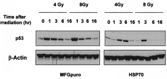
Fig 4. Expression of p53 protein. At the indicated interval after 4 or 8 Gy of radiation, protein extracts (60mg) from growing vector control and inducible hsp70-transfected cells were prepared, separated by SDS-PAGE, and ana-lyzed by Western blot for p53.
creased by radiation, whereas this activity decreased in the inducible hsp70 transfected cells, suggesting a less ra-diation-induced cell cycle arrest by inducible hsp70 than the control vector cells (Fig 3B).
p53 protein level
Since p53 protein is known to be an inducer of G1 or G2/ M phase arrest, p53 protein expression after irradiation was also examined. Induction of p53 protein both in cell lines increased from the first 1 hour of irradiation and lasted for 6 hours, however, with less and delayed in-crease in the Hsp70-overexpressing cells (Fig 4). These results suggested that p53 protein expression correlated well with the radiation-induced cell cycle regulation, with less arrest in the inducible hsp70-overexpressing cells. Cyclin A, cyclin B, and Cdc2 protein levels and their associated kinase activities
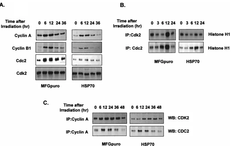
To elucidate the effects of perturbation of S or G2/M phase and premature entry to mitosis before completion of DNA synthesis (Tam and Schlegel 1997), cyclin A and B protein levels were examined. As shown in Figure 5A, irradiated cells showed elevated levels of cyclin A, cyclin B, and Cdc2 expressions all through the G2/M block and the elevated levels persisted for the duration of the block; however, reduced levels of these proteins were shown in the hsp70-transfected cells. When the protein levels of cy-clin A, cycy-clin B, and Cdc2 were elevated, cycy-clin A– or cyclin B–associated kinase activities were also elevated (Fig 5B). Binding activity of cyclin A and Cdk2 or Cdc2 also increased, whereas it was inhibited in the hsp70 transfected cells (Fig 5C).
Cell death
To study whether shortened G2/M phase arrest and al-tered response of cell cycle–related protein expressions by
hsp70 could affect radiation-induced cell death, Hoechst
33258 staining was performed. As shown in Figure 6A, 4 Gy of gamma rays induced an extensive nuclear conden-sation and fragmentation in the vector control cells, whereas overexpression of Hsp70 drastically diminished this phenomenon. The function of inducible Hsp70 in ra-diation-induced mitotic arrest was further studied by us-ing nocodazole, a blockus-ing agent in the M phase. As shown in Figure 6B, about 60% in the control vector cells and 40% in the transfected cells induced mitotic arrest by nocodazole treatment. In addition, when apoptotic cells were counted, nocodazole treatment reduced apoptosis and the reduction was greater in the control vector cells than the hsp70-transfected cell, suggesting the protection of the cells from mitotic cell death by Hsp70.
DISCUSSION
Although induction of Hsp70 protein synthesis may pro-tect cells from lethal effects of various stimuli, the role of
hsp70 genes in the control of cell growth is not well
de-fined. In the present study, we demonstrated that irradi-ation-induced cell death was inhibited by inducible Hsp70, and this inhibition resulted from abrogation of G2/M phase arrest mediated by irradiation.
Inducible hsp70-transfected NIH3T3 cells had delayed cell growth, shown by conventional trypan blue method and [3H]-thymidine incorporation. Flow cytometric
anal-ysis also revealed increased G1 phase, slower cell release after synchronization with serum starvation, reduction of cell cycle–related proteins expression such as cyclin D1 and cdc2, and an increase of dephosphorylated pRb pro-tein in the inducible hsp70-transfected cells (Fig 1). These results suggest that inducible Hsp70 modulates the ex-pression levels of these proteins, which eventually lead to growth delay.
Since radiation-induced DNA damage activates cell cy-cle checkpoints responsible for growth arrest or cell death (Bates and Vousden 1992) and inducible Hsp70 protects
Fig 5. Cyclin A, cyclin B, and Cdc2 protein levels and their associated kinase activities. At the indicated interval after 4 Gy of radiation, protein extracts (60mg) from growing vector control and induciblehsp70-transfected cells were prepared, separated by SDS-PAGE, and analyzed by Western blotting for cyclin A, cyclin B, Cdc2, and Cdk4 (A). Cell lysates (200mg) were immunoprecipitated (IP) with anti-Cdk2 or Cdc2 antibodies, and kinase activity was assayed using histone H1 as a substrate (B). Cell lysates (500mg) were immunoprecipitated (IP) with anti–cyclin A antibody, separated on SDS-polyacrylamide gels, transferred to nitrocellulose, and incubated with Cdk2 or anti-Cdc2 antibodies followed by enhanced chemiluminescence detection (C).
Fig 6. Cell death. At 24 or 48 hours after 4 Gy of radiation, DNA fragmen-tation of vector control and inducible hsp70-transfected cells was obtained by Hoechst 33258 staining (A). Imme-diately after gamma rays, both vector control and induciblehsp70-transfected cells were treated with nocodazole (400 ng/mL), and the mitotic index was determined at indicated times of post-irradiation as described in Materials and Methods (B). Error bar indicates the mean6SD from 3 independent ex-periments.
cells from radiation (Park et al 2000), we explored a ques-tion of whether cell growth delay mediated by inducible Hsp70 was correlated with radioresistance and found that excessive expression of inducible Hsp70 resulted in an alleviation of the radiation-induced G2/M block (Fig 2), which could be explained by the fact that fewer Hsp70-overexpressed cells were arrested in the G2/M phase and
they were released from the G2/M block sooner. To in-vestigate molecular mechanisms, we first checked the p21Waf expression that is known as a broad range CDK
inhibitor. Radiation-induced p21Wafexpression was
inhib-ited by inducible Hsp70 (Fig 3), and its binding efficiency with cyclin A, cyclin B, or cdc2 was also reduced by in-ducible Hsp70. Also, expressions of cyclin D and E were
280 Lee et al
well correlated with p21Waf induction (data not shown).
On the other hand, radiation-induced expressions of cy-clin A and B and their kinase activities, which were as-sociated with inductions of premature mitosis and apo-ptosis, were also inhibited by inducible Hsp70 (Fig 3). In fact, mitotic index was much less in the hsp70 transfected cells when treated with nocodazole. In addition, radia-tion-induced apoptosis was inhibited by nocodazole treatment, and this inhibition was greater in the control vector cells (Fig 6). These results suggest that inducible Hsp70 protects the cells from the radiation-induced cell death, especially mitotic cell death, resulting in reduced radiation-induced G2/M phase arrest. In addition, radi-ation-induced p53 protein induction was also inhibited in
hsp70 transfected cells when compared with vector
con-trol cells (Fig 4). Expression of p53 after radiation showed G2/M phase arrest and induction of apoptosis (Schwartz et al 1997), and Hsp70 attenuated these phenomena.
The fact that radiation blocks cell cycle is well known (Hartwell and Kastan 1994), and delays of cells at G1 and G2/M phases and a small but quantifiable radiation-in-duced S-phase delay have been demonstrated (Wang and Iliakis 1992). Usually, G2/M delay has been suggested to be due to radioresistance (Warenius et al 1996). However, as shown in the present study, reduction of radiation-in-duced G/M phase arrest occurred in the hsp70 transfect-ed cells, which are usually due to p21Wafinduction. There
are reports to suggest a correlation between radiosensi-tivity and G2/M phase delay (McKenna et al 1990; War-enius et al 1996) and a longer delay in cells of more ra-diosensitive cell line with the same radiation dose (Li et al 1998), which are associated with premature mitotic en-try before completion of DNA synthesis. However, there exists another possibility that increased levels of the stress proteins are responsible for development of resistance to stress agents (Li and Werb 1982; Subjeck et al 1982), be-cause protein kinase C activity increased in the hsp70-transfected cells (Park et al 2000). Therefore, it is likely that induced stress proteins can protect cells from stress-induced damage by preventing protein denaturation and/or by repairing such damages (Schroeder et al 1993). In conclusion, we attempted to define the role of in-ducible Hsp70 in radiation-induced cell cycle arrest. Ra-diation may alter signaling mechanisms, control the cell cycle regulation, especially G2/M phase regulators, and consequently promote kinase activity even in the pres-ence of DNA damage. Alternatively, inducible Hsp70 may alter the membrane signaling cascade, which relays the DNA damage signal to the control mechanisms that block the irradiated cells in G2/M, thus effectively preventing the cells to progress through G2 and mitosis. These data may suggest that the Hsp70 induction could enhance tu-morigenesis and limit the efficacy in cancer therapy, be-cause Hsp70 is highly expressed in many tumor cells
(Mivichi and Rossi 1990; Kaur and Ralhan 1995), trans-genic mice expressing the hsp70 gene develop T-cell lym-phomas (Seo et al 1996), and the expression of Hsp70 is an indicator of poor therapeutic outcome in breast cancer (Ciocca et al 1993).
ACKNOWLEDGMENTS
We thank K. J. Kim for his excellent technical assistance. This work was supported by the Nuclear Research and Development Program of the Korean Ministry of Science and Technology.
REFERENCES
Bates S, Vousden KH. 1992. p53 in signaling checkpoint arrest or apoptosis. Curr Opin Genet Dev 6: 12–18.
Ciocca DR, Clark GM, Tandon AK, Fuqua SA, Welch WJ, McGuire WL 1993. Heat shock protein hsp70 in patients with axillary lymph node-negative breast cancer: prognostic implications. J
Natl Cancer Inst 85: 570–574.
Gabai VL, Meriin AB, Mosser DD, Caron AW, Rits S, Shifrin VI, Sherman, MY. 1997. Hsp70 prevents activation of stress kinases.
J Biol Chem 272: 18033–18037.
Gething MJ, Sambrook J. 1992. Protein folding in the cell. Nature 355: 33–45.
Hartwell LH, Kastan MB. 1994. Cell cycle control and cancer. Science 266: 1821–1828.
Hung JJ, Cheng TJ, Lai YK, Chang MD. 1998. Differential activation of p38 mitogen activated protein kinase and extracellular sig-nal-regulated protein kinases confers cadmium-induced Hsp70 expression in 9L rat brain tumor cells. J Biol Chem 273: 31924– 31931.
Ichijo H, Nishida E, Irie K, et al. 1997. Induction of apoptosis by ASK1, a mammalian MAPKKK that activates SAPK/JNK and p38 signaling pathways. Science 275: 90–94.
Jaattela MD, Wissing D. 1993. Heat-shock proteins protect cells from monocyte cytotoxicity: possible mechanisms of self-protection.
J Exp Med 177: 231–236.
Jaattela M, Wissing D, Bauer PA, Li GC. 1992. Major heat shock protein hsp70 protects tumor cells from tumor necrosis factor cytotoxicity. EMBO J 11: 3507–3512.
Jacquier-Sarlin MR, Fuller K, Dinh-Xuan T, Richard MJ, Polla BS. 1994. Protective effect of hsp70 in inflammation. Experientia 50: 1031–1038.
Kaur J, Ralhan R. 1995. Differential expression of 70-kDa heat shock protein in human oral tumorigenesis. Int J Cancer 63: 774–779. Kwak HJ, Jun CK, Pae HO, et al. 1998. The role of inducible 70-kDa heat shock protein in cell cycle control differentiation and ap-optotic cell death of the human myeloid leukemic HL-60 cells.
Cell Immunol 187: 1–12.
Li GC, Werb Z. 1982. Correlation between synthesis of heat shock proteins and development of thermotolerance in Chinese ham-ster fibroblasts. Proc Natl Acad Sci U S A 79: 3218–3222. Li YX, Weber-Johnson K, Sun LQ, Paschoud H, Mirimanoff RO,
Coucke PA. 1998. Effect of phentoxifyllin on radiation-induced G2-phase delay and radiosensitivity of human colon and cer-vical cancer cells. Radiat Res 149: 338–342.
McDonald ER, Wu GS, Waldman T, El-Deiry WS. 1996. Repair De-fect in p21 WAF1/CIP12/2 human cancer cells. Cancer Res 56: 2250–2255.
McKenna WG, Weiss MC, Endlich B, Ling CC, Bakanaustas VJ, Kel-sten ML, Muschel RJ. 1990. Exit from G2 phase after 2 Gy gam-ma irradiation is faster in radiosensitive hugam-man cells with high expression of RAF1 proto-oncogene. Cancer Res 50: 97–102. Milarski KL, Morimoto RI. 1986. Expression of human Hsp70
dur-ing the synthetic phase of the cell cycle. Proc Natl Acad Sci U S
A 83: 9517–9521.
Mivichi NF, Rossi JJ. 1990. Use of polymerase chain reaction to detect the expression of the Mr 70,000 heat shock genes in control or heat shock leukemic cells as correlated to their heat response.
Cancer Res 50: 2877–2884.
Park SH, Lee SJ, Chung HY, Kim TH, Cho CK, Yoo SY, Lee YS. 2000. Inducible heat shock protein 70 involved in the radioadaptive response. Radiat Res 153: 318–326.
Plumier JC, Ross BM, Curie RW, Angelidis CE, Kazlaris H, Kollias G, Pagoulatos GN. 1995. Transgenic mice expressing the human heat shock protein 70 have improved post-ischemic myocardial recovery. J Clin Invest 95: 1854–1860.
Schroeder H, Langer T, Hartl FU, Bukau B. 1993. DnaK, DnaJ and
GrpE form a cellular chaperone machinery capable of repairing heat-induced protein damage. EMBO J 12: 4137–4144. Schwartz D, Almog N, Peled A, Goldfinger N, Rotter V. 1997. Role
of wild type p53 in the G2 phase: regulation of the gamma-irradiation-induced delay and DNA repair. Oncogene 15: 2597– 2607.
Seo JS, Park YM, Kim EH, Shim CW, Jang JJ, Kim SH, Lee WH. 1996. T cell lymphoma in transgenic mice expressing the human hsp70 gene. Biochem Biophys Res Commun 218: 582–587. Subjeck JR, Sciandra JJ, Johnson RJ. 1982. Heat shock proteins and
thermotolerance; a comparison of induction kinetics. Br J Radiol 55: 579–584.
Tam SW, Schlegel R 1997. Staurosporine overrides checkpoints for mitotic onset in BHK cells. Cell Growth Differ 3: 811–817. Wang Y, Iliakis G. 1992. Cell cycle control and cancer. Cancer Res. 52:
508–514.
Warenius HM, Jones MD, Thompson CCM. 1996. Exit from G2 phase after 2 Gy gamma irradiation is faster in radiosensitive human cells with high expression of the RAF1 proto-oncogene.