저작자표시 2.0 대한민국 이용자는 아래의 조건을 따르는 경우에 한하여 자유롭게 l 이 저작물을 복제, 배포, 전송, 전시, 공연 및 방송할 수 있습니다. l 이차적 저작물을 작성할 수 있습니다. l 이 저작물을 영리 목적으로 이용할 수 있습니다. 다음과 같은 조건을 따라야 합니다: l 귀하는, 이 저작물의 재이용이나 배포의 경우, 이 저작물에 적용된 이용허락조건 을 명확하게 나타내어야 합니다. l 저작권자로부터 별도의 허가를 받으면 이러한 조건들은 적용되지 않습니다. 저작권법에 따른 이용자의 권리는 위의 내용에 의하여 영향을 받지 않습니다. 이것은 이용허락규약(Legal Code)을 이해하기 쉽게 요약한 것입니다. Disclaimer 저작자표시. 귀하는 원저작자를 표시하여야 합니다.
Master’s Thesis
5-Fluorouracil Resistance Mechanism and Resistance
Reversal in SNU-C5/5-FU, Fluorouracil-Resistant
Human Colon Cancer Cells
Eun-Ji Kim
Department of Medicine
Graduate School
Jeju National University
i
CONTENTS
CONTENTS...i
LIST OF ABBREVIATIONS ...iv
LIST OF TABLES...v
LIST OF FIGURES...vi
1. ABSTRACT………...………..1
2. INTRODUCTION ……….…...3
3. MATERIALS AND METHODS……….…….……..8
3.1. Reagents………...……8
3.2. Cell culture………...9
3.3. Cell viability assay (Trypan blue staining)………..…9
3.4. Morphological analysis of apoptosis by Hoechst 33342 staining.………….…10
3.5. Western blot analysis ………..………..……10
3.6. Co-immunoprecipitation assay……….……….……12
ii
3.8. Statistical analysis………..13
4. RESULTS………..…….16
4.1. SNU-C5/5-FU stably acquired resistance on 5-fluorouracil………….…...…..16
4.2. SNU-C5/5-FU has the characteristic of overexpression of phospho-AKT.…...18
4.3. SNU-C5/5-FU down-regulates E-cadherin and up-regulates phospho-GSK-3β………..21
4.4. Phosphorylated AKT led to down-regulation of E-cadherin and phosphorylation of GSK-3β in SNU-C5/5-FU……….……….24
4.5. SNU-C5/5-FU has characteristic to translocation of β-catenin in cytoplasm and nucleus………..……….………....27
4.6. Phospho-AKT led to the nuclear translocation of β-catenin……….29
4.7. SNU-C5/5-FU has characteristic to overexpression of cyclooxygenase-2……....34
4.8. Over-activated NF-κB signaling led to overexpression of COX-2……...……....36
4.9. Phospho-AKT led to activation of NF-κB signaling in SNU-C5/5-FU………….39
4.10. Phospho-AKT led to stabilization of survivin via overexpression of COX-2...41
4.11. Phospho-AKT led to expression of survivin in cytoplasm via up-regulation of COX-2………45
iii
4.13. phospho-AKT contributes resistance on 5-fluorouracil in SNU-C5/5-FU..…....…52
4.14. Loss of PTEN was cause of the over-phosphorylated AKT in SNU-C5/5-FU…...55
5. DISCUSSION………..……….…. 58
iv
LIST OF ABBREVIATIONS
Abbreviations Full name
5-FU 5-fluorouracil
TS Thymidylate synthase
PTEN Phosphatase and tensin homolog deletion on chromosome ten
PI3K Phosphatidylinositol 3-kinase
GSK-3β Glycogen synthase kinase-3β
Wnt Wingless-type MMTV integration site family member
Cox-2 Cyclooxygenase-2
NF-κB Nuclear factor- kappa B
IκB Inhibitory kappa B
IKK IκB kinase
PGs Prostaglandins
IAP Inhibitor of apoptosis proteins
PARP Poly-(ADP-ribose) polymerase
v
LIST OF TABLES
vi
LIST OF FIGURES
Figure 1. Cytotoxicity of 5-fluorouracil on SNU-C5/WT and SNU-C5/5-FU……17 Figure 2. Comparison of SNU-C5/WT, SNU-C5/5-FU, and SNU-C5/OXT on AKT and
p-AKT………...……….………....19 Figure 3. Comparison of SNU-C5/WT, SNU-C5/5-FU, and SNU-C5/OXT on mTor and
p-mTor……….………..20 Figure 4. Comparison of SNU-C5/WT, SNU-C5/5-FU, and SNU-C5/OXT on
E-cadherin………...22 Figure 5. Comparison of SNU-C5/WT, SNU-C5/5-FU, and SNU-C5/OXT on GSK-3β and p-GSK-3β………...……….23 Figure 6. The Effect of LY294002 on the expression of E-cadherin in
SNU-C5/5-FU...25 Figure 7. The effect of LY294002 on the expression of GSK-3β, phospho-GSK-3β, cyclin D1, and β-catenin in SNU-C5/5-FU.…………...………..26 Figure 8. Comparison of SNU-C5/WT, SNU-C5/5-FU, and SNU-C5/OXT on
β-catenin……….28 Figure 9. The effect of LY294002 on E-cadherin, β-catenin, GSK-3β, and p-GSK-3β in SNU-C5/5-FU……….30 Figure 10. The effect of LY294002 on β-catenin interacts with E-cadherin in
SNU-C5/5-FU………..31 Figure 11. The characteristics of SNU-C5/5-FU on E-cadherin, GSK-3β, and
β-catenin……….32
vii
Figure 13. Comparison of SNU-C5/WT, SNU-C5/5-FU, and SNU-C5/OXT on IκB-α and p-NF-κB………37
Figure 14. The effect of TPCK on the expression of COX-2in SNU-C5/5-FU…...38
Figure 15. The effect of LY294002 on the expressions of COX-2and p-NF-κB in SNU-C5/5-FU………...………40 Figure 16. Comparisons of SNU-C5/WT, SNU-C5/5-FU, and SNU-C5/OXT on survivin……….42 Figure 17. The effect of LY294002 and Vioxx on the expression of survivin in
SNU-C5/5-FU………..43 Figure 18. The effect of LY294002 and Vioxx on the alteration of survivin expression in SNU-C5/5-FU………...46 Figure 19. The effect of LY294002 and Vioxx on the survivin interacts with caspase-3 in SNU-C5/5-FU……….……..48 Figure 20. The characteristics of SNU-C5/5-FU on NF-κB signaling and survivin….…..50 Figure 21. The effect of LY294002 or Vioxx on induction of apoptosis in
SNU-C5/5-FU………..53 Figure 22. Comparisons of SNU-C5/WT, SNU-C5/5-FU, and SNU-C5/OXT on PTEN………56 Figure 23. The 5-fluorouracil resistance mechanism and resistance reversal in
- 1 -
1. ABSTRACT
Recent studies reported that overexpression of phospho-AKT leads to resistance of
apoptosis via phospho-AKT modulation of a variety of cellular processes. In this study, we
investigated the resistance mechanism of SNU-C5/5-FU, fluorouracil-resistant human colon
cancer cells, and the possible involvement of phospho-AKT for acquired resistance to
5-fluorouracil (5-FU). First, we confirmed that SNU-C5/5-FU overexpress phospho-AKT
compared with SNU-C5/WT and SNU-C5/OXT. Interestingly, the loss of PTEN induced
phosphorylation of AKT in SNU-C5/5-FU. When treated with LY294002 (PI3 kinase
inhibitor), we could observe the expression of E-cadherin increased and the
phospho-GSK-3β decreased, which are known proteins downstream of phospho-AKT. Thereby, it decreased
the nuclear translocation of β-catenin. Furthermore, the NF-κB signaling was activated by
the increased AKT phosphorylation and promoted the formation of COX-2. In cytoplasm,
COX-2 contributes to the stabilization of survivin, an anti-apoptotic protein that directly
interacts with procaspase-3. Also, the treatment of LY294002 down regulated NF-κB
signaling and the COX-2. Treatment with LY294002 or Vioxx (COX-2inhibitor) reduced the
- 2 -
and 5-FU, the apoptotic characteristics, such as apoptotic bodies, cleavage of procaspase-9,
cleavage of procaspase-3 and cleavage of poly(ADP-ribose) polymerase (PARP), were
observed in SNU-C5/5-FU. Furthermore, apoptosis was also induced by combination
treatment with Vioxx and 5-FU. These findings provide evidence that overexpression of
phosphorylated AKT is an important mechanism of resistance in SNU-C5/5-FU. The results
suggest that inhibition of phosphorylated AKT may overcome fluorouracil-resistant in the
SNU-C5/5-FU cells.
Keywords: SNU-C5/5-FU, 5-Fluorouracil, resistant, PI3K/Akt, E-cadherin, GSK-3β,
- 3 -
2. INTRODUCTION
Colon cancer is one of the most prevalent cancers in the United States, and incidence rates
of colon cancer have been increasing steadily worldwide (Jemal et al. 2008). Moreover,
because of the change to Westernized dietary pattern, incidence rates of colon cancer also
have steadily increased in Korea (Park et al. 2012).
In recent years, because of increased colon cancer incidence, a variety of treatments have
been developed. However, 80% of cancer-related deaths were related to resistance to
anticancer-drugs (Luqmani 2005, Ichihashi, Kitajima 2001). Thus, we need to examine
closely the mechanisms of resistance.
5-Fluorouracil (5-FU) is an anti-cancer drug that prevents DNA synthesis by targeting
thymidylate synthase (TS). TS catalyzes the conversion of deoxyuridine monophosphate
(dUMP) to deoxythymidine monophosphate (dTMP) in DNA synthesis. The well-known
resistance mechanism of 5-FU is increased TS (Jette et al. 2008). But recent studies report
that SNU-C5/5-FU colorectal resistance cancer cells which have acquired resistance to 5-FU
did not increase TS (Jung 2006, Kim et al. 2005).
- 4 -
processes, such as cell growth and cell survival (Kim, Chung 2002). In addition, it has been
reported that this signal promotes the proliferation and survival of colon cancer cells (Bao et
al. 2004). Phosphorylation of AKT inhibits GSK-3β (Do et al. 2008) and induces
instability of E-cadherin expression by activating mdm2, an anti-apoptotic protein (Zhou et
al. 2009). Also, activation of AKT modulated NF-κB signaling promotes survival and
resistance apoptosis in cancer cells, via activation of IκB kinase (IKK) (Ahn, Aggarwal 2005,
Dan et al. 2008, Wang et al. 2009). The activation of AKT is strongly related to inactivation
of PTEN, tumor suppressor gene, in breast cancer cells (Clark et al. 2002). Therefore, the
activation of AKT contributes to maintaining immortality in cancer cells via regulating the
various apoptosis factors, which involved apoptosis, even they were exposed to stressful
conditions such as anticancer drug treatment (Clark et al. 2002, Seal et al. 2012).
Phosphatase and tensin homolog deletion on chromosome ten (PTEN) is a
well-recognized negative regulator of PI3K/AKT pathway (Gupta, Dey 2012, Kerr et al. 2006).
PTEN suppresses the PI3K/AKT cell survival pathway via dephosphorylation of
phosphatidylinositol 3,4,5-triphosphate (PIP3)(Tamura et al. 1999). Unfortunately, recent
studies reported that it showed the loss of PTEN expression in colon cancer (Colakoglu et al.
2008, Sawai et al. 2008). In addition, loss of PTEN expression led not only to increasing risk
- 5 -
and poor survival in colon cancer patients (Sawai et al. 2008). Interestingly, low expression
of PTEN was associated the resistance of treatment with anticancer drug (Oki et al. 2005,
Sherbakova et al. 2008).
E-cadherin is a transmembrane glycoprotein which has an important function involving
cell-cell adhesion. Generally, the cytoplasmic domain of E-cadherin is combined with
β-catenin, α-catenin and several ancillary proteins (Fanelli et al. 2008, Mohamet, Hawkins &
Ward 2011, Huber, Weis 2001). When E-cadherin was loosed, β-catenin was translocated to
the nucleus and activated the capacity of transcription factor (Huber, Weis 2001).
In the nucleus, β-catenin forms a transcription factor complex with Tcf/Lef proteins
(Sparks et al. 1998, Akiyama 2000). The transcription factor complex activates the
expression of target genes such as cyclin D1 and c-myc, which have important roles of cell
growth, proliferation and survival (Sparks et al. 1998, Akiyama 2000, Li et al. 2005, Gotoh
et al. 2003). Interestingly, recent studies reported that β-catenin was associated with
resistance to apoptosis by modulating the c-myc, cyclin D1 and other proteins, which are
involved with cell cycle and apoptosis, in various cancer cells (Yeung et al. 2010, Woodward
et al. 2007, Cui et al. 2012).
In wnt/β-catenin signaling, glycogen synthase kinase-3β (GSK-3β) led to ubiquitination
- 6 -
signal, β-catenin accumulated at a high-level in cytoplasm because of inactivated GSK-3β
and translocated to the nucleus (Akiyama 2000, Woodward et al. 2007, Cui et al. 2012).
Nuclear factor kappa B (NF-κB) is a protein transcription factor that is associated with
oncogenesis (Ahn, Aggarwal 2005, Mayo, Baldwin 2000), cell survival and proliferation
(Ahn, Aggarwal 2005, Yang et al. 2012). In addition, it has been reported that activated
NF-κB contributed to resistance for anti-cancer drugs in colon cancer cells (Wang et al. 2009).
Interestingly, NF-κB was activated by phosphorylated AKT that promoted activation of IκB
kinase (IKK). Thereby, inhibition of NF-κB (IκB), which is combined with a NF-κB, was
phosphorylated and then degraded by activated IKK. Sequentially, activated NF-κB was
translocated to the nucleus and acted as a transcription factor that induced gene expression
of anti-apoptotic proteins and enzymes including COX-2 (Ahn, Aggarwal 2005, Dan et al.
2008).
Cyclooxygenase-2 (COX-2) is an enzyme that forms prostaglandins (PGs) from
arachidonic acid. Generally, COX-2 is an inducible form generated by inflammation and
tumors (Abrahao et al. 2010, Asting et al. 2011). Various studies showed that overexpression
of COX-2 was involved in cell survival, development of tumors (Grosch et al. 2006,
Cervello, Montalto 2006) and resistance to apoptosis (Redondo et al. 2011, Chen et al. 2010).
- 7 -
(Choi et al. 2011). Furthermore, when co-treated 5-FU with COX-2 inhibitor, the viability of
SNU-C5/5-FU cells decreased by co-treatment of 5-FU in a dose dependent manner (Choi et
al. 2011). Recent studies reported that survivin was stabilized by PGE2, which suppressed
the ubiquitination of survivin that is anti-apoptotic protein (Krysan et al. 2004).
Survivin, one of the IAP (inhibitor of apoptosis proteins) family, suppressed apoptosis
and promoted development of tumor via modulated microtubule dynamics (Pennati, Folini &
Zaffaroni 2007). Survivin is mainly present in the nucleus, but is simultaneously present in
the nucleus and cytoplasm in carcinogenic cells (Samuel et al. 2005). In cytoplasm, survivin
directly inhibits caspase-3 and caspase-7 (Shin et al. 2001, Chai et al. 2001). Over-expressed
survivin shows resistance to anti-cancer drug and radiation therapy (Pennati, Folini &
Zaffaroni 2007).
Here, we demonstrated the characteristic of SNU-C5/5-FU, fluorouracil resistant
human colon cancer cells, on 5-FU resistance mechanism. We examined the resistance
- 8 -
3. MATERIALS AND METHODS
3.1. Reagents
5-Fluorouracil (5-FU), Oxaliplatin (OXT) and trypan blue were purchased from Sigma
(Sigma Chemical Co., St. Louis, MO, USA). Mouse monoclonal anti-E-cadherin, anti-PTEN,
anti-α Tubulin and anti-Ub, rabbit polyclonal anti-caspase-3, anti-β-catenin and anti-IκB-α
and goat polyclonal anti-COX-2 and anti-survivin antibodies were purchased from Santa
Cruz Biotechnology (Santa Cruz Biotech, CA, USA); rabbit monoclonal p-NF-κB,
GSK-3β and cleaved caspase-3, Rabbit polyclonal phospho-GSK-3β, Akt,
anti-phospho-Akt, anti-cleaved caspase-9, anti-phospho-mTOR, anti-mTOR and anti-poly
(ADP-ribose) polymerase (PARP) antibodies were purchased from Cell Signaling Technology (Cell
signaling Technology, Beverly, MA, USA); mouse monoclonal anti-Cyclin D1 antibodies
were purchased form BD biosciences (BD biosciences, USA); mouse monoclonal β-actin
was purchased from Sigma; PI3 kinase inhibitor (LY294002) was purchased from
Calbiochem (Merck KGaA, Germany); COX-2 inhibitor (Vioxx) was purchased from Santa
Cruz Biotechnology (Santa Cruz Biotech, CA, USA); Dynabeads® Protein G was purchased
- 9 -
from Roche (Roche Applied Science, Indianapolis, IN); Western blotting reagent, West-zol
enhanced chemilumin, was obtained from Intron (iNtROn Biotechnology, Korea).
3.2. Cell culture
SNU-C5/WT, a human colon cancer cell line, was obtained from the Korean Cell Line
Bank (KCLB). SNU-C5/5-FU and SNU-C5/OXT, a human resistant colon cancer cell line,
were obtained the Research Center for Resistant Cells. SNU-C5/WT,SNU-C5/5-FU and
SNU-C5/OXT cells cultured in RPMI 1640 (Hyclone, UT, USA) medium supplemented with
10% heat inactivated fetal bovine serum (Hyclone, UT, USA), 100 U/mL penicillin and 100
mg/mL streptomycin (GIBCO Inc, Grand Island, NY, USA) at 37°C in a humidified
atmosphere with 5% CO2. After 2 days, SNU-C5/5-FU and SNU-C5/OXT cells were
changed Medium, RPMI 1640 medium supplemented with 10% heat inactivated fetal bovine
serum, 100 U/mL penicillin, 100 mg/mL streptomycin and 140 μM of 5-FU or 7.14 μM of
OXT.
3.3. Cell viability assay (trypan blue staining)
The effect of 5-FU, oxaliplatin or combined treatment of LY294002 (PI3 kinase inhibitor)
- 10 -
evaluated using the trypan blue staining (Comes et al. 2007).Cells were seeded at 2×10⁵
cells/mL in 1 ㎖ on 24-well plates at 37°C in 5% CO2 gas to allow cell attachment. After 24
h, cells were treated with 5-FU (1, 10, 50, 100 and 200 μM) or combined treatment of
LY294002 (20μM)and 5-FU (1, 10, 50, 100 and 200 μM)for 72 h. At the end of
experimental incubation, cells were detached using 0.25% trypsin-EDTA. Cell pellets were
then suspended with PBS. 100 μL of suspended cells were mixed with identical volume of
0.01% trypan blue solutions for 4 min. Unstained cells (viable cells) in the mixture were
counted using a hemacytometer.
3.4. Morphological analysis of apoptosis by Hoechst 33342 staining
SNU-C5/5-FU cells were seeded at 2×10⁵ cells/mL in 1 mL on 24-well microplates. After
24 h of incubation, cells were treated with LY294002 (20 μM) and/or 5-FU (100 μM) for 24
h. The cells were incubated in a Hoechst 33342 (10 μg/ml medium at final) at 37°C for 30
min. SNU-C5/5-FU cells were observed with an inverted fluorescent microscope equipped
with an IX-71 Olympus camera and photographed (magnification ×200).
3.5. Western blot analysis
- 11 -
After 24h, cells were lysed with lysis buffer (50 mMTris-HCl [pH 7.5], 150 mMNaCl, 2 mM
EDTA, 1 mM EGTA, 1 mM NaVO3, 10 mMNaF, 1 mMdithiothreitol, 1 mM
Phenylmethylsulfonylfluoride, 25 μg/mL aprotinin, 25 μg/mL leupeptin, 1 mM DTT, 1%
Nonidet P-40) for 30min at 4°C. SNU-C5/5-FU cells were seeded 2×10⁵ cells/mL for 24h
and treated with LY294002 (20μM) and/or 5-FU (10, 50 and 100μM) for 15min ~ 24h. After
treatment, SNU-C5/5-FU cells were lysed with lysis buffer for 30min at 4°C. The lysates
were centrifuged at 15,000 rpm, 4°C for 15min. Protein content was determined according to
the method of Bradford assay (Bradford 1976). The cell lysates were separated by 6~15%
SDS-PAGE gels and then transferred used to polyvinylidene fluoride (PVDF) membrane
(BIO-RAD, Hercules, CA, USA) by glycine transfer buffer (192 mM glycine, 25 mM
Tris-HCl [pH 8.8], and 20% MeOH [v/v]) at 200mA for 2 h. After blocking with 5% skim milk
solution, the membrane was incubated with primary antibody against PARP (1:2000),
caspase-3 (1:1000), cleaved caspase-3 (1:1000), caspase-9 (1:1000), cleaved caspase-9
(1:1000), Akt (1:1000), phospho-Akt (1:1000), GSK-3β (1:1000), phosphor-GSK-3β
(1:1000), β-catenin (1:2000), E-cadherin (1:1000), mTor (1:1000), phospho-mTor (1:1000),
phohpho-NF-κB (1:1000), IκB (1:1000), COX-2 (1:1000), Survivin (1:1000), PTEN
(1:1000), Ub (1:1000), Cyclin D1 (1:1000), α-Tubulin (1:1000) and β-actin (1:5000)
- 12 -
Laboratories, Burlingame, VT, USA) at room temperature for 1h. Protein bands were
detected using a WEST-ZOL® plus Western Blot Detection System (iNtRON., Gyeonggi-do,
Korea) with subsequent exposure to X-ray films (AGFA, Belgium).
3.6. Co-immunoprecipitation assay
SNU-C5/5-FU cells were seeded 2×10⁵ cells/mL for 24h and treated with LY294002
(20μM) for 24h. After treatment, SNU-C5/5-FU cells were lysed with lysis buffer for 30min
at 4°C. The lysates were centrifuged at 15,000 rpm, 4°C for 15min. 50 μL of Dynabeads®
Protein G transfer to tube and remove the supernatant by placing the tube on the magnet to
separate the beads. Separated beads were added directly to antibody in 200 μL PBS with
0.02% Tween-20 and incubated with rotation for 10 min at room temperature. The
supernatant was then removed. The beads - antibody complex was washed using 200 μL PBS
with 0.02% Tween-20 and remove the supernatant. The beads - antibody complex was added
directly to the cell lysates and incubated with rotation for 10 min at room temperature. The
supernatant was removed and the beads - antibody - Ag complex was washed using 200 μL
PBS with 0.02% Tween-20 at 3 times and the supernatant removed. The beads – antibody –
Ag complex was added 20 μL of elution buffer (50 mM Glycine [Ph 2.8]) and 10 μL of
- 13 -
separated from the beads using a magnet and loaded onto an SDS-PAGE gel.
3.7. Confocal microscopy
SNU-C5/WT, SNU-C5/5-FU and SNU-C5/OXT cells were fixed in 3.5% formaldehyde for
30 min. The fixed cells permeabilized in 0.1% triton X-100. Cells were blocked in 3% BSA
for 1h at room temperature. Cells were treated with primary antibodies (1:100) overnight at
4°C. Immunofluorescences stain of primary antibodies was stained with Alexa Fluor 488
goat rabbit IgG, Alexa Fluor 488 goat mouse IgG and Alexa Fluor 594 rabbit
anti-goat IgG secondary antibody. The fluorescence was identified using confocal microscopy
(FV500, OLYMPUS) and the images were acquired at constant PMT, gain, offset,
magnification (40X oil immersion objective with zoom factor of 4) and resolution.
3.8. Statistical analysis
Results are shown as means ± standard deviation (SD) from three independent
experiments. Student’s t-test was used to determine the data with the following significance
levels: *p<0.05, **p<0.01, ***<0.001. All assays were performed with at least three
- 14 - Table 1. Antibodies used in Western blot analysis.
Antibody Origin Company
Akt rabbit polyclonal Cell signaling Technology
phospho-Akt rabbit polyclonal Cell signaling Technology
mTor rabbit polyclonal Cell signaling Technology
phospho-mTor rabbit polyclonal Cell signaling Technology
GSK-3β rabbit monoclonal Cell signaling Technology
phospho-GSK-3β rabbit polyclonal Cell signaling Technology
phospho-NF-κB rabbit monoclonal Cell signaling Technology
caspase-9 rabbit polyclonal Cell signaling Technology
Cleaved caspase-9 rabbit polyclonal Cell signaling Technology
Cleaved caspase-3 rabbit monoclonal Cell signaling Technology
poly-(ADP-ribose)
polymerase (PARP)
rabbit polyclonal Cell signaling Technology
E-cadherin mouse monoclonal Santa Cruz Biotechnology
β-catenin rabbit polyclonal Santa Cruz Biotechnology
- 15 -
IκB-α rabbit polyclonal Santa Cruz Biotechnology
survivin goat polyclonal Santa Cruz Biotechnology
Caspase-3 rabbit polyclonal Santa Cruz Biotechnology
Ub mouse monoclonal Santa Cruz Biotechnology
PTEN mouse monoclonal Santa Cruz Biotechnology
α-Tubulin mouse monoclonal Santa Cruz Biotechnology
Cyclin D1 mouse monoclonal BD biosciences, USA
- 16 -
4. RESULTS
4.1. SNU-C5/5-FU stably acquired resistance on 5-fluorouracil
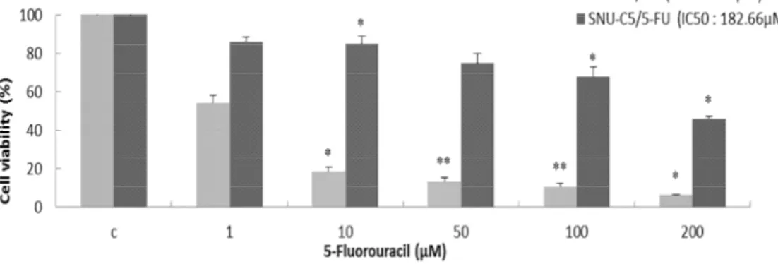
To evaluate the effect of 5-FU on the viability in SNU-C5/WT and SNU-C5/5-FU, cell
viability was demonstrated using trypan blue staining assay. SNU-C5/WT and
SNU-C5/5-FU were treated with 5-SNU-C5/5-FU (1, 10, 50, 100 and 200μM) for 72h,SNU-C5/WT cells were
decreased significantly cell viability (Figure 1; 1μM, 54.3%; 10μM, 18.5%; 50μM, 13.3%;
100μM, 11.09% and 200μM, 6.8%), but SNU-C5/5-FU cells did not decrease significantly
compared to SNU-C5/WT cells (Fig.1; 1μM, 85.9%; 10μM, 84.7%; 50μM, 74.8%; 100μM,
67.8% and 200μM, 46.1%). IC50 of SNU-C5/5-FU was more than 40 times higher (Fig.1;
- 17 -
Figure 1. Cytotoxicity of 5-Fluorouracil on C5/WT and C5/5-FU SNU-C5/WT and SNU-C5/5-FU cells were plated in 6-well (2Χ105cells/ml) and treated with 5-FU (1, 10, 50, 100 and 200 μM) for 72h. The results are expressed as percentages of viable cells compared with control by trypan blue staining. The data are presented as the mean ± SD from experiments representative of three independent trials. *p<0.05, **p<0.01, and ***p<0.001 compared with the control.
- 18 -
4.2. SNU-C5/5FU has the characteristic of overexpression of phospho-AKT.
To determine that exceptional characteristic of C5/5-FU cells, we compared with
SNU-C5/WT, SNU-C5/5-FU and SNU-C5/OXT. Interestingly, phospho-AKT was overexpressed
in SNU-C5/5-FU (Figure 2A). Furthermore, over-phosphorylated AKT of SNU-C5/5-FU
cells were identified at confocal microscopy (Figure 2B). Also, mTor, a protein modulated
by phospo-AKT (LoPiccolo et al. 2008), was over-phosphorylated in SNU-C5/5-FU (Figure
- 21 -
4.3. SNU-C5/5-FU down-regulates E-cadherin and up-regulates phospho-GSK-3β Recent studies reported that phosphorylated AKT leads to decreased E-cadherin (Zhou et
al. 2009) and phosphorylation of GSK-3β (Son et al. 2012). Therefore, we investigated
expression of E-cadherin and phospho-GSK-3β in SNU-C5/5-FU cells, which has
over-phosphorylated AKT. As a result, expression of E-cadherin was suppressed (Figure 4) and
expression of phospho-GSK-3β was increased (Figure 5) in SNU-C5/5-FU compared with
- 24 -
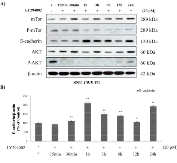
4.4. Phosphorylated AKT led to down-regulation of E-cadherin and phosphorylation of GSK-3β in SNU-C5/5-FU
To establish whether expression of E-cadherin (Figure 4) and phospo-GSK-3β (Figure 5)
depended on overexpression of phospho-AKT, SNU-C5/5-FU cells were treated with
LY294002 (PI3 kinase inhibitor). As a result, E-cadherin expression was increased (Figure
6) and phospho-GSK-3β expression was decreased (Figure 7) by treatment of LY294002 in
time dependent manner. Furthermore, treatment of LY294002 induced down-regulation of
β-catenin and cyclin D1 (Figure 7). These results suggested that phospho-AKT regulates
- 25 -
Figure 6. The effect of LY294002 on the expression of E-cadherin in SNU-C5/5-FU (A) The SNU-C5/5-FU cells (2 x 10⁵ cells/mL) were pre-incubated for 24h and then cells were treated with LY294002 (20 μM) for the indicated time. Lysates were prepared from these cells, and the expression of AKT, Phospho-AKT, mTor, phospho-mTor and E-cadherin measured by Western blot analysis using specific antibodies. (B) Data represent the percentage of E-cadherin expression in SNU-C5/5-FU cells. The data are presented as the mean ± SD from experiments representative of three independent trials. *p<0.05, **p<0.01, and ***p<0.001 compared with the control.
- 26 -
Figure 7. The effect of LY294002 on the expression of GSK-3β, phospho-GSK-3β, cyclin
D1, and β-catenin in SNU-C5/5-FU (A) The SNU-C5/5-FU cells (2 x 10⁵ cells/mL) were
pre-incubated for 24h and then cells were treated with LY294002 (20 μM) for the indicated time. Lysates were prepared from these cells, and the expression of AKT, Phospho-AKT, phospho-GSK-3β, GSK-3β, cyclin D1, and β-catenin measured by Western blot analysis using specific antibodies. (B) Data represent the percentage of β-catenin, phospho-GSK-3β, and GSK-3β expression in SNU-C5/5-FU cells. The data are presented as the mean ± SD from experiments representative of three independent trials. *p<0.05, **p<0.01, and ***p<0.001 compared with the control.
- 27 -
4.5. SNU-C5/5-FU has characteristic to translocation of β-catenin in cytoplasm and nucleus
Various studies showed that β-catenin was involved the cell-cell adhesion by interaction of
E-cadherin in plasma membrane (Fanelli et al. 2008, Mohamet, Hawkins & Ward 2011). But,
loss of E-cadherin led to discharge of catenin into the cytoplasm (Huber, Weis 2001).
catenin was degraded by activated GSK-3β in cytoplasm. If GSK-3β was inactivated,
β-catenin acted as a transcription factor in the nucleus (Akiyama 2000). In other words,
activated GSK-3β and stabilization of E-cadherin prevents β-catenin from acting on
transcription factor. Thus, we determined the expression and location of β-catenin. As a
result, we found no significant difference on expression of β-catenin (Figure 8A). However,
β-catenin located the cytoplasm and nuclear in the C5/5-FU comported with
- 29 -
4.6. Phospho-AKT led to the nuclear translocation of β-catenin
We determined whether localization of β-catenin in cytoplasm and nucleus were associated
with down-regulation of E-cadherin and inactivated GSK-3β by over-phosphorylated AKT.
E-cadherin was increased in plasma membrane and phospho-GSK-3β was decreased by
treatment of LY294002 (Figure 9). Interestingly, treatment of LY294002 induced not only
decreased expression of β-catenin in the cytoplasm and nucleus but also increased expression
of β-catenin in plasma membrane (Figure 9). Therefore, we determined that the interaction of
E-cadherin and β-catenin were regulated by phospho-AKT. β-catenin was decreased but the
interaction of β-catenin and E-cadherin was increased by treatment of LY294002 in a dose
dependent manner (Figure 10). This result indicated that phospho-AKT led to the nuclear
translocation of β-catenin via loss of E-cadherin and inactivated GSK-3β (Figure 11A). As a
resultant, inhibition of phosphorylated AKT prevents β-catenin from entering the nucleus for
- 31 -
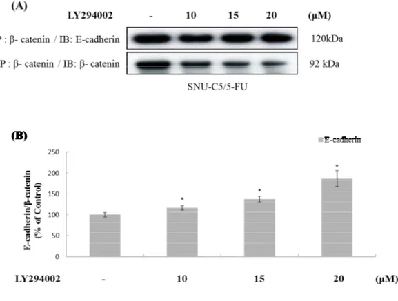
Figure 10. The effect of LY294002 on β-catenin interacts with E-cadherin in
SNU-C5/5-FU (A) The SNU-C5/5-SNU-C5/5-FU cells (2 x 10⁵ cells/mL) were pre-incubated for 24h and then cells
were treated with LY294002 (10, 15 and 20 μM) for 24h. The lysate of SNU-C5/5-FU cells was immunoprecipitated with catenin antibody, and was immunoblotted with anti-β-catenin and anti-E-cadherin antibody. (B) Data represent the percentage of E-cadherin expression in SNU-C5/5-FU cells. The data are presented as the mean ± SD from experiments representative of three independent. *p<0.05, **p<0.01, and ***p<0.001 compared with the control.
- 33 -
Figure 11. The characteristics of SNU-C5/5-FU on E-cadherin, GSK-3β, and β-catenin (A) phosphorylated AKT led to nuclear translocation of β-catenin via loss of E-cadherin and inactivated GSK-3β. (B) Inhibition of phospho-AKT prevents nuclear translocation of β-catenin
- 34 -
4.7. SNU-C5/5FU has characteristic to overexpression of Cyclooxygenase-2
To find other properties of C5/5-FU cells, we compared with C5/WT,
SNU-C5/5-FU and SNU-C5/OXT. SNU-SNU-C5/5-FU cells markedly overexpress COX-2 compared to
SNU-C5/WT and SNU-C5/OXT (Figure 12A). Moreover, overexpression of COX-2 was
identified by confocal microscopy (Figure 12B).
- 36 -
4.8. Over-activated NF-κB signaling led to overexpression of COX-2
NF-κB signaling modulated COX-2 expression (Abrahao et al. 2010). Therefore, we
examined whether that COX-2 was overexpressed by NF-κB signaling in SNU-C5/5-FU.
Interestingly, IκB-α was suppressed and phospho-NF-κB was overexpressed in
SNU-C5/5-FU (Figure 13). Thus, we investigated the involvement of over-activated NF-κB signaling
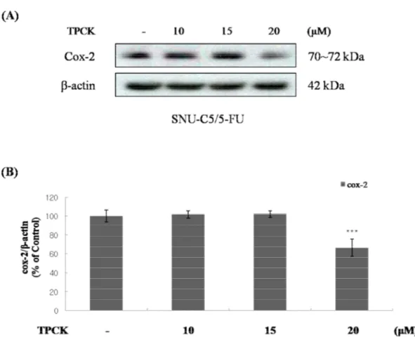
and over-expressed COX-2. The treatment of TPCK (IκB protease inhibitor) decreased the
COX-2 level in dose dependent manner (Figure 14). These data suggest that over-activated
- 38 -
Figure 14. The effect of TPCK on the expression of COX-2 in SNU-C5/5-FU (A) The SNU-C5/5-FU cells (2 x 10⁵ cells/mL) were pre-incubated for 24h and then cells were treated with TPCK (10, 15 and 20 μM). Lysates were prepared from these cells, and the expression of COX-2 measured by Western blot analysis using specific antibodies. (B) Data represent the percentage of COX-2 expression in SNU-C5/5-FU cells. The data are presented as the mean ± SD from experiments representative of three independent. *p<0.05, **p<0.01, and ***p<0.001 compared with the control.
- 39 -
4.9 phospho-AKT led to activation of NF-κB signaling in SNU-C5/5-FU
IκB kinase (IKK) was activated by phospho-AKT and it act NF-κB signaling by
phosphorylation of IκB (Ahn, Aggarwal 2005, Dan et al. 2008). Thus, we examined whether
that phosphorylation of AKT modulates the NF-κB signaling in SNU-C5/5-FU. As a result,
COX-2 and phospho-NF-κB were decreased by treatment of LY294002 in a dose and time
dependent manner (Figure 15). These data indicated that phospho-AKT modulates NF-κB
- 41 -
4.10. phospho-AKT led to stabilization of survivin via overexpression of COX-2
Previous studies reported that SNU-C5/5-FU was showed with overexpression of PGE2 by
overexpression of COX-2 (Choi et al. 2011). In addition, it has been reported that PGE2
affect the resistance to apoptosis via inhibiting ubiquitination of survivin in lung cancer cells
(Krysan et al. 2004). Thus, we confirmed that COX-2 contributes to the stabilization of
survivin expression in SNU-C5/5-FU. Survivin showed overexpression in SNU-C5/5-FU
(Figure 16). Also, it was decreased by treatment with LY294002 or Vioxx (COX-2 selective
inhibitor) in a dose and time dependent manner (Figure 17A and B). Furthermore, treatment
of LY294002 or Vioxx led to the ubiquitination of survivin in a dose dependent manner
(Figure 17C). These results indicated that phospho-AKT induces the stabilization of survivin
- 44 -
Figure 17. The effects of LY294002 and Vioxx on the expression of survivin in
SNU-C5/5-FU (A and B) The SNU-SNU-C5/5-FU cells (2 x 10⁵ cells/mL) were pre-incubated for 24h
and then cells were treated with LY294002 and Vioxx (10, 15 and/or 20 μM) for the indicated time or 24h. Lysates were prepared from these cells, and the expression of survivin measured by Western blot analysis using specific antibodies. (C) The SNU-C5/5-FU cells (2 x 10⁵ cells/mL) were pre-incubated for 24h and then cells were treated with LY294002 and Vioxx (10, 15 and 20 μM) for 24h. The lysate of SNU-C5/5-FU cells was immunoprecipitated with anti-survivin antibody, and was immunoblotted with anti-survivin and anti-ubiquitin antibody.
- 45 -
4.11 phospho-AKT led to expression of survivin in cytoplasm via up-regulation of COX-2
Recent studies reported that nucleus expression of survivin regulated cell cycle but,
cytoplasm expression of survivin induced resistance on apoptosis ((Krysan et al. 2004)).
Interestingly, we identified that survivin expression was increased in the cytoplasm (Figure
15) and that it was stabilized via COX-2 (Figure 17). Thus, we observed that cytoplasm
expression of survivin was involved with COX-2. When treated with 5-FU, we could not
observe a change in expression on survivin. But treatment with Vioxx or LY294002
decreased expression of survivin in cytoplasm (Figure 18). These results indicated that
cytoplasm expression of survivin was modulated by COX-2 and phospho-AKT.
Consequentially, phospho-AKT induces the cytoplasm expression of survivin via
- 47 -
4.12 Phospho-AKT led to resistance on apoptosis via stabilization of survivin
In cytoplasm, stabilized survivin contributes the resistance on apoptosis by interaction
with caspase-3 or caspase-7 (Shin et al. 2001, Chai et al. 2001). Thus, we examined whether
survivin was associated with caspase-3. While treatment of 5-FU did not increase the
activation of caspase-3, co-treatment with Vioxx and 5-FU increased the activation of
caspase-3 (Figure 19A). In addition, treatment of LY294002 or Vioxx reduced the
interaction of porcaspase-3 with survivin and increased the interaction of cleaved-caspase-3
with survivin (Figure 19B). This indicated that survivin avoids apoptosis by direct
interaction of caspase-3 in SNU-C5/5-FU. Consequentially, phospho-AKT induced the
expression of COX-2 via activation of NF-κB signaling. COX-2 contributes to stabilization
of survivin, leading to resistance of apoptosis by direct interaction of caspase-3 in
SNU-C5/5-FU. kinase (Figure 20A). Therefore, inhibition of COX-2 or inhibition of
- 48 -
Figure 19. The effect of LY294002 and Vioxx on survivin interacts with caspase-3 in SNU-C5/5-FU (A) The SNU-C5/5-FU cells (2 x 10⁵ cells/mL) were pre-incubated for 24h and then cells were treated with 5-FU (10, 50 and 100 μM) and/or Vioxx (20 μM) for the indicated time or 24h. Lysates were prepared from these cells, and the expression of AKT, p-AKT, COX-2, and caspase-3 measured by Western blot analysis using specific antibodies.
- 49 -
(B) The SNU-C5/5-FU cells (2 x 10⁵ cells/mL) were pre-incubated for 24h and then cells were treated with LY294002 or Vioxx (10, 15 and 20 μM) for 24h. The lysate of SNU-C5/5-FU cells was immunoprecipitation with anti-survivin antibody, and was immunoblotted with anti-survivin and anti-caspase-3 antibody.
- 51 -
Figure 20. The characteristics of SNU-C5/5-FU on NF-κB signaling and survivin (A) phosphorylated AKT led to activation of NF-κB signaling via activation of IκB kinase. COX-2, which is formation by NF-κB signaling, contributes stabilization of survivin. (B) Inhibition of phospho-AKT prevents stabilization of survivin in cytoplasm.
- 52 -
4.13 phospho-AKT contributes resistance on 5-fluorouracil in SNU-C5/5-FU
We examined whether that overexpression of phospho-AKT contributes the resistance on
5-FU in SNU-C5/5-FU. Treatment of 5-FU did not showed characteristics of apoptosis
(Figure 21A, B and C). However, co-treatment with 5-FU and LY294002 showed the
features of apoptosis, for example, nuclear morphologic changes (Figure 21A), increased
apoptosis related proteins such as cleavage of proaspase-9, cleavage of procaspase-3 and
cleavage of poly(ADP-ribose)polymerase (PARP) (Figure 21B and C). Furthermore, when
treated 5-FU with LY294002, the viability of SNU-C5/5-FU cells decreased by
co-treatment of 5-FU in a dose dependent manner (Fig.21D; 5-FU 1μM, 85.3%; 10μM, 63.3%;
50μM, 54.12%; 100μM, 45.87% and 200μM, 38.35%) compared with treatment of 5-FU
(Figure 1). In addition, combination treatment of 5-FU and Vioxx also were decreased cell
viability (Fig.21D; 5-FU 20 1μM, 90.17%; 10μM, 83.23%; 50μM, 66.14%; 100μM, 49.69%
and 200μM, 44.53%). In addition, IC50 of combination treatments in SNU-C5/5-FU was
significantly reduced (IC50 of co-treatments with LY294002 and 5-FU, 76.342 μM and IC50
of co-treatments with Vioxx and 5-FU, 97.427 μM). These data indicated that
over-expressed phospho-AKT contributes resistance on 5-FU. Moreover, survivin, which was one
of the downstreams of phospho-AKT, was a major resistance mechanism on 5-FU.
- 54 -
Figure 21. The effect of LY294002 or Vioxx on induction of apoptosis in SNU-C5/5-FU (A) The SNU-C5/5-FU cells (2 x 10⁵ cells/mL) were pre-incubated for 24h and then cells were treated with 5-FU(10, 50 and 100 μM) and/or LY294002 (20 μM) for the indicated time or 24h. The cells were stained with Hoechst 33342 (10 μg/ml medium at final) apoptotic bodies were observed with an inverted fluorescent microscope equipped with an IX-71 Olympus camera and photographed (magnification ×200). (B) The SNU-C5/5-FU cells (2 x 10⁵ cells/mL) were pre-incubated for 24h and then cells were treated with LY294002 (20 μM) and/or 5-FU(10, 50 and 100 μM) for 24h. Lysates were prepared from these cells, and the expression of caspase-9, caspase-3, PARP, AKT and phospho-AKT measured by Western blot analysis using specific antibodies. (C) The SNU-C5/5-FU cells (2 x 10⁵ cells/mL) were pre-incubated for 24h and then cells were treated with LY294002 (20 μM) and/or 5-FU (100 μM) for 24h. Immunofluorescent stain of cleaved caspase-3 and cleaved caspase-9 was performed with Alexa Fluor 488 goat anti-rabbit IgG secondary antibody and the fluorescence was identified using confocal microscopy (FV500, OLYMPUS). (D) SNU-C5/5-FU cells were plated in 6-well (2 Χ 105cells/ml) and treated LY294002 or Vioxx (20 μM) and/or 5-FU(1, 10, 50, 100 and 200 μM) for 72h. The results are expressed as percentages of viable cells compared with control by trypan blue staining. The data are presented as the mean ± SD from experiments representative of three independent trials. *p<0.05, **p<0.01, and ***p<0.001 compared with the control.
- 55 -
4.14 Loss of PTEN was cause of the over-phosphorylated AKT in SNU-C5/5-FU Previous studies reported that loss of PTEN promotes the phosphorylation of AKT in
colon cancer (Colakoglu et al. 2008, Sawai et al. 2008). Therefore, we confirmed the PTEN
level of SNU-C5/5-FU. As a result, PTEN expression was down-regulated in SNU-C5/5-FU
compared with SNU-C5/WT and SNU-C5/OXT (Figure 22). This result suggested that loss
of PTEN led to over-phosphorylation of AKT in SNU-C5/5-FU.
Finally, in SNU-C5/5-FU, overexpression of phospho-AKT, which by loss of PTEN,
modulates a variety of downstream effects such as nuclear translocation of β-catenin via loss
of E-cadherin, inactivation of GSK-3β and it led to stabilization of survivin in cytoplasm via
activated NF-κB signaling. Thus, stabilized survivin inhibits apoptosis by interaction of
caspase-3 (Figure 23A). Consequently, inhibition of phospho-AKT was increased sensitive
- 58 -
5. DISCUSSION
In this study, we investigated the resistance mechanism and resistance reversal in
SNU-C5/5-FU, 5-fluorouracil (5-FU) resistance human colon cancer cells. SNU-C5/5-FU has
characteristic that over-phosphorylation of AKT. In SNU-C5/5-FU, phospho-AKT regulates
a variety of cellular processes such as down-regulation of E-cadherin, inactivation of
GSK-3β and activation of NF-κB signaling. Moreover, treatment of LY294002 (PI3 kinase
inhibitor) led to not only reversal of these cellular processes but also increased apoptosis by
5-FU. 5-FU is an anti-cancer drug that prevents DNA synthesis by inhibiting the
biosynthesis of thymine. Variety studied showed that increased thymidylate synthase (TS)
led to resistance of 5-FU (Jette et al. 2008, Peters et al. 2002, Van der Wilt et al. 1992).
Interestingly, TS expression did not increase in SNU-C5/5-FU (Jung 2006, Kim et al. 2005).
The PI3K/AKT pathway regulates cell growth and survival in cancer cells (Kim, Chung
2002). Moreover, phospho-AKT was induced resistance of apoptosis via modulate other
cellular processes (Kim, Chung 2002). The phospho-AKT led to unstable E-cadherin by
activation of mdm2 that known is anti-apoptotic protein (Zhou et al. 2009). E-cadherin is a
transmembrane protein, which is involved in cell-cell adhesion. Cytoplasmic domain of
- 59 -
Hawkins & Ward 2011, Huber, Weis 2001). Thus, loss of E-cadherin led to release of
β-catenin and it could act as transcription factor on cell survival and proliferation (Huber, Weis
2001). However, activated GSK-3β induced degradation of β-catenin in cytoplasm.
Unfortunately, phospho-AKT led to inactivation GSK-3β (Akiyama 2000, Woodward et al.
2007, Cui et al. 2012). In other words, phospho-AKT induced that down-regulation of
E-cadherin and inactivation of GSK-3β. Resultantly, phosphorylated AKT induced activation
to β-catenin transcription factor via down-regulation of E-cadherin and inactivation of
GSK-3β. Actually, we confirmed the previous result that β-catenin was discharged by loss of
E-cadherin (Figure 4 and 8). In addition, because of inactivated GSK-3β (Figure 5), it did not
degrade β-catenin in cytoplasm, translocates into the nucleus and acts as a trans-factor on
cell survival (Figure 8 and 9). The upper mechanism on these series of events was excessive
phosphorylation of AKT (Figure 9 and 11A). Therefore, inhibition of phosphorylation of
AKT can suppresses that β-catenin discharge to the cytoplasm by increased of E-cadherin
and it also promoted the degradation of β-catenin via activated GSK-3β. Conclusively,
inhibition of phosphorylated AKT suppressed β-catenin action as a trans-factor in
SNU-C5/5-FU cells (Figure 9 and 11B).
Various studies show that phospho-AKT promotes the activation of NF-κB signaling via
- 60 -
Activated NF-κB induced enzymes such as COX-2 and variety proteins (Ahn, Aggarwal
2005). Our result indicated the overexpression of cox-2 in SNU-C5/5-FU compared with
SNU-C5/WT and SNU-C5/OXT (Figure 12). As expected, NF-κB signaling was
over-activated in SNU-C5/5-FU and blocked by treatment of LY294002 as well as expression of
COX-2 (Figure 13 and 15). It has been reported that COX-2 modulates the resistance to
apoptosis by inhibition to ubiquitination of survivin, anti-apoptotic protein (Krysan et al.
2004). Therefore, activation of NF-κB by phospho-AKT presumably regulated resistance of
apoptosis via stabilization of survivin. We confirmed the overexpression of survivin (Figure
16). Thus, we identified whether that survivin is regulated by COX-2 and phospho-AKT.
Treatment with LY294002 or Vioxx decreases survivin expression in time- and dose-
dependent manner (Figure 17A and B). Moreover, the ubiquitination of survivin was induced
by LY294002 and Vioxx (Figure 17C). Previous studies showed that survivin modulates the
cell cycle in the nucleus. However, in carcinogenic cells, survivin is located in the nucleus
and cytoplasm (Samuel et al. 2005). In the cytoplasm, survivin directly suppresses the
caspase-3 and caspase-7 leading to resistance of apoptosis (Shin et al. 2001, Chai et al. 2001).
Unfortunately, PGE2 contributes the remaining survivin after half-life in cytoplasm (Krysan
et al. 2004). SNU-C5/5-FU has over-expressed survivin in cytoplasm (Figure 16). Therefore,
over-- 61 over--
expressed COX-2 that was modulated by phospho-AKT. The over-expressed survivin in the
cytoplasm was decreased by LY294002 or Vioxx compared with treatment of 5-FU (Figure
18). These results suggested that COX-2 and phospho-AKT led to stabilization of survivin.
Thus, we examined whether survivin was associated with caspase-3 expression. Inhibition of
COX-2 was induced the cleavage of caspase-3 (Figure 19A). Moreover, treatment of Vioxx
not only decreased the interaction of survivin and procaspase-3 but also increased the
interaction of survivin and cleaved caspase-3. Also, treatment of LY294002 decreased the
interaction of survivin and procaspase-3 (Figure 19B). This result indicates that inhibition of
COX-2 induces the cleavage of caspase-3 via ubiquitination of survivin. In SNU-C5/5-FU,
phospho-AKT indirectly modulates COX-2 expression via activation of NF-κB signaling.
Over-expressed COX-2 regulated PGE2 expression and contributed the stabilization of
survivin. In cytoplasm, accumulated survivin controlled the resistance of apoptosis through a
correction with procaspase-3 (Figure 20A) Therefor the inhibition of phospho-AKT
prevented the resistance to apoptosis by promotes to degradation of survivin (Figure 20B).
Eventually, SNU-C5/5-FU avoids apoptosis that was induced by 5-FU. Thus, we
investigated whether that overexpression of phospho-AKT is associated with the resistance
of 5-FU. Combination treatment of LY294002 and 5-FU led to characteristics of apoptosis
- 62 -
proteins (Figure 21B and C). Moreover, combination treatment of LY294002 and 5-FU
significantly inhibited cell viability (Figure 21D). Interestingly, combination treatment of
Vioxx and 5-FU showed the inhibited cell viability (Figure 21D). These results suggested
that over-expressed AKT contributes to resistance to apoptosis by 5-FU in SNU-C5/5-FU. In
addition, survivin, which was one of the downstream of phospho-AKT, was an important
mechanism of resistance by phospho-AKT.
Previous studies reported that loss of PTEN could explain the phosphorylation of AKT in
colorectal cancer cells (Tamura et al. 1999, Colakoglu et al. 2008, Sawai et al. 2008) and in
this study, we confirmed that expression of PTEN was down-regulated in SNU-C5/5-FU
compared with SNU-C5/WT and SNU-C5/OXT (Figure 22). This result indicated that loss
of PTEN led to over-phosphorylation of AKT in SNU-C5/5-FU.
In summary, SNU-C5/5-FU has over-expressed phospho-AKT by loss of PTEN. Thus,
over-phosphorylation of AKT contributes to resistance to 5-FU by regulating a variety of
downstream such as E-cadherin, GSK-3β and NF-κB signaling (Figure 23A). Therefore,
inhibition of phospho-AKT increase sensitivity on 5-FU (Figure 23B). Taken together, these
results demonstrate that 5-FU resistance mechanism in SNU-C5/5-FU and the inhibition of
phospho-AKT may be a therapeutic target in SNU-C5/5-FU, 5-fluorouracil resistance colon
- 63 -
6. REFERENCES
Abrahao, A.C., Castilho, R.M., Squarize, C.H., Molinolo, A.A., dos Santos-Pinto, D.,Jr & Gutkind, J.S. 2010, "A role for COX2-derived PGE2 and PGE2-receptor subtypes in head and neck squamous carcinoma cell proliferation", Oral oncology, vol. 46, no. 12, pp. 880-887.
Ahn, K.S. & Aggarwal, B.B. 2005, "Transcription factor NF-kappaB: a sensor for smoke and stress signals", Annals of the New York Academy of Sciences, vol. 1056, pp. 218-233.
Akiyama, T. 2000, "Wnt/beta-catenin signaling", Cytokine & growth factor reviews, vol. 11, no. 4, pp. 273-282.
Asting, A.G., Caren, H., Andersson, M., Lonnroth, C., Lagerstedt, K. & Lundholm, K. 2011, "COX-2 gene expression in colon cancer tissue related to regulating factors and promoter methylation status", BMC cancer, vol. 11, pp. 238.
Bao, S., Ouyang, G., Bai, X., Huang, Z., Ma, C., Liu, M., Shao, R., Anderson, R.M., Rich, J.N. & Wang, X.F. 2004, "Periostin potently promotes metastatic growth of colon cancer by augmenting cell survival via the Akt/PKB pathway", Cancer cell, vol. 5, no. 4, pp. 329-339.
- 64 -
quantities of protein utilizing the principle of protein-dye binding", Analytical Biochemistry, vol. 72, pp. 248-254.
Cervello, M. & Montalto, G. 2006, "Cyclooxygenases in hepatocellular carcinoma", World journal of gastroenterology : WJG, vol. 12, no. 32, pp. 5113-5121.
Chai, J., Shiozaki, E., Srinivasula, S.M., Wu, Q., Datta, P., Alnemri, E.S. & Shi, Y. 2001, "Structural basis of caspase-7 inhibition by XIAP", Cell, vol. 104, no. 5, pp. 769-780.
Chen, W., Bai, L., Wang, X., Xu, S., Belinsky, S.A. & Lin, Y. 2010, "Acquired activation of the Akt/cyclooxygenase-2/Mcl-1 pathway renders lung cancer cells resistant to apoptosis", Molecular pharmacology, vol. 77, no. 3, pp. 416-423.
Choi, C.H., Lee, T.B., Lee, Y.A., Choi, S. & Kim, K.J. 2011, "Up-regulation of cyclooxygenase-2-derived prostaglandin E(2) in colon cancer cells resistant to 5-fluorouracil", Journal of the Korean Surgical Society, vol. 81, no. 2, pp. 115-121.
Clark, A.S., West, K., Streicher, S. & Dennis, P.A. 2002, "Constitutive and inducible Akt activity promotes resistance to chemotherapy, trastuzumab, or tamoxifen in breast cancer cells", Molecular cancer therapeutics, vol. 1, no. 9, pp. 707-717.
Colakoglu, T., Yildirim, S., Kayaselcuk, F., Nursal, T.Z., Ezer, A., Noyan, T., Karakayali, H. & Haberal, M. 2008, "Clinicopathological significance of PTEN loss and the phosphoinositide 3-kinase/Akt pathway in sporadic colorectal neoplasms: is PTEN loss predictor of local recurrence?", American Journal of Surgery, vol. 195, no. 6, pp.
719-- 65 719--
725.
Comes, F., Matrone, A., Lastella, P., Nico, B., Susca, F.C., Bagnulo, R., Ingravallo, G., Modica, S., Lo Sasso, G., Moschetta, A., Guanti, G. & Simone, C. 2007, "A novel cell type-specific role of p38alpha in the control of autophagy and cell death in colorectal cancer cells", Cell death and differentiation, vol. 14, no. 4, pp. 693-702.
Cui, J., Jiang, W., Wang, S., Wang, L. & Xie, K. 2012, "Role of Wnt/beta-catenin signaling in drug resistance of pancreatic cancer", Current pharmaceutical design, vol. 18, no. 17, pp. 2464-2471.
Dan, H.C., Cooper, M.J., Cogswell, P.C., Duncan, J.A., Ting, J.P. & Baldwin, A.S. 2008, "Akt-dependent regulation of NF-{kappa}B is controlled by mTOR and Raptor in association with IKK", Genes & development, vol. 22, no. 11, pp. 1490-1500.
Do, T.V., Kubba, L.A., Antenos, M., Rademaker, A.W., Sturgis, C.D. & Woodruff, T.K. 2008, "The role of activin A and Akt/GSK signaling in ovarian tumor biology", Endocrinology, vol. 149, no. 8, pp. 3809-3816.
Fanelli, M.A., Montt-Guevara, M., Diblasi, A.M., Gago, F.E., Tello, O., Cuello-Carrion, F.D., Callegari, E., Bausero, M.A. & Ciocca, D.R. 2008, "P-cadherin and beta-catenin are useful prognostic markers in breast cancer patients; beta-catenin interacts with heat shock protein Hsp27", Cell stress & chaperones, vol. 13, no. 2, pp. 207-220.
- 66 -
correlates with beta-catenin activation, but not with H-ras mutations, and phosphorylation of Akt, GSK3 beta and ERK1/2 in mouse hepatic carcinogenesis", Carcinogenesis, vol. 24, no. 3, pp. 435-442.
Grosch, S., Maier, T.J., Schiffmann, S. & Geisslinger, G. 2006, "Cyclooxygenase-2 (COX-2)-independent anticarcinogenic effects of selective COX-2 inhibitors", Journal of the National Cancer Institute, vol. 98, no. 11, pp. 736-747.
Gupta, A. & Dey, C.S. 2012, "PTEN, a widely known negative regulator of insulin/PI3K signaling, positively regulates neuronal insulin resistance", Molecular biology of the cell, vol. 23, no. 19, pp. 3882-3898.
Huber, A.H. & Weis, W.I. 2001, "The structure of the beta-catenin/E-cadherin complex and the molecular basis of diverse ligand recognition by beta-catenin", Cell, vol. 105, no. 3, pp. 391-402.
Ichihashi, N. & Kitajima, Y. 2001, "Chemotherapy induces or increases expression of multidrug resistance-associated protein in malignant melanoma cells", The British journal of dermatology, vol. 144, no. 4, pp. 745-750.
Jemal, A., Siegel, R., Ward, E., Hao, Y., Xu, J., Murray, T. & Thun, M.J. 2008, "Cancer statistics, 2008", CA: a cancer journal for clinicians, vol. 58, no. 2, pp. 71-96.
Jette, L., Bissoon-Haqqani, S., Le Francois, B., Maroun, J.A. & Birnboim, H.C. 2008, "Resistance of colorectal cancer cells to 5-FUdR and 5-FU caused by Mycoplasma
- 67 -
infection", Anticancer Research, vol. 28, no. 4B, pp. 2175-2180.
Jung, K. 2006, Synergistic effect of combined betulinic acid with anticancer drugs in anticancer drug-resistant colon cancer cells, Chosun university.
Kerr, F., Rickle, A., Nayeem, N., Brandner, S., Cowburn, R.F. & Lovestone, S. 2006, "PTEN, a negative regulator of PI3 kinase signalling, alters tau phosphorylation in cells by mechanisms independent of GSK-3", FEBS letters, vol. 580, no. 13, pp. 3121-3128.
Kim, K.J., Min, Y.D., Choi, C.H. & Lee, T.B. 2005, "The role of selective COX-2 blocker on the 5-FU, irinotecan and oxaliplatin resistant colon cancer cell line", J Kor Soc Colpoproctol, vol. 21, pp. 93.
Kim, D. & Chung, J. 2002, "Akt: versatile mediator of cell survival and beyond", Journal of biochemistry and molecular biology, vol. 35, no. 1, pp. 106-115.
Krysan, K., Merchant, F.H., Zhu, L., Dohadwala, M., Luo, J., Lin, Y., Heuze-Vourc'h, N., Pold, M., Seligson, D., Chia, D., Goodglick, L., Wang, H., Strieter, R., Sharma, S. & Dubinett, S. 2004, "COX-2-dependent stabilization of survivin in non-small cell lung cancer", FASEB journal : official publication of the Federation of American Societies for Experimental Biology, vol. 18, no. 1, pp. 206-208.
Li, Y.J., Wei, Z.M., Meng, Y.X. & Ji, X.R. 2005, "Beta-catenin up-regulates the expression of cyclinD1, c-myc and MMP-7 in human pancreatic cancer: relationships with carcinogenesis and metastasis", World journal of gastroenterology : WJG, vol. 11, no.
- 68 -
14, pp. 2117-2123.
LoPiccolo, J., Blumenthal, G.M., Bernstein, W.B. & Dennis, P.A. 2008, "Targeting the PI3K/Akt/mTOR pathway: effective combinations and clinical considerations", Drug resistance updates : reviews and commentaries in antimicrobial and anticancer chemotherapy, vol. 11, no. 1-2, pp. 32-50.
Luqmani, Y.A. 2005, "Mechanisms of drug resistance in cancer chemotherapy", Medical principles and practice : international journal of the Kuwait University, Health Science Centre, vol. 14 Suppl 1, pp. 35-48.
Mayo, M.W. & Baldwin, A.S. 2000, "The transcription factor NF-kappaB: control of oncogenesis and cancer therapy resistance", Biochimica et biophysica acta, vol. 1470, no. 2, pp. M55-62.
Mohamet, L., Hawkins, K. & Ward, C.M. 2011, "Loss of function of e-cadherin in embryonic stem cells and the relevance to models of tumorigenesis", Journal of oncology, vol. 2011, pp. 352616.
Oki, E., Baba, H., Tokunaga, E., Nakamura, T., Ueda, N., Futatsugi, M., Mashino, K., Yamamoto, M., Ikebe, M., Kakeji, Y. & Maehara, Y. 2005, "Akt phosphorylation associates with LOH of PTEN and leads to chemoresistance for gastric cancer", International journal of cancer.Journal international du cancer, vol. 117, no. 3, pp. 376-380.
- 69 -
Park, B., Choi, K.S., Lee, Y.Y., Jun, J.K. & Seo, H.G. 2012, "Cancer screening status in Korea, 2011: results from the Korean National Cancer Screening Survey", Asian Pacific journal of cancer prevention : APJCP, vol. 13, no. 4, pp. 1187-1191.
Pennati, M., Folini, M. & Zaffaroni, N. 2007, "Targeting survivin in cancer therapy: fulfilled promises and open questions", Carcinogenesis, vol. 28, no. 6, pp. 1133-1139.
Peters, G.J., Backus, H.H., Freemantle, S., van Triest, B., Codacci-Pisanelli, G., van der Wilt, C.L., Smid, K., Lunec, J., Calvert, A.H., Marsh, S., McLeod, H.L., Bloemena, E., Meijer, S., Jansen, G., van Groeningen, C.J. & Pinedo, H.M. 2002, "Induction of thymidylate synthase as a 5-fluorouracil resistance mechanism", Biochimica et biophysica acta, vol. 1587, no. 2-3, pp. 194-205.
Redondo, S., Ruiz, E., Gordillo-Moscoso, A., Navarro-Dorado, J., Ramajo, M., Rodriguez, E., Reguillo, F., Carnero, M., Casado, M. & Tejerina, T. 2011, "Overproduction of cyclo-oxygenase-2 (COX-2) is involved in the resistance to apoptosis in vascular smooth muscle cells from diabetic patients: a link between inflammation and apoptosis", Diabetologia, vol. 54, no. 1, pp. 190-199.
Samuel, T., Okada, K., Hyer, M., Welsh, K., Zapata, J.M. & Reed, J.C. 2005, "cIAP1 Localizes to the nuclear compartment and modulates the cell cycle", Cancer research, vol. 65, no. 1, pp. 210-218.
Sawai, H., Yasuda, A., Ochi, N., Ma, J., Matsuo, Y., Wakasugi, T., Takahashi, H., Funahashi, H., Sato, M. & Takeyama, H. 2008, "Loss of PTEN expression is associated with
- 70 -
colorectal cancer liver metastasis and poor patient survival", BMC gastroenterology, vol. 8, pp. 56.
Seal, S., Chatterjee, P., Bhattacharya, S., Pal, D., Dasgupta, S., Kundu, R., Mukherjee, S., Bhattacharya, S., Bhuyan, M., Bhattacharyya, P.R., Baishya, G., Barua, N.C., Baruah, P.K., Rao, P.G. & Bhattacharya, S. 2012, "Vapor of Volatile Oils from Litsea cubeba Seed Induces Apoptosis and Causes Cell Cycle Arrest in Lung Cancer Cells", PloS one, vol. 7, no. 10, pp. e47014.
Sherbakova, E.A., Stromskaia, T.P., Rybalkina, E.I., Kalita, O.V. & Stavrovskaia, A.A. 2008, "Role of PTEN protein in multidrug resistance of prostate cancer cells", Molekuliarnaia biologiia, vol. 42, no. 3, pp. 487-493.
Shin, S., Sung, B.J., Cho, Y.S., Kim, H.J., Ha, N.C., Hwang, J.I., Chung, C.W., Jung, Y.K. & Oh, B.H. 2001, "An anti-apoptotic protein human survivin is a direct inhibitor of caspase-3 and -7", Biochemistry, vol. 40, no. 4, pp. 1117-1123.
Son, Y.O., Wang, L., Poyil, P., Budhraja, A., Hitron, J.A., Zhang, Z., Lee, J.C. & Shi, X. 2012, "Cadmium induces carcinogenesis in BEAS-2B cells through ROS-dependent activation of PI3K/AKT/GSK-3beta/beta-catenin signaling", Toxicology and applied pharmacology, vol. 264, no. 2, pp. 153-160.
Sparks, A.B., Morin, P.J., Vogelstein, B. & Kinzler, K.W. 1998, "Mutational analysis of the APC/beta-catenin/Tcf pathway in colorectal cancer", Cancer research, vol. 58, no. 6, pp. 1130-1134.
- 71 -
Tamura, M., Gu, J., Danen, E.H., Takino, T., Miyamoto, S. & Yamada, K.M. 1999, "PTEN interactions with focal adhesion kinase and suppression of the extracellular matrix-dependent phosphatidylinositol 3-kinase/Akt cell survival pathway", The Journal of biological chemistry, vol. 274, no. 29, pp. 20693-20703.
Van der Wilt, C.L., Pinedo, H.M., Smid, K. & Peters, G.J. 1992, "Elevation of thymidylate synthase following 5-fluorouracil treatment is prevented by the addition of leucovorin in murine colon tumors", Cancer research, vol. 52, no. 18, pp. 4922-4928.
Wang, S., Liu, Z., Wang, L. & Zhang, X. 2009, "NF-kappaB signaling pathway, inflammation and colorectal cancer", Cellular & molecular immunology, vol. 6, no. 5, pp. 327-334.
Woodward, W.A., Chen, M.S., Behbod, F., Alfaro, M.P., Buchholz, T.A. & Rosen, J.M. 2007, "WNT/beta-catenin mediates radiation resistance of mouse mammary progenitor cells", Proceedings of the National Academy of Sciences of the United States of America, vol. 104, no. 2, pp. 618-623.
Yang, P., Guo, L., Duan, Z.J., Tepper, C.G., Xue, L., Chen, X., Kung, H.J., Gao, A.C., Zou, J.X. & Chen, H.W. 2012, "Histone methyltransferase NSD2/MMSET mediates constitutive NF-kappaB signaling for cancer cell proliferation, survival, and tumor growth via a feed-forward loop", Molecular and cellular biology, vol. 32, no. 15, pp. 3121-3131.
- 72 -
Yeung, J., Esposito, M.T., Gandillet, A., Zeisig, B.B., Griessinger, E., Bonnet, D. & So, C.W. 2010, "beta-Catenin mediates the establishment and drug resistance of MLL leukemic stem cells", Cancer cell, vol. 18, no. 6, pp. 606-618.
Zhou, Q., Yan, B., Hu, X., Li, X.B., Zhang, J. & Fang, J. 2009, "Luteolin inhibits invasion of prostate cancer PC3 cells through E-cadherin", Molecular cancer therapeutics, vol. 8, no. 6, pp. 1684-1691.