INTRODUCTION
Chronic myeloid leukemia (CML) is characterized by the continuous proliferation and abnormal circulation of malig-nant hematopoietic progenitors (1). Effective purging agents used in autologous transplantation in CML must be not only the most specific to leukemic cells but also be the least toxic to normal stem cells (2). Unfortunately, a potential obstacle of autotransplantation for CML is the ineffective removal of residual tumor cells. Various techniques have been devised for tumor cell purging. Until now, the most commonly used purging agents in CML have been 4-hydroperoxycyclophos-phamide, mafosfamide and interferon- (INF- ). However, complete cytogenetic remission rate of the agents is low, approximately from 20% to 40% (2).
Antisense oligodeoxyribonucleotides (AS-ODNs), com-plementary strands of small segments of mRNA, work at the genetic level to interrupt the process by which disease-causing proteins are produced (1). Bcr-abl AS-ODNs have provided evidence of antileukemic effects against Philadel-phia chromosome-positive cells. The PhiladelPhiladel-phia chromo-some creates bcr-abl fusion genes, which encode two abnor-mal mRNAs, b3a2 and b2a2 (3).
Each antisense drug is designed to bind to a specific se-quence of nucleotides in its mRNA target in order to inhibit production of the protein encoded by the target mRNA. How-ever there are large differences in antitumor effects observed among AS-ODNs for the same oncogene according to the size, chemical structure, and special treatment for enhanced permeability (4).
We therefore aimed to develop effective purging agents for use in autotransplantation in CML. We ventured to find meth-ods to increase intracellular uptake of AS-ODNs. We studied the purging effects of various kinds of the AS-ODNs according to their length, chemical structure, breakpoint specific or not specific, and special treatment for enhanced permeability such as streptolysin-O (SL-O) and lipofectin. We also com-pared the in vitro sensitivities of AS-ODNs with those of INF- using MTT assay, clonogenic activities, and RT-PCR.
MATERIALS AND METHODS
Materials
The human bcr-abl containing CML cell line, K562 cell Kyung Ha Ryu, Ju Young Seoh*, Pil Sang Jang�
, Chul Woo Kim� , Sang Hyeok Koh�
, Hee Young Shin� , Hyo Seop Ahn�
Department of Pediatrics and Microbiology*, Ewha Womans University College of Medicine, Seoul; Department of Pediatrics�
, Catholic University of Korea College of Medicine, Seoul; Department of Pathology�
and Pediatrics�
, Seoul National University College of Medicine, Seoul, Korea
Address for correspondence Hyo Seop Ahn, M.D.
Department Pediatrics, College of Medicine, Seoul National University Children’s Hospital, 28 Yongon-dong, Chongno-gu, Seoul 110-744, Korea
Tel : +82.2-760-3625, FAX : +82.2-743-3455 E-mail : hsahn@snu.ac.kr
*This work was supported by a Korea Research Foundation Grant (KRF-97-F00018).
184
Myeloablative Treatment Supported by Autologous Stem Cell Infusion
with Neuroblastoma
Bcr-abl antisense oligodeoxynucleotides (AS-ODNs) have provided evidence of
an antileukemia effect when tested in vitro against Philadelphia-positive cells. In order to investigate the efficacy of AS-ODNs as purging agents in chronic myeloid leukemia (CML) patients, K562 cells, a human CML cell line, were treated in vitro with various types of AS-ODNs and interferon- . Cells were treated in vitro for 0 and 36 hr with 40 g/mL of AS-ODNs, respectively, and incubated at 37℃for 36 hr. Cytotoxic effects were measured by counting the number of viable cells as well as by MTT test. Clonogenic activities were evaluated by methylcellulose culture for 2 weeks. The effects of purging agents on the rearrangement of bcr-abl gene were evaluated by RT-PCR. AS-ODNs inhibited the proliferation of K562 cells with time in cell count assay and MTT test. AS-ODNs were superior to INF-in INF-inhibitINF-ing clonogenic activity (recovery rate; 26.3% vs 64.0%). After INF-incubation with bcr-abl AS-ODNs primers and mRNA isolated from K562 cells, positive bands were abolished, especially of b3a2 type and phosphorothioate type. Our results suggest that AS-ODNs mediated purging may be one of the efficient methods and that autograft may be an alternative treatment for allograft in high-risk group patients of CML if they do not have a stem cell donor.
Key Words : Oligonucleotides, Antisense; Leukemia, Myeloid, Chronic; Philadephia Chromosome
Received : 7 November 2002 Accepted : 27 January 2003
line was obtained from the American type culture collection (Manassas, VA, U.S.A.). K562 CML cells are commonly used for investigating purging effects because the AS-ONDs used in the experiments were derived from antisense sequences directed at the CML-related bcr-abl oncogene. The break-point of the bcr-abl oncogene in K562 cell is b3a2.
ODN structure and
INF-AS-ODNs were purchased from Bioneer (Okchun, Korea). All AS-ODNs were 18-mers or 24-mers spanning either the b3a2 or the b2a2 bcr-abl fusion breakpoint (Table 1). In addi-tion to AS-ODNs specific to the breakpoint, two to four nucleotides substituted missense ODNs were also studied. All AS-ODNs but two were phosphodiester-linked structure (PO), and the two AS-ODNs were phosphorothioate-linked (PS).
K562 cells (105) were seeded into a 24-well cell culture plate in RPMI-1640 (Gibco BRL, Gaithersburg, U.S.A.) medium supplemented with 10% fetal bovine serum (FBS). The cells were incubated with 40 g/mL of each AS-ODN at the start of the culture period and the same dose was added to the medium 24, 48, and 72 hr. The control groups were incubated in the same condition in the absence of AS-ODNs.
INF- was incubated at a concentration of either 300,000 U/mL or 30,000 U/mL. At intervals, part of the nonadhesive cells was harvested and the viable cell number was determined by trypan blue elusion test, then used for the next experiment. Flow cytometric analysis for intracellular uptake of AS-ODN
A total of 1.5×10⁶cells in 60 L of RPMI/FBS were incu-bated with 40 g/mL FITC-labeled b3a2 AS-ODN at 37℃ for up to 6 hr. Ten L aliquots were removed at 0, 1, 2, 4, and 5 hr into 1 mL of RPMI/FBS containing 10 g/mL pro-pidium iodide at 4℃for 10 min and washed three times in RPMI/FBS.
Fluorescence was measured on a FACStar plus (Becton Dickinson Immunocytometry systems, San Jose, CA, U.S.A.) and uptake was recorded as the mean fluorescence intensity (MFI).
The permeabilization enhancing techniques using SL-O
was essentially that of Barry et al. (5). Briefly, after washing in serum-free RPMI medium, 1.5×10⁶ K562 cells were exposed to dithiothreitol (DTT)-activated SL-O at 1,000 IU/mL or 100 IU/mL concentration at 37℃.Thirty minutes later, cells were washed with RPMI/FBS twice and used for flow cytometric analysis as previously mentioned.
We also used the liposome formation technique to enhance permeabilization. FITC-labeled AS-ODNs and lipofectin (Gibco BRL, Gaitherburg, U.S.A.) were mixed for 15 min at 37℃, using sufficient AS-ODNs to achieve a final con-centration of 2 mol/L after addition to the cell suspensions. 1.5×10⁶K562 cells were suspended in 200 L RPMI/FBS and incubated with AS-ODN/lipofectin mixture for 15 min at 37℃. Subsequently, the intracellular uptake of this mix-ture was analyzed by FACS. No attempt was made to remove the lipofectin.
MTT assay
Thirty thousand cells pretreated with AS-ODNs or IFN-were seeded into a 96-well cell culture plate in 200 L RPMI/ FBS. Two g/mL MTT [3-(4, 5-dimethylthiazol-2-yl)-2,5-diphenyl tetrazolium bromide; Sigma, St Louis, MO, U.S.A.] solubilized in 0.15 M PBS was added and incubated for 4 hr at 37℃. After solubilizing the formazan with 100 L 0.04N HCl-isopropanol, the absorbance at 540 nm was measured by using a microplate reader (Molecular Devices, Gaither-burg, U.S.A.).
Colony-forming unit for culture (CFU-C)
CFU-C assays were performed using a complete methyl-cellulose medium with recombinant cytokines for colony assays of human cells (Methocult GF-H4434; StemCell Tech-nologies, Vancouver, BC, Canada). It contains 1.0% methyl-cellulose, 30% fetal bovine serum, 1% BSA, 10 M 2-mer-captoethanol, 2 mM L-glutamine, 10 ng/mL recombinant human (rh) interleukin-3, 10 ng/mL rh granulocyte-mono-cyte colony stimulating factor, 50 ng/mL rh stem cell factor, 3 units/mL rh erythopoietin in Iscove’s Modified Dulbecco’s medium (IMDM; GibcoBRL, Gaithersburg, MD, U.S.A.).
MNCs (2×10⁵) were resuspended in plastic 35 mm tis-sue culture dishes containing 1 mL methylcellulose medium and cultured at 37℃in 100% humidified 5% CO2in air. After 2 weeks, CFU-Cs were counted according to color, size, and marginal shape.
Reverse transcription polymerase chain reaction (RT-PCR) analysis
Total RNAs were extracted by acid-guanidinium phenol chloroform method using RNAzol B (TEL-TEST, Friends-wood, U.S.A.).
Two g RNAs were reverse transcribed in a mixture of 1× 18-mers
b3a2 antisense 5′-GAA-GGG-CTT-TTG-AAC-TCT-3′ B3a2 missense(TAT) 5′-GAA-GTG-CTG-TTG-CAC-TAT-3′ b3a2 missense 5′-GAA-GTG-CTG-TTG-CAC-TAT-3′ b2a2 antisense 5′-GAA-GGG-CTT-CTT-CCT-TAT-3′ b2a2 missense 5′-GAA-CGG-CAT-CTA-CGT-TAT-3′ 24-mers
b3a2 antisense 5′-GCT-GAA-GGG-CTT-TTG-AAC-TCT-GCT-3′ b2a2 antisense 5′-GCT-GAA-GGG-CTT-CTT-CCT-TAT-TGA-3′ Table 1.AS-ODN sequence used
RT buffer [50 mM Tris-HCl (pH 8.3), 75 mM KCl, 10 mM DTT, 3 mM MgCl2], 1 M oligo dT primer (Pharmacia, Uppsala, Sweden), 100 U Moloney murine leukemia virus reverse transcriptase (Gibco BRL, Gaithersburg, U.S.A.), 0.5 mM each dNTPs (Boehringer Mannheim, Germany), and 20 U RNase inhibitor (Boehringer Mannheim) at 37℃ for 90 min.
For the detection of bcr-abl mRNA, 200 ng cDNA was amplified in a mixture of 1×Taq DNA polymerase buffer [20 mM Tris-HCl (pH 7.5), 100 mM KCl, 1 mM DTT, 0.1 mM EDTA, 0.5% Tween-20, 0.5% Nonidet P40, 50% glycerol, 1.5 mM MgCl2], 1U Taq DNA polymerase, 250 M each dNTPs, and 250 nM each primers for 30 cycles of 1 min denaturation at 94℃, 1 min annealing at 61℃, and 2 min extension at 72℃. For the nested PCR, another 30 cycles at 68℃annealing temperature was added. In order to verify the quantity of mRNA in each sample,
-
actin mRNA was also amplified to the same concentration for 35 cycles of 1 min denaturation at 94℃, 1 min annealing at 65℃, and 2 min extension at 72℃. After confirming bcr-abl mRNA expression, the amount of expression was measured by densitometer (GS-700; BioRad, Hercules, CA, U.S.A.)The primer sequences used were as follows (Table 1); for bcr-abl, 5′-GAAGTGTTTCAGAAG CTT CTCC-3′(outer sense; B1), 5′-TGATTATAGCCTAAGACCCGGA-3′(outer antisense; A3), 5′ -TGGAGCTGCAGATGCTGACCAAC-TCG-3′(inner sense; B2), 5′-ATCTCCACTGGC CACAA-AATCATACA-3′(inner antisense; A4), for -actin, 5′ -GT-GGGGCGCCCCA GGCACCA-3′(sense), 5′ -GTCCTTA-ATGTCACGCACGATTTC-3′(antisense).
Statistical Analysis
Data were represented by mean±SD. A two-group
com-parison was based on a 2-tailed paired sample t test. A p value <0.05 was considered statistically significant.
RESULTS
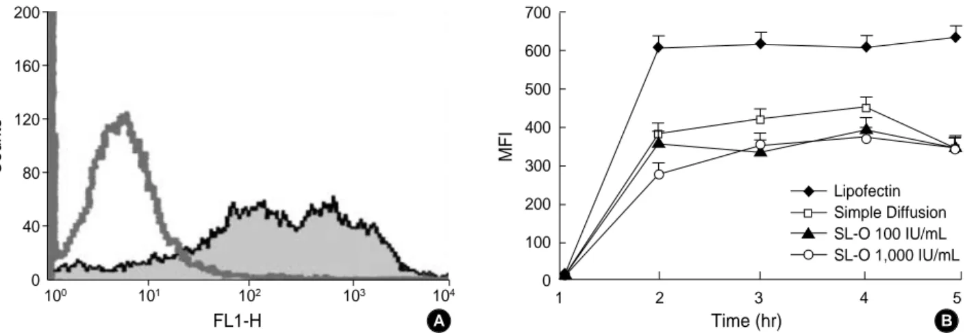
FACS analysis for the detection of AS-ODNs uptake The frequency distribution of fluorescent AS-ODNs into K562 cells was compared to various purging agents.
Fig. 1 illustrates the AS-ODNs uptake into K562 CML cells exposed continuously to b3a2-directed ODN of various treatments for 5 hr. The uptake is expressed as the mean flu-orescence intensity (MFI) of fluorescein-labeled AS-ODNs per cell (Fig. 1A). AS-ODNs uptakes increased sharply with time to 1 hr following incubation and then maintained a plateau until 5 hr in all groups. There were no differences in AS-ODNs uptake between the simple diffusion group and the SL-O treatment group. Additionally, no differences were seen in the concentration of SL-O. Transfection by lipofectin treatments was more efficient than the other groups in intra-cellular uptake of AS-ODNs (p<0.05) (Fig. 1B).
Cytotoxic effects of various types of purging agents measured by cell count assay
Cell numbers were increased until 72 hr and then decreased in untreated control group. The cytotoxic effects of AS-ODNs groups were superior to those of INF- groups at 48 hr, 72 hr, and 96 hr (p<0.05). AS-ODNs groups were examined for their cytotoxic effects, divided into three aspects accord-ing to the breakpoint specific or not specific for target gene, chemical structure linked to PO or PS form, and the length of AS-ODNs for 18-or 24-mers. The purging effects of
Counts 200 160 120 80 40 0 100 101 102 103 104 FL1-H
Fig. 1.Antisense oligodeoxyribonucleotides (AS-ODN) uptakes over a 5-hr incubation period. (A) Representative histogram of intracellar uptake of FITC-labeled AS-ODNs with simple diffusion method after 4 hr of incubation. (B) (◆) transfection by lipofectin; (□) simple dif-fusion without any manipulation; (▲) conjugaton into streptolysin O (SL-O) with 100 IU/mL conc. and (○) with 1,000 IU/mL conc. AS-ODN uptakes were increased sharply with time to 1 hr after incubation and maintained a plateau until 5 hr. Transfection by lipofectin treatments was superior to other groups. Data represent mean+SD of these separate experiments.
MFI 700 600 500 400 300 200 100 0 1 2 3 4 5 Time (hr) A B Lipofectin Simple Diffusion SL-O 100 IU/mL SL-O 1,000 IU/mL ◆ ◆ ◆ ◆ ◆ ◆ ▲ ▲ ▲ ▲ ▲ ▲
AS-ODNs were more superior in the PS type, long chain and breakpoint specific, but there were no statistically significant differences (Fig. 2).
Cytotoxic effects of various types of purging agents measured by MTT assay
We also used an MTT assay to measure the cytotoxic effects of various kinds of AS-ODNs. As in cell count assay, in MTT test, the cytotoxic effects of the AS-ODNs groups were also superior to those of INF- groups (p<0.05). AS-ODNs in-hibited the proliferation of K562 cells in a time-dependent manner, especially the PS type, long chain and b3a2, but there were no significant differences among various types of AS-ODNs (Fig. 3).
Cytotoxic effects of various types of purging agents measured by clonogenic assay
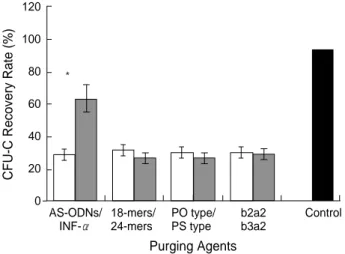
The actual number of colony formation from 105
mononu-clear K562 cells was 5,290±2,110 in the untreated control group. Incubation with purging agents decreased the num-ber of colonies significantly in all groups. As expected in the AS-ODN group, a significant inhibition of CML clonogenic cells was found.
Although INF- inhibited CML clonogenic cells, AS-ODN was a more stronger inhibitor of CML cells. The recovery rate of CFU-C as an untreated control was 64.0% in the INF- group and 26.3% in the AS-ODNs (p<0.05). But we could not find a morphologic variation of colony form according to the purging agents (Fig. 4).
Inhibitory effects on the expression of bcr-abl mRNA of various purging agents
The effects of various AS-ODNs on the expression of bcr-abl mRNA has been examined by RT-PCR. As shown in Fig. 5, positive bands were abolished in AS-ODN groups, whereas they were not inhibited in the INF- groups with RT-PCR using bcr-abl primer and mRNA isolated from K562 cells. Fig. 2.Cytotoxic effects of various purging agents measured by viable cell number. Viable cell numbers were decreased with time and the peak effects were visible after 96 hr of incubation in all groups. The cytotoxic effects of the antisense oligodeoxyribonucleotides (AS-ODNs) groups were superior to those of the interferon- (INF- ) groups (p<0.05) (A). Among AS-ODNs groups, cytotoxic effects were higher in the phosphorothioate (PS) type (B), long chain type (C), and breakpoint specific b3a2 type (D), but there were no statistically significant differences.
Viable cell number (
× 10 5 cells/mL) 2.5 2.0 1.5 1.0 0.5 0.0 -0.5 0 24 48 72 96 Time (hr) A AS-ODNs Control INF-◆ ◆ ◆ ◆ ◆ ◆ ▲ ▲ ▲ ▲ ▲ ▲
Viable cell number (
× 10 5 cells/mL) 2.5 2.0 1.5 1.0 0.5 0.0 -0.5 0 24 48 72 96 Time (hr) C 18-mers Control 24-mers ◆ ◆ ◆ ◆ ◆ ◆ ▲ ▲ ▲ ▲ ▲ ▲
Viable cell number (
× 10 5 cells/mL) 2.5 2.0 1.5 1.0 0.5 0.0 -0.5 0 24 48 72 96 Time (hr) B PO type Control PS type ◆ ◆ ◆ ◆ ◆ ◆ ▲
Viable cell number (
× 10 5 cells/mL) 2.5 2.0 1.5 1.0 0.5 0.0 -0.5 0 24 48 72 96 Time (hr) D b2a2 Control b3a2 ◆ ◆ ◆ ◆ ◆ ◆ ▲ ▲ ▲ ▲ ▲ ▲ ▲ ▲ ▲ ▲ ▲
Treatment with INF- did not affect the gene expression in K562 cells. However, treatment with AS-ODNs reduced the bcr-abl mRNA expression. In particular, b3a2, (PS) type showed the most profound effects (Fig. 5).
DISCUSSION
AS-ODN directed to leukemia-specific transcripts has the potential to specifically target the genes with a pathologic role in leukemogenesis while sparing normal hematopoietic cells. The use of bcr-abl AS-ODN may be considered one of the newest approaches for the treatment of CML, and the in vitro treatment before autograft, at the moment, appears to be a clinical application more likely to produce favorable thera-peutic outcomes (6).
Although leukemic cells take up AS-ODN more readily than normal cells for AS-ODN in order to have a worthwhile biologic effect, some means of enhancing cellular uptake is essential (7). Therefore, we examined different strategies for improving the effectiveness of AS-ODN uptake in CML cells
(8, 9).
Liposome formation has been commonly used for effective intracellular delivery of AS-OND. This kind of packaging leads to the formation of micromolecular complexes with a positive charge on the surface, permitting binding to the negatively charged membrane. Following attachment to the cell, the complexes are presumably taken up via endocytosis (2, 4, 10, 11). In the present study, we discerned that lipo-some formation with lipofectin of AS-ODNs increased the intracellular delivery of AS-ODN, achieving levels of approxi-mately double.
SL-O permeabilization was also enough to deliver AS-ODNs into the cells. Especially SL-O permeabilization translates into increased biologic effectiveness of AS-ODN in cells, as evidenced by the decreased target mRNA levels (12, 13). Although treatment with lipofectin resulted in higher intra-cellular uptake of AS-ODNs than otherwise would have been resulted from simple diffusion, simple diffusion method is enough to deliver AS-ODNs to K562 cells. Furthermore, diffusion is the easiest mode by which purging agents pass across the biologic membrane in vitro. Taking all these into Fig. 3.Cytotoxic effects of various purging agents measured by MTT assay. In the MTT assay, the cytotoxic effects of antisense oligodeoxyri-bonucleotides (AS-ODNs) groups were also superior to those of the interferon- (INF- ) groups (p<0.05) (A). AS-ODNs inhibited the prolif-eration of K562 cells with time, particularly in phosphorothioate (PS) type (B), long chain type (C), and breakpoint-specific b3a2 type (D), although there were no significant differences among the various types of AS-ODNs.
Viability (%) 120 100 80 60 40 20 0 24 48 72 96 Time (hr) A AS-ODNs Control INF-◆ ◆ ◆ ◆ ◆ ▲ ▲ ▲ ▲ ▲ Viability (%) 120 100 80 60 40 20 0 24 48 72 96 Time (hr) B PO type Control PS type ◆ ▲ ▲ ▲ ▲ ▲ ◆ ◆ ◆ ◆ Viability (%) 120 100 80 60 40 20 0 24 48 72 96 Time (hr) C 18-mers Control 24-mers ◆ ▲ ▲ ▲ ▲ ▲ ◆ ◆ ◆ ◆ Viability (%) 120 100 80 60 40 20 0 24 48 72 96 Time (hr) D b2a2 Control b3a2 ◆ ▲ ▲ ▲ ▲ ▲ ◆ ◆ ◆ ◆
account, we used simple diffusion as a mode to transfer AS-ODNs into K562 cells.
The breakpoint of the Philadelphia chromosome in the K562 cells is b3a2. Thus breakpoint specific AS-ODNs were prepared for the b3a2 type (2, 3). Breakpoint-specific AS-ODN may have a greater purging effectiveness than a non-specific one. In our study, the breakpoint-non-specific AS-ODNs of the b3a2 type show most effective purging effects than nonspecific one although there were no statistically signifi-cant differences.
The PO types of AS-ODNs are rapidly degraded in vivo by the ubiquitously distributed DNases before they reach their intracellular target RNA. Chemical modifications were therefore made to increase their stability by protection against enzymatic cleavage (14). The changes made in the chemical structure of the AS-ODNs should not be associated with a decrease in their RNA-binding ability. The most common modified versions of AS-ODNs include the PS type, in which one of the oxygens of the phosphate backbone is substituted by a sulfuration. In mRNA expression of bcr-abl, we demon-strated the differences between chemical structure of AS-ODNs. The PS type is more effective than the PO type (15). However, there were no differences between the PS and PO types in the cell count assay and MTT methods.
The length of AS-ODNs is another factor of anti-tumor effects of AS-ODNs. However, we could not find any differ-ences according to the length using 18-mers and 24-mers.
There may be significant differences in the antitumor effects if the size variation is greater (15).
The clonogenic activities of CML cells were reduced in the AS-ODN b3a2 PS groups but not in the control and INF-groups.
As expected, in methods of analysis, the AS-ODNs group exhibited a superior purging effect to the INF group. There-fore, AS-ODNs provide new anti-CML effects over conven-tional chemotherapy (13).
Gewirtz et al. first tried autologous transplantations in CML patients with purged MNCs using AS-ODNs (16). One of the problems associated with the systemic adminis-tration of AS-ODNs in patients with CML may relate to the nonspecific effects of these compounds and possible toxicity toward normal marrow progenitors that could limit their use as in vitro purging agents and as drugs for systemic therapy (17). Additionally, due to the short living nature of AS-ODNs, we should develop methods of more stable and sustained inhibition of target genes. Moreover, further investigation on the degradation kinetics and biological mechanisms of AS-ODNs are needed further before clinical trials (14, 18).
The development of more specific and active AS-ODNs and the optimization of in vitro and in vivo treatment con-ditions may yield improved results in autografted CML patients (19).
REFERENCES
1. Bhatia R, Verfaillie CM. Inhibition of BCR-ABL expression with
CFU-C Recovery Rate
(%) 120 100 80 60 40 20 0 AS-ODNs/ INF-18-mers/ 24-mers PO type/ PS type b2a2 b3a2 Control Purging Agents
Fig. 4. Recovery rate of colony formation in methylcellulose cul-ture for 2 weeks. The actual number of colony formation from 105 mononuclear K562 cells was 5,290±2,110 in the untreated con-trol group. Incubation with purging agents decreased the num-ber of colonies significantly in all groups. The recovery rate of colony forming unit-culture (CFU-C) was 64.0% in the interferon-(INF- ) treated group and 26.3% in the antisense oligodeoxyri-bonucleotide (AS-ODN) group (p<0.05). AS-ODNs inhibited the recovery of CFU-C from K562 cells, particularly in the long chain type, phosphorothioate (PS) type, and breakpoint-specific b3a2 type, although there were no significant differences. Data repre-sent mean±SD of these separate experiments.
* 1,000 600 500 400 200 bp -actin bcr-abl 1 2 3 4 5 6 7 8 9
Fig. 5. Inhibitory effects on the expression of bcr-abl mRNA of various purging agents. Positive bands were abolished in the antisense oligodeoxyribonucleotides (AS-ODNs) groups, where-as they were not inhibited in the interferon- (INF- ) groups by RT-PCR using bcr-abl primers and mRNA isolated from K562 cells. -actin was used as a internal control, appearing at 550 bp. 1: Size marker. 2: Control. 3: b2a2 18 AS (PO). 4: b3a2 18 AS (PO). 5: b2a2 24 MS (PO). 6: b3a2 24 MS (PO). 7: b2a2 24 AS (PS). 8: b3a2 24 AS (PS). 9: IFN 30,000 IU.
antisense oligodeoxynucleotides restores beta1 integrin-mediated adhesion and proliferation inhibition in chronic myelogenous leukemia hematopoietic progenitors. Blood 1998; 91: 3414-22.
2. De Fabritiis P, Petti MC, Montefusco E, De Propris MS, Sala R, Bellucci R, Mancini M, Lisci A, Bonetto F, Geiser T, Calabretta B, Mandelli F. BCR-ABL antisense oligodeoxynucleotide in vitro purg-ing and autologous bone marrow transplantation for patients with chronic myelogenous leukemia in advanced phase. Blood 1998; 91: 3156-62.
3. Warashina M, Kuwabara T, Nakamatsu Y, Taira K. Extremely high and specific activity of DNA enzymes in cells with a Philadelphia chromosome. Chem Biol 1998; 6: 237-50.
4. Kronewett R, Hass R. Antisense strategies for the treatment of hema-tologic malignancies and solid tumors. Ann Hematol 1998; 77: 1-12. 5. Barry ELR, Gesek FA, Friedman PA. Induction of antisense oligo-deoxynucleotides into cells by permeabilisation with Streptolysin-O. Biotechniques 1993; 15: 1016-8.
6. Pich A, Rancourt C. A role for intracellular immunization in chemo-sensitization of tumor cells? Gene Ther 1999; 6: 1202-9.
7. Agrawal S, Kandimalla ER. Antisense therapeutics: is it as simple as complementary base recognition? Mol Med Today 2000; 6: 72-81. 8. Vaerman JL, Moureau P, Deldime F, Lewalle P, Lammineur C, Morschhauser F, Martiat P. Antisense oligonucleotides suppress hematologic cell growth through stepwise release of deoxyribonu-cleotides. Blood 1997; 91: 331-9.
9. Cotter FE. Antisense therapy of hematologic malignancies. Semin Hematol 1999; 36(S): 9-14.
10. Zabner J, Fasbender AJ, Monibger T, Poellinger K, Welsh MJ. Cel-lular and molecular barriers to gene transfer by a cationic lipid. J Biol Chem 1995; 270: 18997-9007.
11. Zhou X, Huang L. DNA transfection mediated by cationic liposomes containing lipolysin: characterization and mechanism of action.
Biochim Biophys Acta 1994; 1189: 195-203.
12. David GS, Richard VG, Grzybowski J, David MT, Richard EC. Improving the intracellular delivery and molecular efficacy of anti-sense oligonucleotide in chronic myelgenous leukemia cells: a com-parison of streptolysin-O permeabilization, electroporation, and lipophilic conjugation. Blood 1998; 91: 4738-46.
13. Lee JT, Park S, Lee JH, Kim BK, Kim NK. Efficacy of in vitro treatment of CML cell line, K562 cells, using 4-hydroxycyclophos-phamide, -interferon and -interferon. J Korean Med Sci 1996; 11: 26-32.
14. Kronewett R, Steidl U, Kirsch M, Sczakiel G, Hass R. Oligonu-cleotides uptake in primary human hematopoietic cells is enhanced by cationic lipids and depends on the hematopoietic cell subset. Blood 1997; 91: 852-62.
15. Towatari M, Adachi K, Marunouchi T, Saito H. Evidence for a crit-ical role of DNA topoisomerase II alpha in drug sensitivity revealed by inducible antisense RNA in a human leukaemia cell line. Br J Haematol 1998; 101: 548-51.
16. Gewirtz AM, Luger S, Sokol D, Gowdin B, Stadtmauer E, Reccio A, Ratajczak MZ. Oligonucleotide therapeutics for human myel-ogenous leukemia: interm results. Blood 1996; 88: 1069a. 17. Guo JQ, Lian JY, Xian YM, Lee MS, Deisseroth AB, Stass SA,
Champlin RE, Talpas M, Wang JYJ, Arlinghaus RB. BCR-ABL protein expression in peripheral blood cells of chronic myelogenous leukemia patients undergoing therapy. Blood 1994; 12: 3629-37. 18. Gauchez AS, Du Moulinet D’Hardemare A, Lunardi J, Vuillez JP,
Fagret D. Potential use of radiolabeled antisense oligonucleotides in oncology. Anticancer Res 1999; 19: 4989-97.
19. Ryu KH, Koh SH, Yoo KH, Chang PS, Shin HY, Kim CW, Ahn HS. Ex vivo purging effects of bcr-abl antisense oligodeoxynu-cleotide in human chronic myeloid leukemia cell line. Korean J BRM 2000; 10: 267-75.