Mutational spectrum of NF1 gene
in Korean patients
with neurofibromatosis type 1
Chul-Ho Lee
Department of Medical Science
The Graduate School, Yonsei University
Mutational spectrum of NF1 gene
in Korean patients
with Neurofibromatosis type1
Directed by Professor Jin-Sung Lee
The Master's Thesis
submitted to the Department of Medical Science,
the Graduate School of Yonsei University
in partial fulfillment of the requirements for the
degree of Master of Medical Science
Chul-Ho Lee
This certifies that the Master's Thesis
of Chul-Ho Lee is approved.
Thesis Supervisor : Jin-Sung Lee
---Dong-Seok Kim
---Kee-Yang Chung
The Graduate School
Yonsei University
ACKNOWLEDGEMENTS
This work was carried out at the Department of Medical Science, the Graduate School, Yonsei University. I wish to express my sincere gratitude to those who have helped me throughout the work and made this thesis possible. In particular, I would like to thank:
Professor Jin-Sung Lee, my adviser, for accepting me as his student and teaching me how to be a scientist, for his guidance, invaluable support and encouragement throughout the course of this investigation.
The member of may graduate committee, Drs. Dong-Seok Kim, Kee-Yang Chung for their assistance, creative suggestions and interest in my work.
All the staff in Department of Clinical Genetics, Drs. Hwan-Seok Lee, Eun-Sook Park, Bo-Bae Park for their help and suggestions. Hyoung-Won Lee for his support and helping me with administrative matters. Min Jung Lee, Dr.Jung-Hee Ahn, Won-Woo Kim, Hae-Jin Oh, Jung-Won Yeom for their technical help and for being so nice and friendly to me.
Finally, I would like to share the accomplishment of this work with my parents. their love, understanding and support encouraged me throughout all this study.
July 2005 Chul-Ho Lee
TABLE OF CONTENTS
I. INTRODUCTION···3
II. MATERIALS AND METHODS···6
1. Subject···6
2. Polymerase Chain Reaction···7
3. DHPLC analysis···8
4. Sequencing analysis···12
III. RESULTS
1. Results of DHPLC analysis···13
2. Results of Sequencing···14
3. Mutation spectrum···14
4. Polymorphisms···16
IV. DISCUSSION···19
V. CONCLUSION···22
REFERENCES···24
ABSTRACT(IN KOREAN)···29
LIST OF FIGURES
Figure 1. Genetic map of NF1 gene. The NF1 gene is located on
the chromosome 17q11.2 and consist of 60 exons···4
Figure 2. DHPLC chromatograms for NF1 mutations identified···13
Figure 3. Results of direct sequencing···15
Figure 4. Histogram of number of mutations···23
LIST OF TABLES
Table 1. PCR Annealing Temperature for the NF1 Amplicons···8Table 2. Primer sequences, PCR product size and their fragment analysis conditions···9
Table 3. Summary of Mutations Identified in the NF1 gene···17
ABSTRACT
Mutational spectrum of NF1 gene in Korean patients with neurofibromatosis type 1
Chul-Ho Lee
Department of Medical Science The Graduate School, Yonsei University
(Directed by Professor Jin-Sung Lee)
Neurofibromatosis type 1 (NF1) is one of the most common autosomal dominant disorder with an incidence of 1 : 3,500 which is caused by mutations in the NF1 gene. NF1 is characterized particularly by café-au-lait spots and fibromatous tumors of the skin. The NF1 gene is located on the chromosome 17q11.2 and spans approximately 350 kb of genomic DNA. It consist of 60 exons which translates into neurofibromin.
Screening of mutations in NF1 gene is complicated because of the large size of the gene, the presence of pseudogenes, the great variety of possible lesions, and the lack of significant mutational clustering.
We screened for mutations in 36 patients who are clinically diagnosed as neurofibromatosis type 1. The whole coding sequences and all splice sites were examined for mutations using DHPLC followed by
direct sequencing of PCR products. Disruptive mutations were identified in 31 individuals with an overall mutation detection rate of 86%. The mutations included one indel (nt.4079), three insertions (nt.1233, 4159, 4630), seven deletions (nt.953, 1017, 1418, 1541, 2679, 2816, 3525), sixteen missense / nonsense mutations (192, 384, 386, 465, 467, 489, 616, 1403, 1619, 2157, 2197, 2237, 2426, 2429, 2483, 2496 codon) and two splicing error (IVS 25, 34). Sixteen unclassified polymorphisms were also detected. Twenty one (72.4%) of the identified disruptive mutations are novel. Eight mutations have been previously reported. It appeared that mutational spectrum of NF1 gene in patients is heterogeneous as previously shown in other populations. By using strategies for mutation screening in NF1 gene used in this study can easily be applied for clinical purpose.
Key Words: NF1, Neurofibromatosis type1, neurofibromin, mutational analysis.
Mutational spectrum of NF1 gene in Korean patients with neurofibromatosis type 1
Chul-Ho Lee
Department of Medical Science The Graduate School, Yonsei University
(Directed by Professor Jin-Sung Lee)
I. IN TROD U CTION
Neurofibromatosis type 1 (NF1; MIM# 162200), formerly known as Von Recklinghausen neurofibromatosis, is a common autosomal dominant disorder with an incidence of 1 : 3,500, characterised by cafe-au-lait spots, peripheral neurofibromas, Lisch nodules and flexural freckling. Other features found in a minority of patients include scoliosis, macrocephaly, pseudarthrosis, short stature, malignancies and learning disabilities.1
The mutation rate of the neurofibromatosis type 1 (NF1) gene is one of the highest in the human genome, with about 50 percent of cases being due to de novo mutations.2
The NF1 gene is located on the chromosome 17q11.2 and spans approximately 350 kb of genomic DNA. It consist of 60 exons and
translates into neurofibromin3,4,5(Fig 1.).
FIG 1. Genetic map of NF1 gene. The NF1 gene is located on the chromosome 17q11.2 and consist of 60 exons.
The neurofibromin comprises 2818 amino acids and has an estimated molecular weight of 327kDa. The central region of neurofibromin (encoded by exons 2127a) possesses marked homology to Ras-GTPase activation proteins (GAPs), or NF1 GRD, that is able to down-regulate p21ras6 by stimulating its intrinsic GTPase. GTP is a major regulator of growth and differentiation and mutant neurofibromins might interfere with ras signaling pathways and contribute to the development of tumors.7,8
Mutation screening in NF1 gene has been made difficult because of the large size of the gene, the existence of a number of homologous pseudogene sequences spread throughout the genome,9,10 and the lack of defined mutational hotspots. To overcome these problems, various techniques have been employed for screening mutations within the NF1 gene. Most studies have been based on single strand conformation
polymorphism, heteroduplex analysis, temperature gradient gel electrophoresis and denaturing gradient gel electrophoresis. In the largest study to date, involving 500 patients used a protein truncation test, temperature gradient gel electrophoresis, and direct genomic sequencing to examine all of the individual exons, finding sequence variants in 301 patients. Within these variants 278 mutations were considered pathogenic.11 In two more papers published recently, the same methodologies were used sequentially to raise mutation detection rates. In the other study, using cDNA single strand conformation polymorphism and heteroduplex analysis, a detection rate of 70-80% of mutations was achieved (22 of 80 patients).12 Messiaen et al. used a protein truncation test, fluorescence in situ hybridization, southern blot and cytogenetic analysis with 67 patients, and reported a detection rate of 95%, including a high frequency of unusual splicing defects.13 The sensitivity of each technique is hard to establish, as mutation analysis reports have either concentrated on groups of exons, small number of patients included in their studies, or used a combination of techniques. In reviews of known NF1 mutations, several mutation types are described, but no correlation with phenotype was documented. Most of the fully characterised NF1 mutations are either nonsense or frameshift mutations, which presumably lead to premature truncation of neurofibromin synthesis. Large deletions of the NF1 gene are thought to account for less than 10% of cases. A relationship between whole gene deletions and a more severe NF1 phenotype has been reported.14,15
fast and highly sensitive technique based on the detection of heteroduplexes in PCR products by ion pair reverse-phase HPLC under partially denaturing conditions, is in many ways ideally suited to mutation detection in these conditions17. A preliminary study with a basic DHPLC system detected all known mutations within exon 16 of the NF1 gene.18 Recently, the sensitivity of DHPLC was evaluated in the retrospective study of a cohort of 111 unrelated NF1 patients with known germline mutations, which detected 97% of mutations.17 In a subsequent prospective analysis of 50 unrelated NF1 individuals , germline mutations were identified in 34 (68%), including 22 novel alterations. This figure represents the highest rate of mutation detection in the NF1 gene, so far reported using a single screening technique with genomic DNA as a target.17
We screened for mutations in 36 patients who are clinically diagnosed as neurofibromatosis. The whole coding sequences and all splice sites were examined for mutations using DHPLC followed by direct sequencing of PCR products. Disruptive mutations were identified in 31 individuals with an overall mutation detection rate of 86%.
II. MA TERIA LS A N D METHOD S
1. Subject
presence of two or more of the diagnostic criteria proposed by the NIH Consensus Statement in 1988 (Stumpf et al. 1988).
2. Polymerase Chain Reaction
Genomic DNA was extracted using phenol-chloroform extraction method from peripheral blood leukocytes.
The 60 exons of NF1 gene were amplified in 60 PCR fragments. Sixty primer pairs were designed according to Han et al.17 Genomic polymerase chain reaction (PCR) was carried out in 50 ㎕ reaction volumes containing 100 ng genomic DNA, 20 ρM primers, 75 mM dNTP, 5 ㎕ reaction buffer and 1U Ex-Taq Polymerase (TaKaRa Shuzo Co., Ltd., Otsu, Japan) with the following cycling profile; 5 min denaturation at 94℃ and 35 cycles of denaturation at 94℃ for 30 sec, specific annealing temperature for 30 sec, extension at 72℃ for 30 sec, followed by a 5 min final extension step at 72℃ (Table 1). All thermal cycles were run on a Takara PCR thermocycler (TaKaRa Shuzo Co., Ltd., Otsu, Japan). Amplicons were checked by DHPLC sizing method before DHPLC analysis.
An additional denaturation and re-annealing step was required after standard PCR amplification for heteroduplex formation prior to DHPLC analysis.19 Samples were denatured at 95℃ for 10 minutes and then allowed to reanneal for over 30 min (-0.1℃/4sec).
3. DHPLC analysis
DHPLC was performed on a WAVE MD DNA fragment analysis system by using a DNASep column.18 (Transgenomic Inc., Omaha, NE) DNASep columns contain non-porous alkylated polystyrene-divinylbenzene particles that are both electrically neutral and hydrophobic; thus, the negatively charged phosphate ions of DNA molecules cannot bind to the Table 1. PCR Annealing Temperature for the NF1 Amplicons.
Fragment Annealing
Temp.(℃) Fragment Temp.(Annealing ℃). Fragment Temp.(Annealing ℃) Fragment Temp.(Annealing ℃)
1 65 11 60 23-2 58 36 62 2 58 12a 55 23a 62 37 58 3 60 12b 58 24 58 38 60 4a 55 13 64 25 53 39 62 4b 64 14 58 26 60 40 59 4c 58 15 65 27a 60 41 65 5 57 16 60 27b 53 42 60 6 58 17 56 28 64 43 53 7 64 18 58 29 57 44 64 8 62 19a 65 30 57 45 64 9 56 19b 56 31 64 46 60 9a 63 20 64 32 58 47 60 10a 58 21 58 33 64 48 66 10b 54 22 58 34 57 48a 65 10c 57 23-1 57 35 65 49 62
column unaided. Triethylammonium acetate (TEAA) is a positively charged reagent that facilitates interaction between the stationary matrix and DNA molecules. DNA fragments are eluted from the column by reducing the hydrophobic interaction between the alkyl chains of TEAA and the stationary phase of the column. This is achieved by altering the
ratio of TEAA to acetonitrile. The DNA molecules eluted from the column are detected by scanning with a UV-C detector. The successful resolution of heteroduplexes from homoduplexes requires an elution gradient at partially denaturing temperature. At this temperature, only heteroduplexes are destabilised by the mismatched bases and are therefore slightly more melted than the homoduplexes, resulting in earlier elution than the homoduplexes. This special resolution temperature can be predicted by use of DHPLCMelt software (http://insertion.stanford.edu/melt.html).20
Re-annealed PCR products19 after denaturation were injected onto the column and eluted with a linear acetonitrile gradient at a flow rate of 0.9ml/min, with a mobile phase consisting of a mixture of buffers A (0.1 mol/l TEAA and 1mM EDTA) and B (25% acetonitrile in 0.1 mol/l TEAA).
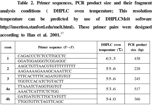
Table 2. Primer sequences, PCR product size and their fragment analysis conditions ( DHPLC oven temperature; This resolution temperature can be predicted by use of DHPLCMelt software http://insertion.stanford.edu/melt.html). These primer pairs were designed according to Han et al. 2001.17
exon Primer sequence (5'→3') DHPLC oven
temperature (℃℃℃℃) PCR product size (bp) 1 CAGACCCTCTCCTTGCCTC 63.3 438 GGATGGAGGGTCGGAGGC 2 AAGCTGTTAACGTGTTTTTTTTT 55.6 228 AAGAAAAGAAAGCAAATTCC 3 TTTCACTTTTCAGATGTGTGT 55.6 245 TGGTCCACATCTGTACTT 4a TTAAATCTAGGTGGTGT 53.6 517 AAACTCATTTCTCTGG 4b GATGATGTCTTGCTATGTT 54.0 366 TTGGTGTTCTAGTTCAGC
exon Primer sequence (5'→3') DHPLC oven temperature (℃℃℃℃) PCR product size (bp) 4c TTTCCTAGCAGACAACTATC 54.8 308 AGGATGCTAACAACAGCAA 5 GAAGGAAGTTAGAAGTTTGTGA 55.2 172 CAATCGTATCCTTACCAGCC 6 CATGTTTATCTTTTAAAAATCTTG 55.0 301 ATAATGGAAATAATTTTGCCCT 7 ACATCTGGAATAGAAGAAACTT 53.9 377 CAGTAACAACAAAAGCAAGT 8 GGATTTTACTGCCATTTGTG 55.7 276 TAACAGCATCAGTAAATATAGTTAGA 9 TTGAAGTTCGTTTCAAGA 53.5 272 ACGCAAAGAAAAGAAAGAA 9a CTGTGGCTCAGAACACTAA 54.1 308 CACATGCAGTGCTCATTA 10a ACGTAATTTTGTACTTTTTCTT 57.0 222 CAATAGAAAGGAGGTGAGAT 10b ATTATCCTGAGTCTTACG 54.4 229 TAACTTAGTGTGATAATTTTGA 10c ATTGAAGTTTCCTTTTTTTCCTT 57.0 275 GTATAGACATAAACATACCATTT 11 CCAAAAATGTTTGAGTGAGT 52.9 256 ACCATAAAACCTTTGGAAG 12a AAACCTTACAAGAAAACTAAG 53.7 303 ATTACCATTCCAAATATTCTTC 12b CTCTTGGTTGTCAGTGCT 55.8 261 CAGAAAACAAACAGAGCAC 13 GTCTTCCACCCTTGACTC 57.2 387 GCTACTTGAATTTCCCCT 14 GCTCTTCCTACTCCTTTT 60.3 191 TTTCTGTTGCTAAGGGCA 15 ACTTGGCTGTAGCTGATT 56.6 247 ACTTTACTGAGCGACTCTTG 16 ACTTTACTGAGCGACTCTTG 56.2 549 TAGAGAAAGGTGAAAAATAAG 17 TCTCTAGGGGGTCTGTCT 55.6 326 CACCCTAGTTTGTGTGCA 18 AGAAGTTGTGTACGTTCTTTT 53.2 367 CTCCTTTCTACCAATAACC 19a TCATGTCACTTAGGTTATCT 55.6 242 TAAAACCCACTAATACTTGAA 19b TGAGGGGAAGTGAAAGAA 54.5 236 GGCTTTATTTGCTTTTTG
exon Primer sequence (5'→3') DHPLC oven temperature (℃℃℃℃) PCR product size (bp) 20 CCACCCTGGCTGATTAT 57.0 402 TAATTTTTGCTTCTCTTACAT 21 TGGTCTCATGCACTCCA 55.6 474 CATCTTTCTTCTGGCTCT 22 TGCTACTCTTTAGCTTCCT 56.7 331 CCTTAAAAGAAGACAATCAG 23-1 TTTGTATCATTCATTTTGTGTG 56.6 282 AAAAACACGGTTCTATGTGAAA 23-2 CTTAATGTCTGTATAAGAGTC 53.3 268 ACTTTAGATTAATAATGGTAATC 23a AGCCAGAAATAGTATACATGATTGG 54.0 446 CTATTTTCTGCCAGAATTAGTA 24 TTGAACTCTTTGTTTTCATGTC 54.1 266 GGAATTTAAGATAGCTAGATTA 25 CCTGTTTTATTGTGTAGATACTT 57.8 134 TAAGTGGCAAGAAAATTAC 26 AATTCTAATGACTTTGCATTTT 56.8 226 ATCTAAATTTAAACGGAGAG 27a GTTACAAGTTAAAGAAATGTGT 56.6 298 CTAACAAGTGGCCTGTGTGCAA 27b TTTATTTGTTTATCCAATTATAGAC 54.7 296 TCCTGTTAAGTCAACTGGGAAAA 28 TTTCCTTAGGTTCAAAACT 56.0 517 CTAGGGAGGCCAGGATAT 29 TCACCCCGTCACCACCACT 58.0 411 GCAACAACCCCAAATCAAACT 30 CAACTTCATTTGTGTTTTCTCCT 54.4 282 CTTTGAATTCTCTTAGAATAATTGT 31 ATAATTGTTGATGTGATTTTCAT 56.5 424 AATTTTGAACCAGATGAAG 32 ATCTAGTATTTTTGAGGCCTC 53.3 312 CAGATATGCTATAGTACAGAA 33 TCCTGCTTCTTTACAGGTTA 56.2 409 AAGTAAAATGGAGAAAGGAACT 34 TTTTCTGTCTTTACTTGTTCCTT 52.3 384 CAGTCCATGCAAGTGTTT 35 GCATGGACTGTGTTATTGG 52.2 319 TGCAATTAAAAGATCCACA 36 GTTCTGTGGATCTTTTAATT 55.1 238 CATTGACCTCAAATTTAAA 37 CATTCCGAGATTCAGTTTAGG 54.1 236 AAGTAACATTCAACACTGATAC
3. Sequencing
Those PCR products displaying a heterozygous pattern were purified and were directly sequenced in both directions using a BigDye 3.1 Terminator Cycle Sequencing Kit (PE Applied Biosystem, Foster City, CA) and an ABI-PRISM3100 Genetic Analyzer (PE Applied Biosystem), according to the manufacturer's instructions.
exon Primer sequence (5'→3') DHPLC oven
temperature (℃℃℃℃) PCR product size (bp) 38 CTATGTCATGATTCATCTTACTA 57.3 233 CTAAATTTGAGTAATCTAGGAACC 39 CTACTGTGTGAACCTCATCAA 53.4 284 GTAAGACATAAGGGCTAACTTACT 40 TCAGGGAAGAAGACCTCAGGAGAT 55.9 328 TGAACTTTCTGCTCTGCCACGCAA 41 GTGCACATTTAACAGGTACT 55.6 373 CTTCCTAGGCCATCTCTAG 42 CTTGGAAGGAGCAAACGATGGT 52.0 356 CAAAAACTTTGCTACACTGACAT 43 TTTTCTTTTTAGTGTATTCCCA 54.8 287 GATTCTAAGAAATGGCTGG 44 CACGTTAATTCCCTATCTT 56.1 268 TGAGAAGTAGAAGACTGTAT 45 CATGAATAGGATACAGTCTTCT 56.7 269 CACATTACTGGGTAAGCATTTA 46 AAATGTTCCTCTGTTGAC 56.0 211 CATCAACCATCCTTCTCCA 47 CTGTTACAATTAAAAGATACCTT 55.4 185 TGTGTGTTCTTAAAGCAGGCAT 48 TTTTGGCTTCAGATGGGGATTT 55.1 351 AAGGGAATTCCTAATGTTGGTG 48a ATTCCTTCTGAAAACCAA 54.2 280 AAGGCAGACTGAGCTTAC 49 CTGGGAGAAACAGGCTAT 57.3 363 AGCAAGCTTCACACGAT
III. RESU LTS
1. DHPLC analysis
We screened for mutations in 36 patients who were clinically diagnosed as neurofibromatosis type 1. The whole coding sequences and all splice sites were examined for mutations using DHPLC. PCR fragments
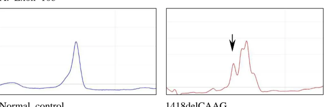
FIG 2. Different DHPLC chromatograms for NF1 mutations identified. Arrow indicates chromatograms originating from a heteroduplex.
A. Exon 10b
Normal control 1418delCAAG DHPLC oven temperature = 54.4℃
B. Exon 21
Normal control 3525delAA DHPLC oven temperature = 55.6℃
C. Exon 27a
Normal control E1619K DHPLC oven temperature = 56.6℃
that showed heteroduplex forms in DHPLC analysis appeared to show abnormal sequences in more than 98%. Fig 2. shows DHPLC chromatograms for NF1 mutations identified in exons 10b, 21 and 27a.
2. DNA Sequencing analysis
Could find twenty-nine different mutations in direct sequencing with PCR fragment that shows heteroduplex pattern in DHPLC analysis (Fig 3.).
3. Mutation spectrum
All 60 exons of the NF1 gene were screened by optimised DHPLC analysis in 36 unrelated NF1 patients. Twenty nine different mutations were identified in thirty one unrelated individuals, including nine missense mutations (exons 8, 10b, 28, 34, 41, 42), seven nonsense mutations (exon 4b, 12b, 24, 35, 36, 41, 42), two splicing errors (intron 25, 34), one indel (exon 23-2), seven deletions (exon 7, 10b, 10c, 16, 21) and three insertions (exon 9, 24, 27a).
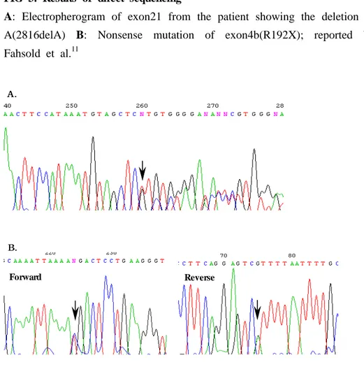
FIG 3. Results of direct sequencing
A: Electropherogram of exon21 from the patient showing the deletion of A(2816delA) B: Nonsense mutation of exon4b(R192X); reported by Fahsold et al.11 Forward Reverse A AA... B B B...
Two small deletions (2679 del C, 2816 del A) and one nonsense mutation (Gln611Term) were located within the putative cysteine/serine-rich domain (exons 11-17). Six different lesions in the NF1 GRD region (exons 20-27a) were identified. All of these lesions are novel and together comprise one nonsense mutation (Tyr 1403 Term), one mutation in splice sites (IVS 25 -1 G>A) and four frameshift mutations (3525delAA, 4079 AA>T, 4159insT, 4630insA).
Two splicing errors were detected (IVS25 -1 G>A and IVS34 +45 T>A). These mutations are predicted to generate truncated neurofibromin protein. (Table 3.)
4. Polymorphisms
A total of sixteen polymorphisms detected, were novel (1458 A>C, 1461 A>G, 1393-9 A>C, 1641+117 G>A, 1721+88 A>C, 3315-106 C>T, 3496+37 A>G, 3871-96 A>G, 5379 C>T, 5886 C>T, 6459 T>G), while five were previously reported (288+41 G>A, 702 G>A, 1641+39 T>C, 5540+19 T>A, 7126+37 C>G ; Table 3). Six silent mutations were also identified. (Leu234Leu, Thr486Thr, Arg487Arg, His1793His, Ile1962Ile, Ala2153Ala). (Table 4.)
Table 3. Summary of Mutations identified in the NF1 Gene. (Nucleotide numbering is based on GeneBank accession no. M82814.)
Location Genomic mutation Amino acid substitution Mutation type References
Exon 4b 574 C>T R192X Nonsense Fahsold et al.11
Exon 7 953 del AAA Frameshift ․
Exon 7 1017 del CT Frameshift Upadhyaya et al.15
Exon 8 1150 T>C F384L Missense ․
Exon 8 1156 A>G I386V Missense ․
Exon 9 1233 ins T Frameshift ․
Exon 10b 1394 G>C S465T Missense ․
Exon 10b 1400 C>T T467I Missense ․
Exon 10b 1418 del CAAG Frameshift ․
Exon 10b 1466 A>G Y489C Missense ․
Exon 10c 1541 del AG Frameshift Robinson et al.23
Exon 12b 1846 C>T Q616X Nonsense ․
Exon 16 2679 del C Frameshift ․
Exon 16 2816 del A Frameshift ․
Exon 21 3525 del AA Frameshift ․
Exon 23-2 4079 AA>T Frameshift ․
Exon 24 4159 ins T Frameshift ․
Exon 24 4209 T>G Y1403X Nonsense ․
IVS 25 IVS25 -1 G>A Miss splicing ․
Exon 27a 4630 ins A Frameshift ․
Exon 28 4855 G>A E1619K Missense ․
Exon 34 6469 T>A F2157I Missense ․
IVS 34 IVS34 +45 T>A Miss splicing ․
Exon 35 6589 A>T R2197X Nonsense ․
Exon 36 6709 C>T R2237X Nonsense Fahsold et al.11
Exon 41 7276 G>C E2426K Missense ․
Exon 41 7286 C>T R2429X Nonsense Fahsold et al.11
Exon 42 7447 C>T L2483F Missense ․
Table 4. Summary of Polymorphisms Identified in the NF1 gene.
(dbSNP ID : NCBI SNP database ID ; http://www.ncbi.nlm.nih.gov/projects/SNP/)
Location Nucleotide change dbSNP ID
IVS 3 288 +41 G>A 2952976 Exon 5 702 G>A 1801052 Exon 10b 1458 A>C ․ Exon 10b 1461 A>G ․ IVS 10b 1393 -9 A>C ․ IVS 10c 1641 +39 T>C 2905880 IVS 10c 1641 +117 G>A ․ IVS 11 1721 +88 A>C ․ IVS 19b 3315 -106 C>T ․ IVS 20 3496 +37 A>G ․ IVS 23-1 3871 -96 A>G ․ Exon 29 5379 C>T ․ IVS 29 5540 +19 T>A 2285894 Exon 31 5886 C>T ․ Exon 34 6459 T>G ․ IVS 39 7126 +37 C>G 7405740
Exon 5 702G>A Leu234Leu ․
Exon 10b 1458A>C Thr486Thr ․
Exon 10b 1461A>G Arg487Arg ․
Exon 29 5379C>T His1739His ․
Exon 31 5886C>T Ile1962Ile ․
IV . D ISCU SSION
Neurofibromatosis type 1 (NF1), formerly known as Von Recklinghausen Neurofibromatosis, is a common genetic disorder affecting approximately 1 in 3,500 people. It is a fully penetrant autosomal dominant disorder. Strict diagnostic criteria that include café au lait spots, neurofibromas, plexiform neurofibromas, freckling in the axillary or inguinal regions, Lisch nodules, optic or chiasma glioma, pseudoarthrosis, and sphenoid dysplasia define NF1 according to NIH diagnostic criteria. Most disease features are present in more than 90% of patients at puberty.1 Further manifestations are known to occur in this disorder, including macrocephaly, short stature, learning difficulties, scoliosis and certain malignancies. There is, however, great intra and interfamilial phenotypic variability. In addition a number of patients who have a clinical picture suspected to be NF1 do not fulfil the diagnostic criteria particularly in the younger age groups and large number of patients are asymptomatic in their lifes except café au lait spots. As a consequence, genetic testing would have a major impact on the diagnosis and management of patient's families. However, mutation detection in the NF1 gene is laborious and complex due to the great variety of possible lesions. These may include chromosomal abnormalities, large deletions or insertions, microdeletions or insertions, splicing mutations, nonsense and missense mutations.9,10 As previously shown , NFI is a very heterogeneous disease on the basis of phenotypes and genotypes. Experimented to observe whether this mutational heterogeneity exists in
Korean patients with neurofibromatosis type1.
The whole coding sequences and all splice sites were examined for mutations using DHPLC followed by direct sequencing of PCR products. Disruptive mutations were identified in 31 individuals with an overall mutation detection rate of 86%. This is the highest mutation detection rate reported so far for the NF1 gene by using a single technique to screen patient genomic DNA samples. Previous to this report, Han et al.17 and Alessandro et al.21 have applied DHPLC method to the screening of the entire NF1 gene for mutation, reporting a detection rate of 68% and 72%. DHPLC is unable to detect the multi-exonic and large deletions that comprise approximately 20% of all NF1 gene mutations.15,22 In addition, some splice site errors will also be missed by DHPLC as the change may be too far into the intronic or too near the end of the amplimers.20 Therefore, splicing error was found two cases in this research though splicing error of NF1 gene is very high than other genetic disorder that is different (26%).12,13 However, we have confirmed the possibility for routine clinical diagnosis in NF1 by direct mutation detection using DHPLC. DHPLC analysis of the entire NF1 gene in the present group of Korean patients disclosed a number of mutations well comparable with the previous studies. In terms of mutation detection, DHPLC based heteroduplex analysis appears to be the efficient method available. Its advantages include the low costs, its potential for automation and the speed and sensitivity of each analytical run, which permits the rapid screening of large numbers of patient samples. The use of DHPLC for mutation identification thus represents a
significant advancement in the clinical diagnosis of NF1. If run parallel experiment method such as PTT to overcome shortcoming of DHPLC, NF1 diagnosis will be more efficient.
In this study, we found twenty nine mutations. Twenty one (72.4%) of the identified pathologic mutations are novel, eight mutations have been previously reported. This is an evidence of the high level of mutational heterogeneity in the NF1 gene. It has also been claimed that missense mutations and single amino acid deletions tend to cluster in two distinct region of NF1 gene. The regions encompassing exons 11-17 (cysteine/serine-rich domain) and exon 21-27a (GAP-related domain). In contrast to the earlier study of Fahsold et al.11, in which 17% of alterations occurred within a CpG dinucleotide, only two of the mutations reported here involved a CpG dinucleotide, the two being small deletions (2679delC, 2816delA).
As in the previous studies, it was not possible to correlate the presence or severity of clinical features, malignancy, and/or mental retardation with the type of the mutations25. In addition, mutational hot spot was not found in Korean patients with neurofibromatosis type 1. However, when the mutation caused protein truncations, such as frameshifts and nonsense mutations, the symptoms were found more severely.
V . CON CLU SION
1. Showed mutation detection ratio of high efficiency, but detection of splicing error is insufficient. If run parallel experiment method such as PTT to overcome shortcoming of DHPLC, NF1 diagnosis will be more efficient.
2. Mutation was found evenly from NF1 gene. Two specification domains (cysteine/serine-rich domain and GAP-related domain) and relation of mutation could not be found, but the corelations should be more closely examined through research of more NF1 patients.
3. As in the previous studies, we were unable to correlate the presence or severity of clinical features, malignancy, and/or mental retardation with the site of the mutation25 and mutational hot spot was not found in Korean patients with neurofibromatosis type 1(Fig 4.). However, when the mutation caused protein truncations, such as frameshifts and nonsense mutations, the symptoms were found more severely.
FIG 4. Histogram of number of mutations. As previously reported, no mutational hotspots within the
REFEREN CES
1. Huson SM, Hughes RAC. The neurofibromatoses: a clinical and pathogenetic overview. Chapman & Hall, London; 1994. 2. Lazaro C, Ravella A, Gaona A, Volpini V, Estivill X.
Neurofibromatosis type 1 due to germ-line mosaicism in a clinically normal father. New Eng. J. Med. 1994; 331: 1403-1407.
3. Cawthorn RM, Weiss R, Xu G, Viskochil D, Culver M, Stevens J, Robertson M, et al. A major segment of the neurofibromatosis type 1 gene: cDNA sequence, genomic structure and point mutations. Cell 1990;62:193-201.
4. Viskochil D, Buchberg AM, Xu G, Cawthorn RM, Stevens J, Wolff RK, et al. Deletions and a translocation interrupt a cloned gene at the neurofibromatosis type 1 locus. Cell 1990;62:187-92.
5. Wallace MR, Marchuk DA, Anderson LB, Letcher R, Odeh HM, Saulino AM, et al. Type 1 neurofibromatosis gene: identification of a large transcript disrupted in three NF1 patients. Science 1990;249:181-186.
6. Martin GA, Viskochil D, Bollag G, McCabe PC, Crosier WJ, Haubruck H, Conroy L, et al. The GAP-related domain of the neurofibromatosis type 1 gene product interacts with ras p21. Cell 1990;63:843-849.
7. Guha A, Lau N, Huvar I, Gutmann D, Provias J, Pawson T, Boss G. Ras-GTP levels are elevated in human NF1 peripheral nerve tumors. Oncogene 1996;12:507-513.
8. Li Y, Bollag G, Clark R, Stevens J, Conroy L, Fults D, et al. Somatic mutations in the neurofibromatosis 1 gene in human tumors. Cell 1992 Apr 17;69(2):275-281.
9. Cummings L, Glatfelter A, Marchuk D. NF1-related loci on chromosomes 2, 12, 14, 15, 20, 21 and 22: a potential role of gene conversion in the high spontaneous mutations rate of NF1. Am J Hum Genet 1993;53 :672A
10. Hulsebos T, Bijleveld E, Riegman P, Smink L, Dunham I. Identification and characterization of NF1-related loci on human chromosomes 22, 14 and 2. Hum Genet 1996;98 :7-11. 11. Fahsold R, Hoffmeyer S, Mischung C, Gille C, Ehlers C,
Kucukceylan N, et al. Minor lesion mutational spectrum of the entire NF1 gene does not explain its high mutability but points to a functional domain upstream of the GAP-related domain. Am J Hum Genet 2000;66:790-818.
12. Ars E, Serra E, Garcia J, Kruyer H, Gaona A, Lazaro C, et al. Mutations affecting mRNA splicing are the most common molecular defects in patients with neurofibromatosis type 1. Hum Mol Genet 2000;9:237-247.
I, Van Roy N, et al. Exhaustive mutation analysis of the NF1 gene allows identification of 95% of mutations and reveals a high frequency of unusual splicing defects. Hum Mutat 2000;15:541-555.
14. Cnossen MH, van der Est MN, Breuning MH, van Asperen CJ, Breslau-Siderius EJ, van der Ploeg AT, et al. Deletions spanning the neurofibromatosis type 1 gene: implications for genotype-phenotype correlations in neurofibromatosis type 1? Hum Mutat 1997;9:458-464.
15. Upadhyaya M, Cooper D, eds. Neurofibromatosis type 1: from genotype to phenotype. Oxford: BIOS Scientific Publishers; 1998.
16. Xiao W and Oefner PJ. Denaturing high-performance liquid chromatography: A review. Hum Mutat 2001;17:439-474. 17. Han SS, Cooper DN, Upadhyaya MN. Evaluation of
denaturing high performance liquid chromatography
(DHPLC) for the mutational analysis of the
neurofibromatosis type 1 (NF1) gene. Hum Genet 2001;109:487-497.
18. O'Donovan MC, Oefner PJ, Roberts SC, Austin J, Hoogendoorn B, Guy C, et al. Blind analysis of denaturing high-performance liquid chromatography as a tool for mutation detection. Genomics 1998;52:44-49.
19. Kuklin A, Munson K, Gjerde D, Haefele R, Taylor P. Detection of single-nucleotide polymorphisms with the WAVE DNA fragment analysis system. Genet Test 1998;1:201-206.
20. Song H, David N. C, Meena U. Evaluation of denaturing high performance liquidchromatography (DHPLC) for the mutational analysis of the neurofibromatosis type 1 ( NF1) gene. Hum Genet 2001;109 :487-497.
21. Alessandro D. L, Anna B, Debora G, Massimo M, Sandra G, Antonio R, et al. NF1 Gene Analysis Based on DHPLC. Human Mutation : Mutation in Brief # 582 Online; 2003
22. Grifa A, Piemontese MR, Melchionda S, Origone P, Zelante L, Coviello D, et al. Screening of neurofibromatosis type 1 gene: Identification of a large deletion and of an intronic variant. Clin Genet 1995;47:281-284.
23. Robinson P, Boddrich A, Peters H, Tinschert S, Buske A, Kaufman D, et al. Two recurrent nonsense mutations and a 4 bp deletion in a quasi-symmetric element in exon 37 of the
NF1 gene. Hum Genet 1995;96 : 95-98.
24. Purandare S, Lanyon WG, Connor JM. Characterisation of inherited and sporadic mutations in neurofibromatosis type-1. Hum Mol Genet 1994;3 : 1109-1115.
ABSTRACT(IN KOREAN)
한국인
한국인
한국인
한국인 제
제
제
제1형
형
형 신경섬유종
형
신경섬유종
신경섬유종(NF1) 환자의
신경섬유종
환자의
환자의
환자의
돌연변이
돌연변이
돌연변이
돌연변이 분석
분석
분석
분석
<<<지지지도도도교교교수수수 이이이 진진진 성성성>>> 연연연세세세대대대학학학교교교 대대대학학학원원원 의의의과과과학학학과과과 이 이 이 철철철 호호호 제 1형 신경섬유종은 가장 흔한 유전성 질환 중의 하나 로NF1
유전자의 돌연변이에 의해 유발되며 상염색체 우성으 로 유전되는 질환이다.약 3,000명당 한명꼴로 나타나며 신경성섬유종,커피색반점,axillary freckling,lisch nodule등의 증상을
동반하고,한 가족 안에서도 임상적 증상이 매우 다양하게 나타 난다고 알려져있다.
NF1
은 17q11.2에 위치한,350kb 크기의 유전자로 60개의 엑손으로 구성되어있으며,11-13kb의 mRNA로 전사된 후
2,818개의 아미노산으로 이루어진 neurofibromin을 합성한다.
neurofibromin은 ras-specificGTPaseactivating protein(GAPs)
과 기능과 구조가 매우 유사한 GAP-related domain(GRD,360
a.a)을 가지고 있으며,이는 ras의 활성조절을 저하시키는 역할 을 한다. 제 1형 신경섬유종의 30~50%가
NF1
유전자의 자연발 생적인 돌연변이에 의해 나타나며 어떠한 유전질환 보다도 높은 돌연변이 발생 빈도를 보인다(~1×10⁴/gamete/generation).그 로인해 유전자가 규명된지 14년이 지났지만 돌연변이 위치에 대한 정보는 매우 한정적이다.그 밖에도 유전자의 크기가 매우
크다는 점,homologous pseudogene의 존재 (10),특정 위치가
아닌 매우 다양한 위치에서 돌연변이가 발생하는 특징으로 인해 돌연변이의 위치를 찾아내는데 어려움을 겪고 있다.
본 연구에서는 PCR,DHPLC,directsequencing 방법을
통하여 36명의 NF1환자의 유전자 돌연변이를 분석한 결과 31명 에게서 발병 원인 유전자를 찾아낼 수 있었다(86%).분석결과
하나의 indel,세종류의 insertion,일곱종류의 deletion,열여섯종
류의 nonsense/misssensemutations,두종류의 splicing error,
열여섯 종류의 polymorphsim이 발견되었으며,그 중 스물한종
류의 돌연변이는 보고되지 않은 새로운 것이었다.
핵심되는 말 : NF1, neurofibromatosis type1, 제 1형 신경섬유종 , neurofibromin.