SHORT REPORT
Direct lineage reprogramming of mouse
fibroblasts to functional midbrain
dopaminergic neuronal progenitors
Han-Seop Kim
a,1, Janghwan Kim
a,b,1, Yeonju Jo
a,
Daejong Jeon
c, Yee Sook Cho
a,b,⁎
a
Stem Cell Research Center, Korea Research Institute of Bioscience and Biotechnology, 125 Gwahak-ro, Yuseong-gu, Daejeon 305-806, Republic of Korea
b
University of Science & Technology, 113 Gwahak-ro, Yuseong-gu, Daejeon 305-333, Republic of Korea
cLaboratory for Brain Behavior and Therapeutics, Department of Bio and Brain Engineering,
Korea Advanced Institute of Science and Technology (KAIST), 291 Daehak-ro, Yuseong-gu, Daejeon 305-701, Republic of Korea
Received 1 May 2013; received in revised form 20 August 2013; accepted 16 September 2013 Available online 27 September 2013
Abstract The direct lineage reprogramming of somatic cells to other lineages by defined factors has led to innovative cell-fate-change approaches for providing patient-specific cells. Recent reports have demonstrated that four pluripotency factors (Oct4, Sox2, Klf4, and c-Myc) are sufficient to directly reprogram fibroblasts to other specific cells, including induced neural stem cells (iNSCs). Here, we show that mouse fibroblasts can be directly reprogrammed into midbrain dopaminergic neuronal progenitors (DPs) by temporal expression of the pluripotency factors and environment containing sonic hedgehog and fibroblast growth factor 8. Within thirteen days, self-renewing and functional induced DPs (iDPs) were generated. Interestingly, the inhibition of both Jak and Gsk3β notably enhanced the iDP reprogramming efficiency. We confirmed the functionality of the iDPs by showing that the dopaminergic neurons generated from iDPs express midbrain markers, release dopamine, and show typical electrophysiological profiles. Our results demonstrate that the pluripotency factors-mediated direct reprogramming is an invaluable strategy for supplying functional and proliferating iDPs and may be useful for other neural progenitors required for disease modeling and cell therapies for neurodegenerative disorders.
© 2013 The Authors. Published by Elsevier B.V.
Introduction
Induced pluripotent stem cells (iPSCs) and cellular repro-gramming technology (Takahashi et al., 2007; Takahashi and Yamanaka, 2006) provide tremendous potential in disease modeling, cell therapy, and regenerative medicine, likely leading to a personalized approach. Recent advances have shown that the fate of a cell type can be directly changed ⁎ Corresponding author at: Stem Cell Research Center, Korea
Research Institute of Bioscience and Biotechnology, 125 Gwahak-ro, Yuseong-gu, Daejeon 305-806, Republic of Korea. Fax: +82 42 860 4608.
E-mail address:june@kribb.re.kr(Y.S. Cho). 1These authors contributed equally to this work.
1873-5061 © 2013 The Authors. Published by Elsevier B.V. http://dx.doi.org/10.1016/j.scr.2013.09.007
A v a i l a b l e o n l i n e a t w w w . s c i e n c e d i r e c t . c o m
ScienceDirect
w w w . e l s e v i e r . c o m / l o c a t e / s c r
Open access under the CC BY-NC-ND license.
from one lineage to another by direct reprogramming using pre-determined reprogramming factors without generating pluripotent cells (Sancho-Martinez et al., 2012; Liu et al., 2012a). The directly reprogrammed cells exhibit equivalent functionality to the differentiated cells from pluripotent cells and their in vivo counterparts and also show no tumorigenicity when they are transplanted in vivo (Liu et al., 2012a; Matsui et al., 2012).
Most direct lineage reprogramming strategies use factors that show specific expression in target cells. In contrast, the pluripotency factor-mediated direct reprogramming (PDR) strategy (Kim et al., 2012) uses the same pluripotency factors as iPSC reprogramming. During PDR, flexible inter-mediate cell types are generated, and those interinter-mediates can be further specified into various tissue-specific target cells under specific conditions (Kim et al., 2011a, 2012; Efe et al., 2011).
Parkinson's disease (PD) is a neurological disorder character-ized by the degeneration of dopaminergic neurons in the midbrain substantia nigra, leading to a reduction of dopamine in the striatum (Gaillard and Jaber, 2011). Currently, dopaminer-gic neurons can be obtained through differentiation from pluripotent cells (Ganat et al., 2012). Recently, the direct conversion of fibroblasts also generates personalized induced dopaminergic neurons (Pfisterer et al., 2011; Caiazzo et al., 2011; Liu et al., 2012b; Kim et al., 2011b). However, the terminally differentiated induced neurons are not adequate for transplantation (Rhee et al., 2011). Progenitors or precursors should be advantageous in handling and obtaining the cells in vitro as well as in proper integration in vivo.
Thus, we hypothesized that dopaminergic progenitors/ precursors (DPs) also can be generated by direct lineage reprogramming. As the PDR approach can generate prolifer-ating neural stem cells (NSCs) under appropriate environ-mental conditions (Liu et al., 2012a; Kim et al., 2011a; Wang et al., 2012; Thier et al., 2012; Han et al., 2012; Lu et al., 2013), we assumed that DPs, which are further specified than general NSCs but not fully differentiated into neurons, can be generated under appropriately modified environ-mental conditions by PDR. Here, we showed that mouse fibroblasts can be directly reprogrammed into midbrain-specific DPs through the transient expression of the four Yamanaka factors under dopaminergic neuron-specific and intermediate cell-enriching conditions. This work demon-strates direct cell fate alteration from fibroblast to specific neural progenitors through PDR strategy and provides another novel route for obtaining useful progenitors for potential therapies and studies on various neural diseases.
Materials and methods
Direct reprogramming and differentiation
Reprogrammable MEFs were prepared as previously described (Kim et al., 2011a) and used for direct reprogramming into dopaminergic progenitors. The cells were plated on Matrigel-coated culture dishes at 2 × 10⁴cells/cm2 in
Dulbecco's modified Eagle's medium (DMEM) containing 10% FBS, 1% nonessential amino acids (NEAA), and 1% penicillin/ streptomycin. On the next day (D1), the cells were cultured in reprogramming initiation medium containing 10%
knock-out serum replacer, 5% FBS, 1% NEAA, 2 mM Glutamax, and 0.055 mMβ-mercaptoethanol in knock-out DMEM for 4 days. Doxycycline (Dox) (Sigma, St. Louis, MO, USA) was included in media from the first day (D0) to day five (D5). For reprogramming to iDPs, the medium was changed to RepM-DP containing 1 × N2, 1 × B27, 0.05% BSA, 2 mM Glutamax, 0.11 mM β-mercaptoethanol, 200 ng/mL SHH (Peprotech, Rocky Hill, NJ, USA), and 100 ng/mL FGF8b (Peprotech) in advanced DMEM/F12 and neurobasal medium (1:1 mix) for the next 8 days. For reprogramming to iNSCs, the cells were cultured in RepM-Neural, as previously reported (Kim et al., 2011a). For neuronal differentiation, NSC-like colonies were selected and dissociated into single cells with Accutase (Millipore, Billerica, MA, USA) and plated on poly-ornithine/laminin-coated culture dishes in differentiation medium containing 1 × N2, 1 × B27, 1.0 mM Glutamax, 0.11 mMβ-mercaptoethanol, 1.0 mM dibutyryl-cAMP (Enzo), 0.2 mM ascorbic acid (Sigma), 10 ng/mL brain-derived neurotrophic factor (BDNF) (Peprotech), and 10 ng/mL glial cell line-derived neurotrophic factor (GDNF) (Peprotech) in DMEM/F12. The medium was changed every 3–4 days. Unless otherwise indicated, all reagents were purchased from Invitrogen (Carlsbad, CA, USA).
Quantitative RT-PCR
Total RNA was extracted from cultured cells using Trizol reagent (Invitrogen). cDNA was synthesized from 1μg of total RNA using the SuperScript III Reverse Transcriptase Kit (Invitrogen) and oligo(dT) primers (Invitrogen) according to the manufacturer's instructions. Quantitative polymerase chain reaction (PCR) was performed with Power SYBR Green Master Mix (Takara Bio Inc., Shiga, Japan) and analyzed using the 7500 Fast Real-Time PCR system (Applied Biosystems). The primers used are listed in supplementary material Table S2.
Immunocytochemistry
The cultured cells were immersed in 4% formaldehyde (Electron Microscopy Sciences, Ft. Washington, PA, USA) in PBS for 10 min and washed with PBS four times. The fixed cells were blocked and permeabilized with 0.3% Triton X-100, 10% FBS, and 1% BSA in PBS for 1 h at room temperature. After washing with PBS three times, the cells were incubated with primary antibody in blocking solution (PBS containing 10% FBS and 1% BSA) for 1 h. The primary antibodies are listed in supplementary material Table S3. After the primary antibody reaction, the cells were washed with PBS three times and incubated for 1 h at room temperature in PBS containing 1% BSA with anti-mouse Alexa 488-conjugated (1:500, Invitrogen), anti-anti-mouse Alexa 546-conjugated (1:500, Invitrogen), anti-rabbit Alexa 488-conjugated (1:500, Invitrogen), or anti-rabbit Alexa 546-conjugated (1:500, Invitrogen) secondary antibodies. Fluores-cent images were obtained using an Axio Vert.A1 microscope (Carl Zeiss, Oberkochen, Germany).
Dopamine enzyme-linked immunosorbent assay
After 14 days of differentiation into dopaminergic neurons from neural precursor cells, the examination of dopamine
release was performed as previously described (Trzaska et al., 2007). Briefly, cells plated on a 24 well dish were washed with PBS then incubated in 200μL of a low-KCl (4.5 mM) solution or 200μL of a high-KCl (56 mM) solution for 10 min. Levels of dopamine in supernatants were quantified by using an enzyme-linked immunosorbent assay kit obtained from Rocky Mountain Diagnostics (Colorado, USA) according to the manufacturer's instructions.
Electrophysiology
The method of whole-cell patch-clamp recording has been described previously (Jung et al., 2012). Cells plated on coverslips were placed in a submerged recording chamber and constantly perfused with oxygenated (95% O2, 5% CO2), artificial
cerebrospinal fluid (ACSF, 124 mM NaCl, 3.0 mM KCl, 1.23 mM NaH2PO4, 2.2 mM CaCl2, 1.2 mM MgCl2, 26 mM NaHCO3, and
10 mM glucose, pH 7.4). Whole-cell recordings were performed at 31 °C using glass pipette electrodes (3–5 MΩ). To measure action potentials (APs), glass pipettes were filled with an internal solution (135 mM K-gluconate, 5 mM KCl, 2 mM MgCl2,
5 mM EGTA, 10 mM HEPES, 0.5 mM CaCl2, 5 mM Mg-ATP, and
0.3 mM Na-GTP) which was buffered to pH 7.4 with KOH. Resting membrane potential was estimated immediately after breaking the membrane and establishing a whole-cell configu-ration. APs were triggered by a step-current injection (10 pA steps) in current clamp mode for 500 ms. The threshold, amplitude, half-width, and after hyperpolarization (AHP) of the 1st AP were analyzed. Spontaneous firings were measured with 0 pA-current injection, and rebound APs were induced by brief injections of hyperpolarizing current (−20 pA). To block APs, 1μM tetrodotoxin (TTX) was used. Patch-clamp recordings were performed using a MultiClamp 700B amplifier and a Digidata 1440 (Axon Instruments), and the acquired data were analyzed using the pCLAMP version 10.2 (Axon Instruments) and the Mini-Analysis Program (Synaptosoft) (Jung et al., 2012; Jeon et al., 2008).
Lentivirus preparation and reprogramming mouse tail tip fibroblasts (TTFs)
Lentiviral infection was performed as described previously (Kim et al., 2011a). Briefly, 293T cells were transfected with 8μg pHAGE2-TetOminiCMV-STEMCCA or FUW-M2rtTA (Addgene) along with a packaging mixture (5μg psPAX2 and 2.5μg pMD2.G) (Addgene) and FuGENE HD transfection reagent (Promega, Madison, WI, USA) according to the manufacturer's instructions. TTF cells were prepared as previously described (Kim et al., 2011a). After transduction with STEMCCA and rtTA-expressing virus, the cells were reprogrammed according to the procedure used for repro-grammable MEFs.
Statistical analysis
The results are presented as the mean ± s.e.m. Student's unpaired t-test was used for statistical evaluation, with p values of 0.01 or 0.001 as the level of significance.
Results and discussion
Dopaminergic progenitors are directly reprogrammed from mouse fibroblasts
During PDR, the lineage-specific commitment to target cells of interest is largely dependent on environmental cues (Liu et al., 2012a; Kim et al., 2011a, 2012; Efe et al., 2011). Thus, to advance and expand the PDR approach to new target cell types, it is crucial to determine the appropriate environmental conditions. Accordingly, we attempted to determine which environmental factors were specifically required for direct reprogramming to DPs, as these factors were expected to be different from those to enable the reprogramming to iNSCs.
Similar to the direct reprogramming to iNSCs (Kim et al., 2011a), the reprogrammable MEFs were used to induced dopaminergic progenitors/precursors (iDPs) to tightly control the expression of the four Yamanaka factors and enhance the homogeneity of the entire process. As a novel approach, we determined whether sonic hedgehog (SHH) and fibroblast growth factor 8 (FGF8), important morphogens for midbrain development that are generally used in differentiation to ventral midbrain dopaminergic neurons (Cho et al., 2008; Yan et al., 2005; Roussa and Krieglstein, 2004; Momcilovic et al., 2012; Swistowski et al., 2010), possessed the potential to specify cell fate, particularly to DPs. At 5 days after Oct4, Sox2, Klf4, and c-Myc induction, the cells were further cultured in the presence of SHH and FGF8 (Fig. 1A). Similar to the iNSC reprogramming in which FGF2, EGF, and FGF4 were used, under SHH- and FGF8-supplemented condition, colonies were emerged from around day 10 and grown continuously until the isolation on day 13 (Fig. 1B). We compared these two approaches to investigate how the different environmental conditions affected the reprogramming. Surprisingly, the marker genes of ventral midbrain dopaminergic precursors, such as Pax2, Lmx1a, Msx1, Ngn2, Foxa2, and Corin (Rhee et al., 2011; Aguila et al., 2012; Roybon et al., 2008; Studer, 2012), were initially detected from day 7, only two days after SHH and FGF8 supplementation, and distinctly increased compared to iNSC reprogramming (Fig. 1C; supplementary material Fig. S1). These results show that a highly specific and rapid cell fate change to DPs can be forced by the newly applied environmental factors SHH and FGF8 (SF) in our PDR approach.
To determine the properties of the reprogrammed cells with SHH and FGF8, we measured the amount of dopaminergic neurons differentiated from the reprogrammed iDPs and iNSCs under a serum-free differentiation condition. After 1–2 weeks of spontaneous differentiation, the reprogrammed iDPs yielded a higher proportion of TH+/TUJ1+ dopaminergic neurons
(26.9 ± 7.2%) than iNSCs (b3%) (Fig. 1D and E). These results demonstrate that the reprogrammed iDPs are significantly more potent than iNSCs in generating dopaminergic neurons. In summary, we were able to obtain iDPs with the PDR approach under the DP-favorable condition containing SHH and FGF8.
Inhibition of Jak and Gsk3β enhanced the reprogramming to iDPs
Although we could reprogram cells to iDPs, the efficiency of differentiation to TH+ neurons from iDPs appeared to be
comparable to the efficiency of differentiation from embry-onic stem cells (ESCs) (Momcilovic et al., 2012). Thus, to increase the efficiency of the process, we inhibited Jak–Stat signaling as in the PDR to induced cardiomyocytes, whereby a Jak–specific small molecule inhibitor increased the repro-gramming efficiency (Efe et al., 2011). We hypothesized that inhibition of the Jak–Stat pathway, which is important for self-renewal of ESCs and reprogramming to pluripotency (Kim et al., 2011a, 2012; Efe et al., 2011; Efe and Ding,
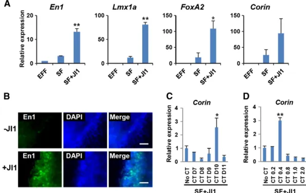
2011a, 2011b; van Oosten et al., 2012; Yang et al., 2010), would enrich the pool of intermediate cells which can be destined to iDPs by blocking the alternative path to the pluripotent state. As expected, temporal Jak inhibitor 1 (JI1) treatment from day 5 to day 7 significantly increased the expression of dopaminergic marker genes, such as En1, Lmx1a, FoxA2, and Corin, compared to untreated controls (Fig. 2A). Using immunocytochemistry, we also determined that En1-expressing cells were markedly increased in the
Figure 1 Direct reprogramming to dopaminergic neuronal progenitors through the temporal expression of pluripotent cell-specific reprogramming factors. (A) A schematic of the direct reprogramming of reprogrammable mouse embryonic fibroblasts to induced dopaminergic neuronal progenitors (iDPs). Induction of the four reprogramming factors for five days and subsequent exposure to SHH and FGF8 enabled the conversion to iDPs. (B) Bright-field images of direct reprogramming to iDPs on the designated day. Scale bars = 100μm. (C) Gene expression analysis showed that the major midbrain dopaminergic neuronal progenitor markers (Foxa2, Lmx1a, and Ngn2) were differentially expressed during the direct reprogramming to iDPs and to induced neural stem cells (iNSCs). All the values are relative to day 0. Mean ± s.e.m. (n = 3–5). (D) Immunocytochemical analysis of terminally differentiated cells from iNSCs or iDPs. The dopaminergic neuronal marker (TH, red)-expressing cells were more abundant in the cells differentiated from iDPs. The neuronal marker TUJ1 is shown in green. Scale bars = 100μm. (E) The percentage of TH-expressing neurons of the TUJ1-expressing neurons was calculated. Mean ± s.e.m. (n = 6), pb 0.01.
JI1-treated iDP population (Fig. 2B). Thus, we concluded that inhibition of Jak–Stat signaling in the intermediate cells is effective in enhancing the efficiency of direct repro-gramming to iDPs.
Second, we considered the contribution of Wnt signaling, which is important in the early (E9.5–E10.5) and late (E11.5– E12.5) stages of ventral midbrain dopaminergic neuronal development (Momcilovic et al., 2012; Prakash et al., 2006) and the differentiation of hESCs or iPSCs to dopaminergic neurons (Kirkeby et al., 2012; Kriks et al., 2011; Chung et al., 2009). We applied a specific inhibitor of Gsk3β, CT99021 (CT), to activate Wnt signaling and assessed with Corin expression, the most reliable marker of midbrain dopaminergic progenitors (Chung et al., 2011; Xi et al., 2012; Jonsson et al., 2009). We found an optimum time-frame and concentration of CT treatment to enhance the efficiency of reprogramming to iDPs (Fig. 2C and D). These results are similar to previous reports, in which both a specific concentration of CT and a particular time window of CT administration were necessary for specification to midbrain cells (Kirkeby et al., 2012; Xi et al., 2012). Additionally, Corin-expressing cells were only detectable in the JI1- and CT-treated cultures on day 13 (supplementary material Fig. S2). In summary, we found that combined inhibition of Jak and Gsk3β significantly enhanced the process of DP specification during our direct reprogramming. In addition, the concentration and
time-window of CT treatment need to be finely optimized to obtain the highest reprogramming efficiency.
iDPs can self-renew and generate functional ventral midbrain dopaminergic neurons
After we optimized iDP generation (Fig. 3A), we analyzed the characteristics of the dopaminergic neurons differentiated from the iDPs to prove the functionality of our reprogrammed iDPs. Most of the TH-expressing dopaminergic neurons co-stained with ventral midbrain precursor markers (FoxA2 and Lmx1a) and midbrain dopaminergic neuronal markers (Nurr1, En1, and Pitx3) (Studer, 2012) (Fig. 3B and supplementary material Fig. S3A), confirming that the differentiated dopami-nergic neurons manifest midbrain specificity. We also showed that the JI1-treated iDPs generated 44.3 ± 8.4% TH+/TUJ1+
neurons and the JI1 and CT co-treated iDPs generated 57.2 ± 7.2% TH+/TUJ1+ neurons (Fig. 3C; supplementary material
Fig. S3B), confirming that the reprogramming efficiency to iDPs is actually increased by the combined treatment. To assess whether the generated TH-expressing neurons are functional dopaminergic neurons, we measured the level of released dopamine in the culture medium. As expected, the neurons from iDPs of higher reprogramming efficiency released even more dopamine than other samples (Fig. 3D) and these
Figure 2 Enhancement of direct reprogramming to iDPs by Jak and Gsk3β inhibition. (A) Quantitative PCR analysis shows that the expressions of DP markers were increased by Jak inhibitor 1 (JI1) treatment from day 5 to day 7. EGF, FGF2, and FGF4 (EFF) were treated for iNSCs and SHH and FGF8 (SF) for iDPs. All the values are relative to iNSCs. (B) Immunocytochemical analysis shows increased En1 (a midbrain DP marker)-expressing cells by the JI1 treatment. Scale bars = 50μm. (C, D) Quantitative PCR analysis of Corin, a representative DP marker, on day 13 to find optimal duration and concentration of CT99021 (CT) treatment over SF + JI1 treatment as in panel A. (C) 0.5μM CT was treated from the indicated day until day 12. (D) Different concentrations (μM) of CT were treated from day 10 to day 12. All the values are relative to basal SF + JI1 treatment. Mean ± s.e.m. (n = 3), pb 0.01 (*), p b 0.001 (**). A statistical analysis was performed using Student's t-test.
dopamine releases were additionally increased by high-KCl solution (HK), a depolarizing condition. These results represent that the generated neurons from iDPs are functional dopami-nergic neurons which show responsive release of dopamine. Finally, we analyzed the electrophysiological properties of the neurons from iDPs. Under a current-clamp configuration, depolarizing current injections with 10-pA steps induced action potentials (APs) in ~ 42% of recorded cells (11/26 cells) (Fig. 3E). The generation of APs were blocked by
TTX treatment, indicating its dependency on voltage-gated sodium channels. A majority of cells (9/11 cells) showing APs also exhibited spontaneous firings (Fig. 3F) and rebound depolarizations resulting in AP generation after short hyper-polarizations (Fig. 3G), which are characteristics of midbrain dopamine neurons (Grace and Onn, 1989). The electrophysi-ological properties of differentiated cells from iDPs were described in supplementary material Table S1. We also tested whether the iDPs could self-renew in vitro. As previously
Figure 3 The iDPs were differentiated into typical ventral midbrain dopaminergic neurons. (A) A schematic of the optimized protocol for direct reprogramming into iDPs. (B) Immunocytochemical analysis of dopaminergic neuronal makers, such as TH and Nurr1, in the differentiated neurons derived from the CT- and JI1-treated iDPs. Scale bars = 50μm. (C) The percentages of TH+ neurons of the total neurons (TUJ1 +) differentiated from the reprogrammed cells under various conditions were analyzed by immunocytochemistry. Mean ± s.e.m. (n = 7–10), p b 0.001 (**). (D) Measurement of released dopamine after the treatment of low (LK) and high (HK) concentration of KCl on the differentiated cells from designated reprogrammed cells. (E) The representative traces of membrane potential changes and action potentials (Aps) elicited by step-current injections before and after an application of TTX. (F) Example trace of a cell exhibiting spontaneous APs at resting membrane potential. (G) Example trace of a cell showing rebound depolarizations.
reported (Chung et al., 2011), under FGF2 containing culture condition, the Corin-expressing progenitors were increased (Fig. S4A) and showed co-expression with Ki67, a marker for proliferating cells (Fig. S4B). The Corin-expressing popula-tion was maintained up to 16.22% by day 18 (Fig. S4C). These results show that our directly reprogrammed iDPs are bona fide progenitors which can generate functional and respon-sive dopaminergic neurons of midbrain. Considering that catecholaminergic neurons also express TH, our direct reprogramming strategy to iDPs endorses a robust protocol for generating the midbrain-specific dopaminergic neurons which are required for potential cell therapy and drug discovery for PD.
Adult mouse tail tip fibroblasts are successfully reprogrammed into iDPs
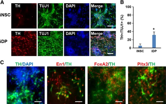
Lastly, we evaluated our direct reprogramming to iDPs with mouse adult tail-tip fibroblasts (TTFs) using the Dox-inducible STEMCCA system (Kim et al., 2011a; Sommer et al., 2009) to prove our reprogramming strategy is also effective with adult cells. As observed in iDPs from reprogrammable MEFs, we found highly efficient differentiation into TH-expressing neu-rons from TTF-iDPs compared to TTF-iNSCs (Fig. 4A and B). These TH-expressing neurons also expressed midbrain-specific markers, such as En1, FoxA2, and Pitx3 confirming the specification to midbrain DA neurons (Fig. 4C). Thus, adult mouse fibroblasts can be efficiently reprogrammed into ventral midbrain iDPs by our PDR strategy.
Conclusions
Recently, several groups reported direct conversion of fibroblasts into dopaminergic neurons (Pfisterer et al., 2011; Caiazzo et al., 2011; Liu et al., 2012b; Kim et al., 2011b), where the resulting cells are non-proliferating terminally differentiated neurons. We demonstrated here that mouse fibroblasts can be directly reprogrammed to functional and proliferating midbrain DPs through cell activation by pluripotency factors and directed specifica-tion by signal factors including SHH and FGF8. The trajectory of iDP reprogramming is traced as different from iNSC reprogramming. We were able to finely tune the process to increase the efficiency by co-inhibition of Jak and Gsk3β. The iDPs are functional and proliferating progenitors which give rise to typical midbrain dopami-nergic neurons. We expect that our PDR strategy is not only applicable for iDP generation but also for direct lineage reprogramming to other progenitors which are required for various neural diseases.
Supplementary data to this article can be found online at http://dx.doi.org/10.1016/j.scr.2013.09.007.
Author contributions
HSK, JK, and YSC designed and performed the experiments, evaluated the data, and wrote the manuscript. DJ performed the patch-clamp experiment and wrote the manuscript. YJ designed the research and analyzed the data.
Figure 4 Adult mouse tail tip fibroblasts were directly reprogrammed into iDPs (A) Immunocytochemical analysis of terminally differentiated cells from TTF-iNSCs or TTF-iDPs. The TH-expressing cells (red) were more abundant in the cells differentiated from TTF-iDPs. Scale bars = 100μm. (B) The percentage of TH-expressing neurons of total neurons (TUJ1+) differentiated from iNSCs and iDPs. A statistical analysis was performed using Student's t-test. Mean ± s.e.m., pb 0.01 (*). (C) Immunocytochemical analysis of midbrain dopaminergic neuronal markers in differentiated neurons. The TH + (green) neurons expressed dopaminergic neuronal markers (red), such as Foxa2, Pitx3, and En1. Scale bars = 50μm.
Acknowledgments
This work was supported by grants through the National Research Foundation of Korea (NRF) funded by the Ministry of Education, Science and Technology (2010-020272(3), 2012M3A9C7050224, NRF-2012R1A1A2043433, and 2011-0014893), and the KRIBB/KRCF research initiative program (NAP-09-3). We thank Dr. Sheng Ding in Gladstone Institute of Cardiovascular Disease for generous gifts of reprogram-mable cells and vector.
References
Takahashi, K., Tanabe, K., Ohnuki, M., Narita, M., Ichisaka, T., Tomoda, K., Yamanaka, S., 2007.Induction of pluripotent stem cells from adult human fibroblasts by defined factors. Cell 131,
861–872.
Takahashi, K., Yamanaka, S., 2006.Induction of pluripotent stem cells from mouse embryonic and adult fibroblast cultures by defined
factors. Cell 126, 663–676.
Sancho-Martinez, I., Baek, S.H., Izpisua Belmonte, J.C., 2012.Lineage conversion methodologies meet the reprogramming toolbox. Nat. Cell Biol. 14, 892–899.
Liu, G.H., Yi, F., Suzuki, K., Qu, J., Izpisua Belmonte, J.C., 2012a. Induced neural stem cells: a new tool for studying neural
development and neurological disorders. Cell Res. 22, 1087–1091.
Matsui, T., Takano, M., Yoshida, K., Ono, S., Fujisaki, C., Matsuzaki, Y., Toyama, Y., Nakamura, M., Okano, H., Akamatsu, W., 2012.Neural stem cells directly differentiated from partially reprogrammed fibroblasts rapidly acquire gliogenic competency. Stem Cells 30,
1109–1119.
Kim, J., Ambasudhan, R., Ding, S., 2012.Direct lineage reprogramming
to neural cells. Curr. Opin. Neurobiol. 22, 778–784.
Efe, J.A., Hilcove, S., Kim, J., Zhou, H., Ouyang, K., Wang, G., Chen, J., Ding, S., 2011.Conversion of mouse fibroblasts into cardiomyocytes using a direct reprogramming strategy. Nat. Cell
Biol. 13, 215–222.
Kim, J., Efe, J.A., Zhu, S., Talantova, M., Yuan, X., Wang, S., Lipton, S.A., Zhang, K., Ding, S., 2011a.Direct reprogramming of mouse fibroblasts to neural progenitors. Proc. Natl. Acad. Sci. U.
S. A. 108, 7838–7843.
Gaillard, A., Jaber, M., 2011.Rewiring the brain with cell
transplan-tation in Parkinson's disease. Trends Neurosci. 34, 124–133.
Ganat, Y.M., Calder, E.L., Kriks, S., Nelander, J., Tu, E.Y., Jia, F., Battista, D., Harrison, N., Parmar, M., Tomishima, M.J., Rutishauser, U., Studer, L., 2012.Identification of embryonic stem cell-derived midbrain dopaminergic neurons for
engraft-ment. J. Clin. Investig. 122, 2928–2939.
Pfisterer, U., Kirkeby, A., Torper, O., Wood, J., Nelander, J., Dufour, A., Bjorklund, A., Lindvall, O., Jakobsson, J., Parmar, M., 2011. Direct conversion of human fibroblasts to dopaminergic neurons.
Proc. Natl. Acad. Sci. U. S. A. 108, 10343–10348.
Caiazzo, M., Dell'Anno, M.T., Dvoretskova, E., Lazarevic, D., Taverna, S., Leo, D., Sotnikova, T.D., Menegon, A., Roncaglia, P., Colciago, G., Russo, G., Carninci, P., Pezzoli, G., Gainetdinov, R.R., Gustincich, S., Dityatev, A., Broccoli, V., 2011.Direct generation of functional dopaminergic neurons from mouse and human
fibroblasts. Nature 476, 224–227.
Liu, X., Li, F., Stubblefield, E.A., Blanchard, B., Richards, T.L., Larson, G.A., He, Y., Huang, Q., Tan, A.C., Zhang, D., Benke, T.A., Sladek, J.R., Zahniser, N.R., Li, C.Y., 2012b. Direct reprogramming of human fibroblasts into dopaminergic neuron-like cells. Cell Res. 22, 321–332.
Kim, J., Su, S.C., Wang, H., Cheng, A.W., Cassady, J.P., Lodato, M.A., Lengner, C.J., Chung, C.Y., Dawlaty, M.M., Tsai, L.H.,
Jaenisch, R., 2011b.Functional integration of dopaminergic neurons directly converted from mouse fibroblasts. Cell Stem
Cell 9, 413–419.
Rhee, Y.H., Ko, J.Y., Chang, M.Y., Yi, S.H., Kim, D., Kim, C.H., Shim, J.W., Jo, A.Y., Kim, B.W., Lee, H., Lee, S.H., Suh, W., Park, C.H., Koh, H.C., Lee, Y.S., Lanza, R., Kim, K.S., 2011.Protein-based human iPS cells efficiently generate functional dopamine neurons and can treat a rat model of Parkinson disease. J. Clin. Investig.
121, 2326–2335.
Wang, L., Huang, W., Su, H., Xue, Y., Su, Z., Liao, B., Wang, H., Bao, X., Qin, D., He, J., Wu, W., So, K.F., Pan, G., Pei, D., 2012. Generation of integration-free neural progenitor cells from cells in
human urine. Nat. Methods 10, 84–89.
Thier, M., Worsdorfer, P., Lakes, Y.B., Gorris, R., Herms, S., Opitz, T., Seiferling, D., Quandel, T., Hoffmann, P., Nothen, M.M., Brustle, O., Edenhofer, F., 2012.Direct conversion of fibroblasts into stably expandable neural stem cells. Cell stem
cell 10, 473–479.
Han, D.W., Tapia, N., Hermann, A., Hemmer, K., Hoing, S., Arauzo-Bravo, M.J., Zaehres, H., Wu, G., Frank, S., Moritz, S., Greber, B., Yang, J.H., Lee, H.T., Schwamborn, J.C., Storch, A., Scholer, H.R., 2012.Direct reprogramming of fibroblasts into neural stem cells by defined factors. Cell Stem Cell 10,
465–472.
Lu, J., Liu, H., Huang, C.T., Chen, H., Du, Z., Liu, Y., Sherafat, M.A., Zhang, S.C., 2013.Generation of integration-free and region-specific neural progenitors from primate fibroblasts.
Cell Rep. 3, 1580–1591.
Cho, M.S., Hwang, D.Y., Kim, D.W., 2008.Efficient derivation of functional dopaminergic neurons from human embryonic stem
cells on a large scale. Nat. Protoc. 3, 1888–1894.
Yan, Y., Yang, D., Zarnowska, E.D., Du, Z., Werbel, B., Valliere, C., Pearce, R.A., Thomson, J.A., Zhang, S.C., 2005.Directed differen-tiation of dopaminergic neuronal subtypes from human embryonic
stem cells. Stem Cells 23, 781–790.
Roussa, E., Krieglstein, K., 2004. Induction and specification of midbrain dopaminergic cells: focus on SHH, FGF8, and TGF-beta.
Cell Tissue Res. 318, 23–33.
Momcilovic, O., Montoya-Sack, J., Zeng, X., 2012. Dopaminergic differentiation using pluripotent stem cells. J. Cell. Biochem. 113,
3610–3619.
Swistowski, A., Peng, J., Liu, Q., Mali, P., Rao, M.S., Cheng, L., Zeng, X., 2010.Efficient generation of functional dopaminergic neurons from human induced pluripotent stem cells under defined
condi-tions. Stem Cells 28, 1893–1904.
Aguila, J.C., Hedlund, E., Sanchez-Pernaute, R., 2012. Cellular programming and reprogramming: sculpting cell fate for the production of dopamine neurons for cell therapy. Stem Cells Int.
2012, 412040.
Roybon, L., Hjalt, T., Christophersen, N.S., Li, J.Y., Brundin, P., 2008.Effects on differentiation of embryonic ventral midbrain progenitors by Lmx1a, Msx1, Ngn2, and Pitx3. J. Neurosci. Off.
J. Soc. Neurosci. 28, 3644–3656.
Studer, L., 2012.Derivation of dopaminergic neurons from pluripotent
stem cells. Prog. Brain Res. 200, 243–263.
Efe, J.A., Ding, S., 2011a.The evolving biology of small molecules: controlling cell fate and identity. Philos. Trans. R. Soc. Lond. Ser. B Biol. Sci. 366, 2208–2221.
Efe, J.A., Ding, S., 2011b.Reprogramming, transdifferentiation and the shifting landscape of cellular identity. Cell cycle 10,
1886–1887.
van Oosten, A.L., Costa, Y., Smith, A., Silva, J.C., 2012.JAK/ STAT3 signalling is sufficient and dominant over antagonistic cues for the establishment of naive pluripotency. Nat.
Commun. 3, 817.
Yang, J., van Oosten, A.L., Theunissen, T.W., Guo, G., Silva, J.C., Smith, A., 2010.Stat3 activation is limiting for reprogramming to
Prakash, N., Brodski, C., Naserke, T., Puelles, E., Gogoi, R., Hall, A., Panhuysen, M., Echevarria, D., Sussel, L., Weisenhorn, D.M., Martinez, S., Arenas, E., Simeone, A., Wurst, W., 2006. A Wnt1-regulated genetic network controls the identity and fate of midbrain-dopaminergic progenitors in vivo.
Develop-ment 133, 89–98.
Kirkeby, A., Grealish, S., Wolf, D.A., Nelander, J., Wood, J., Lundblad, M., Lindvall, O., Parmar, M., 2012. Generation of regionally specified neural progenitors and functional neurons from human embryonic stem cells under defined conditions. Cell
Rep. 1, 703–714.
Kriks, S., Shim, J.W., Piao, J., Ganat, Y.M., Wakeman, D.R., Xie, Z., Carrillo-Reid, L., Auyeung, G., Antonacci, C., Buch, A., Yang, L., Beal, M.F., Surmeier, D.J., Kordower, J.H., Tabar, V., Studer, L., 2011.Dopamine neurons derived from human ES cells efficiently engraft in animal models of Parkinson's disease. Nature 480, 547–551.
Chung, S., Leung, A., Han, B.S., Chang, M.Y., Moon, J.I., Kim, C.H., Hong, S., Pruszak, J., Isacson, O., Kim, K.S., 2009.Wnt1-lmx1a forms a novel autoregulatory loop and controls midbrain
dopami-nergic differentiation synergistically with the SHH–FoxA2 pathway.
Cell Stem Cell 5, 646–658.
Chung, S., Moon, J.I., Leung, A., Aldrich, D., Lukianov, S., Kitayama, Y., Park, S., Li, Y., Bolshakov, V.Y., Lamonerie, T., Kim, K.S., 2011. ES cell-derived renewable and functional midbrain dopaminergic
progenitors. Proc. Natl. Acad. Sci. U. S. A. 108, 9703–9708.
Xi, J., Liu, Y., Liu, H., Chen, H., Emborg, M.E., Zhang, S.C., 2012. Specification of midbrain dopamine neurons from primate
pluripotent stem cells. Stem Cells 30, 1655–1663.
Jonsson, M.E., Ono, Y., Bjorklund, A., Thompson, L.H., 2009. Identification of transplantable dopamine neuron precursors at different stages of midbrain neurogenesis. Exp. Neurol. 219,
341–354.
Grace, A.A., Onn, S.P., 1989. Morphology and electrophysiological properties of immunocytochemically identified rat dopamine neurons recorded in vitro. J. Neurosci. Off. J. Soc. Neurosci. 9,
3463–3481.
Sommer, C.A., Stadtfeld, M., Murphy, G.J., Hochedlinger, K., Kotton, D.N., Mostoslavsky, G., 2009.Induced pluripotent stem cell generation using a single lentiviral stem cell cassette. Stem
Cells 27, 543–549.
Trzaska, K.A., Kuzhikandathil, E.V., Rameshwar, P., 2007. Specifica-tion of a dopaminergic phenotype from adult human mesenchymal
stem cells. Stem Cells 25, 2797–2808.
Jung, S., Yang, H., Kim, B.S., Chu, K., Lee, S.K., Jeon, D., 2012.The immunosuppressant cyclosporin A inhibits recurrent seizures in an experimental model of temporal lobe epilepsy. Neurosci.
Lett. 529, 133–138.
Jeon, D., Song, I., Guido, W., Kim, K., Kim, E., Oh, U., Shin, H.S., 2008.
Ablation of Ca2+channel beta3 subunit leads to enhanced
N-methyl-D-aspartate receptor-dependent long term potentiation and


