(
ⴚ)-Epicatechin Gallate (ECG) Stimulates Osteoblast
Differentiation via Runt-related Transcription Factor 2
(RUNX2) and Transcriptional Coactivator with PDZ-binding
Motif (TAZ)-mediated Transcriptional Activation
*
Received for publication, September 27, 2013, and in revised form, February 6, 2014Published, JBC Papers in Press, February 10, 2014, DOI 10.1074/jbc.M113.522870 Mi Ran Byun‡1, Mi Kyung Sung‡1, A Rum Kim‡, Cham Han Lee‡, Eun Jung Jang§, Mi Gyeong Jeong§, Minsoo Noh¶, Eun Sook Hwang§2, and Jeong-Ho Hong‡3
From the‡Division of Life Sciences, Korea University, Seoul 136-701, Korea, the§College of Pharmacy and Global Top5 Research
Program, Ewha Womans University, Seoul 120-750, Korea, and the¶College of Pharmacy, Seoul National University,
Seoul 151-742, South Korea
Background:Catechins in green tea have a beneficial effect in bone formation, but the detailed mechanism is not fully understood.
Results:ECG, a major compound of green tea, stimulates TAZ- and RUNX2-mediated osteogenic gene transcription through PP1A.
Conclusion:ECG stimulates osteoblast differentiation through a transcriptional activation.
Significance:A novel mechanism for green tea-stimulated osteoblast differentiation is revealed.
Osteoporosis is a degenerative bone disease characterized by low bone mass and is caused by an imbalance between osteoblas-tic bone formation and osteoclasosteoblas-tic bone resorption. It is known that the bioactive compounds present in green tea increase osteogenic activity and decrease the risk of fracture by improv-ing bone mineral density. However, the detailed mechanism underlying these beneficial effects has yet to be elucidated. In this study, we investigated the osteogenic effect of ( ⴚ)-epicat-echin gallate (ECG), a major bioactive compound found in green tea. We found that ECG effectively stimulates osteoblast differ-entiation, indicated by the increased expression of osteoblastic marker genes. Up-regulation of osteoblast marker genes is mediated by increased expression and interaction of the tran-scriptional coactivator with PDZ-binding motif (TAZ) and Runt-related transcription factor 2 (RUNX2). ECG facilitates nuclear localization of TAZ through PP1A. PP1A is essential for osteoblast differentiation because inhibition of PP1A activity was shown to suppress ECG-mediated osteogenic differentia-tion. Taken together, the results showed that ECG stimulates osteoblast differentiation through the activation of TAZ and RUNX2, revealing a novel mechanism for green tea-stimulated osteoblast differentiation.
Osteoporosis is a bone disease that leads to reduced bone mineral density and an increased risk of fracture. Bone mineral
density is maintained by the balance between osteoclastic bone resorption and osteoblastic bone formation. Therefore, com-pounds that increase osteoblastic activity or decrease osteoclas-tic activity have been considered as potential drug candidates for osteoporosis (1, 2).
Green tea is a popular beverage in Asian countries, and it is made from the leaves of Camellia sinensis. Green tea contains several polyphenolic compounds called catechins, including (⫺)-epigallocatechin-3-gallate (EGCG),4(⫺)-epigallocatechin, (⫺)-epicatechin-3-gallate (ECG), and (⫺)-epicatechin (3). Pre-vious studies have reported a correlation between tea con-sumption and the prevention of age-related bone loss in the elderly human population (4). Green tea polyphenols have been shown to improve bone mass and microarchitecture in various animal models, including ovarectomized aged female rats (5). Among the green tea catechins, EGCG is the most abundant, and its biological relevance in osteoblast differentiation and bone formation has been evaluated by several investigators (6). In mesenchymal stem cells, EGCG induces alkaline phospha-tase activity and stimulates the expression of osteoblast marker genes, such as Runx2 (Runt-related transcription factor 2), osterix, and osteocalcin (7). EGCG was shown to increase min-eralized bone module formation in human osteoblast-like SaOS-2 cells (8). Further, studies show that EGCG has a vital role in osteogenic induction and inhibits the SAPK/JNK pathway by suppressing transforming growth factor- (TGF) and the pros-taglandin D2-mediated induction of HSP27 (9, 10). EGCG may also stimulate osteogenesis by increasing the synthesis of prosta-glandin F2-induced vascular endothelial growth factor (6). How-ever, the mechanism of action of other catechin compounds on the osteogenesis of bone has yet to be investigated.
*This work was supported by the Korea Healthcare Technology R&D project grant, Ministry for Health and Welfare Grants A120349 and A120476, and Basic Science Research Program Grant 2011-0022926 through the National Research Foundation of Korea, Republic of Korea. This work was also sup-ported by a Korea University grant (to M. R. B.).
1Both authors contributed equally to this work.
2To whom correspondence may be addressed. Tel.: 82-2-3277-4369; E-mail:
3To whom correspondence may be addressed. Tel.: 82-2-3290-3427; Fax:
82-2-927-9028; E-mail: [email protected].
4The abbreviations used are: EGCG, (⫺)-epigallocatechin-3-gallate; ECG,
(⫺)-epicatechin gallate; hMSCs, human mesenchymal stem cells; PP1A, protein phosphatase 1A; qRT-PCR, quantitative RT-PCR.
at Ewha Medical Library on April 27, 2016
http://www.jbc.org/
Osteoblast differentiation is critical for osteogenesis, and osteoblast-specific gene products regulate the differentiation process (11, 12). RUNX2 is a key transcription factor and a central regulator of bone formation, which mediates the tem-poral activation or repression of cell growth and phenotypic genes that regulate osteoblast-specific target genes, such as osteocalcin, during osteoblast differentiation (13, 14). Runx2 knock-out mice show a complete lack of ossification (13, 14). RUNX2 interacts with a spectrum of transcription factors and coregulatory proteins that modulate its functions (15).
TAZ (transcriptional coactivator with PDZ-binding motif) is a 14-3-3-binding protein that regulates cell differentiation, pro-liferation, and stem cell renewal. TAZ functions as a transcrip-tional co-regulator and interacts with several transcription fac-tors, including RUNX2 and PPAR␥ (16–24). TAZ stimulates osteogenic differentiation through RUNX2-mediated gene transcription while inhibiting adipogenic differentiation through the suppression of PPAR␥-mediated gene transcrip-tion (16). The nuclear localizatranscrip-tion of TAZ is important for its binding with transcription factors and the activation of target genes. Extracellular signals, including Hippo, TGF, and Wnt pathways, regulate the localization and activity of TAZ. The Hippo signal regulates TAZ-mediated cell proliferation and tumorigenesis (25, 26). The canonical Wnt signal regulates osteogenic and adipogenic differentiation through the stabili-zation of TAZ (27). In this study, we report that ECG activates TAZ and stimulates the RUNX2-mediated gene transcription during osteoblast differentiation.
MATERIALS AND METHODS
Reagents and Cell Lines—(⫺)-Epicatechin gallate, ascorbic acid, -glycerophosphate, fast blue BB salt, and naphthol AS-MX phosphate were purchased from Sigma-Aldrich. C3H10T1/2 cells were purchased from the American Type Cul-ture Collection, and bone marrow-derived human mesenchy-mal stem cells (hMSCs) were obtained from Lonza.
Cell Culture and Osteoblast Differentiation—C3H10T1/2 cells were cultured in DMEM supplemented with 10% FBS, 100 units/ml penicillin, and 100g/ml streptomycin. For the osteo-genic differentiation of C3H10T1/2 cells, 8⫻ 104cells/well were seeded in 12-well culture plates. After 2 days, the culture medium was replaced with DMEM containing 50g/ml ascor-bic acid, 10 mM-glycerophosphate, and 10% FBS (osteoblast
differentiation medium). For the osteogenic differentiation of hMSCs, cells were plated in 12-well culture plates at a density of 3⫻ 104cells/cm2, and 48 h later, the medium was replaced with osteoblast differentiation medium containing 0.5M
dexam-ethasone. The differentiation medium was replaced every 2 days for differentiation of C3H10T1/2 and hMSCs.
Alkaline Phosphatase Staining and Enzyme Activity Assay— For alkaline phosphatase staining, differentiated osteoblast cells were fixed with 3.7% formaldehyde at room temperature for 10 min and then stained with 0.1 mg/ml naphthol AS-MX phosphate, 0.5% N,N-dimethylformamide, 2 mM MgCl2, 0.6
mg/ml fast blue BB salt, and 0.1MTris-HCl (pH 8.5) for 30 min
at room temperature. To determine alkaline phosphatase enzyme activity, cells were lysed in 25 mMHEPES (pH 7.6), 0.1%
Triton X-100, and 0.9% NaCl, and the cell lysates were
incu-bated with p- nitrophenyl phosphate substrate solution for 1 h at 37 °C, and 3 MNaOH solution was added to the reaction
mixture to stop the reaction. The absorbance of alkaline phos-phatase activity was measured at 405 nm using a microplate reader (Bio-Rad model 680).
Immunoblot Analysis—Immunoblot analysis was performed using the following primary antibodies: TAZ (28), RUNX2 (MBL, D130-3), HA (Covance), ␣-tubulin (Abfrontier, LF-PA0146), Lamin B1 (Santa Cruz Biotechnology, Inc., sc-20682), Erk1/2 (Cell Signaling Technology, catalog no. 9102), phospho-Erk1/2 (Cell Signaling Technology, catalog no. 9101), JNK (Cell Signaling Technology, catalog no. 9252), phospho-JNK (Cell Signaling Technology, catalog no. 9251), p38 MAPK (Cell Signaling Tech-nology, catalog no. 9212), phospho-p38 MAPK (Cell Signaling Technology, catalog no. 9211), AKT (Santa Cruz Biotechnology, sc-8312), phospho-AKT (Thr-308) (Cell Signaling Technology, catalog no. 9275), GSK-3 (Santa Cruz Biotechnology, sc-9166), phospho-GSK-3 (Ser-9) (Cell Signaling Technology, catalog no. 9323), and-actin (Sigma-Aldrich, A5441).
Stable Cell Lines—Phoenix cells were transfected using the calcium phosphate-mediated transfection method with pBabe-puro (Bp), pBabepBabe-puro-TAZ (T), or pBabepBabe-puro-mTAZ⌬WW (T⌬WW) expression plasmids, which have been described pre-viously (16). Viral supernatants were harvested and added to the C3H10T1/2 cells cultured in DMEM containing 4g/ml Polybrene. The cells were further incubated with 2g/ml puro-mycin for 6 days to eliminate the uninfected cells. The stable cell lines were used in follow-up experiments. The expression of TAZ was analyzed by immunoblot analysis.
Quantitative Real-time PCR Analysis—Total RNA was iso-lated from differentiated osteogenic cells using TRIzol reagent (Invitrogen), and cDNA was generated using RevertAid reverse transcriptase (Thermo Scientific). Expression of osteogenic marker genes, including osteocalcin (Oc), osteopontin (Opn),
TAZ, Runx2, and PP1A, was analyzed by quantitative RT-PCR. Expression of mRNA was normalized using GAPDH. Quantitative RT-PCR was performed using the primers described in Table 1.
Chromatin Immunoprecipitation—Differentiated osteoblast cells were cross-linked with 0.75% formaldehyde and lysed in buffer containing 50 mMHEPES-KOH (pH 7.5), 140 mMNaCl,
1 mMEDTA (pH 8.0), 1% Triton X-100, 0.1% sodium
deoxy-cholate, 0.1% SDS, and protease inhibitors. The immunopre-cipitation, reverse cross-linking, and DNA purification were performed as described previously (29). The promoter region of the osteocalcin gene was amplified using the following primers: osteocalcin promoter F, 5 ⬘-CTGAACTGGGCAAATGAGG-ACA-3⬘; osteocalcin promoter R, 5⬘-AGGGGATGCTGCCA-GGACTAAT-3⬘.
Fractionation—C3H10T1/2 cells were differentiated with or without 10MECG for 6 days and harvested with hypotonic
buffer (20 mMHEPES, 10 mMKCl, 2 mMMgCl2, 1 mMEDTA,
and protease inhibitor) for 5 min, and the lysed cells were cen-trifuged at 10,000⫻ g for 10 min. After that, soluble superna-tants were collected for cytosolic protein, and then the pellets were lysed with radioimmune precipitation buffer for nuclear protein. Fractionated protein was analyzed by immunoblot analysis.
at Ewha Medical Library on April 27, 2016
http://www.jbc.org/
Luciferase Assay—The 293T cells were transfected with 6XOSE2-luciferase reporter, RUNX2, and TAZ plasmids using X-tremeGene 9 DNA transfection reagent (Roche Applied Sci-ence). 6XOSE2-luciferase reporter and RUNX2 plasmids were a gift from R. Derynck (University of California, San Francisco, CA) and Yoshiaki Ito (Institute of Molecular and Cell Biology, Sin-gapore), respectively. The culture medium was replaced with DMEM containing 10MECG 24 h post-transfection. Luciferase
activity was measured with the luciferase assay system (Promega, E1501), using a luminometer (Promega, Glomax威).
PP1A siRNA Transfection—C3H10T1/2 cells were seeded at a density of 4⫻ 104cells/cm2in 12-well culture plates, trans-fected with PP1A siRNA duplex using Lipofectamine 2000 (Invitrogen), and, 24 h after transfection, incubated with osteo-genic differentiation medium in the presence or absence of 10 MECG for 8 days. PP1A siRNA target sequences were as
fol-lows: mouse PP1A siRNA target sequence 1, 5 ⬘-CCATTCT-TCTGGAGCTTGA-3⬘; mouse PP1A siRNA target sequence 2, 5⬘-TCTCCAGACTTGCAATCCA-3⬘.
Immunocytochemistry—C3H10T1/2 cells were plated on 12-mm coverglasses and treated with or without 10MECG for
24 h. Then cells were fixed with 4% paraformaldehyde, permea-bilized with 0.05% Triton X-100, and incubated with TAZ-specific antibody (prepared with the TAZ-TAZ-specific peptide SSGGHPGPRLAGGA at Covance (Boston, MA)) overnight at 4 °C. After primary antibody incubation (1:100 dilution), cells were washed three times with 1⫻ PBS containing 0.05% BSA and further incubated with FITC-conjugated secondary antibody. The green fluorescent TAZ signal was observed by confocal micros-copy (Carl Zeiss LSM 510 Meta confocal microscope).
Mitogen-activated Protein Kinase (MAPK) Activity Assay— For the kinase activity assay, C3H10T1/2 cells were plated at a density of 2⫻ 105cells/well with 6-well plate, and 24 h later, the culture medium was replaced with DMEM containing 0.1% FBS for 16 h. After the serum starvation, the cells were treated with 2 ng/ml EGF (R&D Systems) or 10MECG for 30 min and lysed
with 2⫻ SDS sample buffer (125 mMTris-HCl (pH 6.8), 5%
-mer-captoethanol, 15% glycerol, 0.01% bromphenol blue, 4% SDS). MAPK activities were analyzed by immunoblot analysis.
Endogenous RUNX2 Immunoprecipitation—C3H10T1/2 cells were incubated in osteoblast differentiation medium with or without 10MECG for 6 days. The differentiated cells were
lysed with radioimmune precipitation assay buffer (150 mM
NaCl, 50 mMTris-HCl (pH 7.5), 1% Nonidet P-40, 0.5% sodium
deoxycholate, 0.1% SDS, 1 mMEDTA, and protease inhibitors),
and immunoprecipitated with 1g of immunoglobulin G (IgG)
or TAZ antibody (BD Biosciences, catalog no. 560235) over-night. Endogenous immune complexes were analyzed by immunoblot analysis.
RESULTS
ECG Stimulates Osteoblast Differentiation—To study the effect of ECG (Fig. 1A) on osteoblast differentiation, C3H10T1/2 cells were cultivated in osteogenic differentiation medium with increasing concentrations of ECG. To assess differentiation, alkaline phosphatase activity, an osteogenic marker, was ana-lyzed at 2, 4, and 6 days. As shown in Fig. 1, B and C, a dose-dependent induction of alkaline phosphatase activity was observed. To study the activity of ECG further, expression of osteoblast marker genes, including TAZ, Runx2, and osteopon-tin, was analyzed after 6 days of differentiation using qRT-PCR. As shown in Fig. 1D, a dose-dependent increase in the expres-sion of the marker genes was observed (Fig. 1D). We also observed a significant, dose-dependent increase in the expres-sion of the TAZ and RUNX2 proteins during osteogenic differ-entiation (Fig. 1E). Thus, the results suggest that ECG stimu-lates osteogenic differentiation by inducing the expression of TAZ and RUNX2.
ECG Stimulates RUNX2-mediated Gene Transcription— TAZ physically interacts with RUNX2 and activates RUNX2-mediated gene transcription (16) of osteoblastic marker genes, including osteocalcin. Thus, to study whether ECG stimulates RUNX2-mediated gene transcription through TAZ, luciferase reporter constructs containing RUNX2-binding sites (6xOSE2-luc) were used. The reporter plasmids were transfected into 293T cells with the RUNX2 and TAZ expression plasmids in the presence of ECG. As shown in Fig. 2A, RUNX2 and TAZ stimulated reporter activity, and the presence of ECG further increased reporter activity by 50%, suggesting that ECG stimu-lates RUNX2 and that TAZ mediates the transcription of osteo-blastic genes.
Next, to understand the mechanism of ECG-induced reporter activity, we investigated the physical interaction between TAZ and RUNX2 in the presence of ECG. Tagged TAZ and RUNX2 plas-mids were transfected into 293T cells, and the physical interac-tion of TAZ and RUNX2 was analyzed by immunoprecipita-tion. As shown in Fig. 2B, ECG significantly enhanced the physical interaction of TAZ and RUNX2. We further investi-gated the interaction. Endogenous TAZ proteins were immu-noprecipitated, and the bound proteins were analyzed with RUNX2 antibody. We observed that endogenous TAZ interacts with RUNX2, and ECG facilitates the interaction (Fig. 2C). TABLE 1
qRT-PCR primers
Gene Forward primer Reverse primer
Mouse Oc 5⬘-CTGACCTCACAGATGCCAAGC-3⬘ 5⬘-TGGTCTGATAGCTCGTCACAAG-3⬘ Mouse Opn 5⬘-GATTTGCTTTTGCCTGTTTGG-3⬘ 5⬘-TGAGCTGCCAGAATCAGTCACT-3⬘ Mouse Taz 5⬘-GTCACCAACAGTAGCTCAGATC-3⬘ 5⬘-GGACACTGTAGCACCCTAACCCCA-3⬘ Mouse Runx2 5⬘-CGGCCCTCCCTGAACTCT-3⬘ 5⬘-CGGTGGGGAAGACTGTGCCTG-3⬘ Mouse Pp1a 5⬘-GACAGTTGGTGACACTCTTCTC-3⬘ 5⬘-CTTGAGGATCTGGAAGGAACAC-3⬘ Mouse Gapdh 5⬘-GCTTGTCATCAACGGGAAG-3⬘ 5⬘-GATGTTAGTGGGGTCTCG-3⬘ Human TAZ 5⬘-CCTCTTCAATGATGTAGAGTCTGC-3⬘ 5⬘-AGTGATTACAGCCAGGTTAGAAAG-3⬘ Human MSX2 5⬘-CTACCCGTTCCATAGACCTGT-3⬘ 5⬘-GAGAGGGAGAGGAAACCCTTT-3⬘ Human DLX5 5⬘-CTACAACCGCGTCCCAAG-3⬘ 5⬘-GCCATTCACCATTCTCACCT-3⬘ Human RUNX2 5⬘-AGAGGTACCAGATGGGACTGT-3⬘ 5⬘-GGTAGCTACTTGGGGAGGATT-3⬘ Human GAPDH 5⬘-GACCCCTTCATTGACCTCAAC-3⬘ 5⬘-GGTGCCATGGAATTTGCCATG-3⬘
at Ewha Medical Library on April 27, 2016
http://www.jbc.org/
These results suggest that the increased interaction between TAZ and RUNX2 stimulates RUNX2-mediated osteogenic gene transcription.
Next, to study whether TAZ induces osteogenic marker genes through an endogenous RUNX2-binding site in the pres-ence of ECG, chromatin immunoprecipitation analysis was performed with cells overexpressing FLAG-tagged TAZ or WW domain-deleted TAZ, which cannot interact with RUNX2 (Fig. 2, D and E). The chromatin fragments that co-precipitated with the FLAG-tagged proteins were analyzed with PCR prim-ers spanning the RUNX2-binding site of the osteocalcin pro-moter. As shown in Fig. 2, F and G, FLAG-TAZ was recruited into the RUNX2-binding site of the osteocalcin gene promoter, and a 2-fold increase in the recruitment of FLAG-TAZ was observed in the presence of ECG. However, WW-deleted TAZ was not recruited into the promoter (Fig. 2, F and G). These results showed that ECG stimulates osteocalcin expression through the recruitment of TAZ at the endogenous RUNX2-binding site of osteocalcin promoter in differentiating cells.
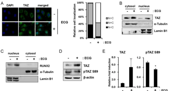
ECG Stimulates Nuclear Localization of TAZ—Increased recruitment of TAZ at the endogenous osteocalcin promoter indicated that TAZ preferentially localizes to the nucleus in the presence of ECG. The cellular distribution of TAZ after ECG treatment was evaluated using immunocytochemistry. As shown in Fig. 3A, ECG facilitated nuclear localization com-pared with the control, and predominant nuclear localization of
TAZ was observed in over 50% of cells. To further analyze the nuclear localization of TAZ, ECG-treated cells were prepared, and cytosolic and nuclear protein fractions were isolated. As in Fig. 3B, a significant increase in TAZ was observed in the nucleus of ECG-treated cells. We also observed that RUNX2 expression was significantly increased in nuclei after ECG treat-ment (Fig. 3C). Thus, these results suggest that ECG increases nuclear localized TAZ and RUNX2 and stimulates TAZ-medi-ated gene transcription.
The subcellular localization of TAZ is determined by its phosphorylation status (28). The phosphorylation of TAZ at serine 89 induces 14-3-3 binding and cytosolic sequestration of TAZ, whereas dephosphorylated TAZ is not subjected to 14-3-3 binding and localizes to the nucleus (16, 28). Therefore, we studied the phosphorylation status of TAZ at serine 89 using a phospho-specific antibody. There was a significant increase in total TAZ protein levels, although phosphorylation at serine 89 was reduced in the presence of ECG (Fig. 3, D and E). These results suggest that ECG may regulate the phosphorylation sta-tus of TAZ at serine 89.
ECG Stimulates PP1A Expression and Stimulates Dephos-phorylation of TAZ—Hippo signal regulates organ size by mod-ulating cell proliferation, cell death, and cell differentiation (30, 31). Lats kinase is a core component of the Hippo signaling pathway and has been reported to phosphorylate human TAZ (25) at serine 311 and the mouse TAZ at serine 306. Casein FIGURE 1. ECG stimulates the differentiation of osteoblasts. A, structure of ECG. B, ECG increases alkaline phosphatase activity in a dose-dependent manner. C3H10T1/2 cells were incubated in osteogenic differentiation medium in the presence of ECG at the indicated concentration. At 6 days after differentiation, alkaline phosphatase activity was visualized by staining to determine the osteogenic potential of ECG. A blue color indicates increased alkaline phosphatase activity. C, alkaline phosphatase activity in B was analyzed at the indicated time points. DMSO was used as a vehicle. **, p⬍ 0.05 by Student’s t test. D, ECG stimulates the expression of osteoblastic marker genes. C3H10T1/2 cells were incubated in osteoblastic differentiation medium in the presence of 10MECG. After 6 days of differentiation, the cells were harvested, and total RNA was obtained. Using qRT-PCR, the expression of TAZ, Runx2, and Opn was analyzed. Their relative expression was calculated after normalization to the GAPDH level. *, p⬍ 0.01; **, p ⬍ 0.05 by Student’s t test. E, C3H10T1/2 cells were treated with ECG and induced to differentiate for 6 days. Whole-cell extracts were harvested, resolved by SDS-PAGE, and analyzed for TAZ, RUNX2, and-actin levels using immunoblotting. Error bars, S.D.
at Ewha Medical Library on April 27, 2016
http://www.jbc.org/
kinase-1 further phosphorylates TAZ and induces its ubiquitin-mediated proteolytic degradation (25). The phosphorylation of TAZ is a reversible process, and dephosphorylation occurs by PP1A phosphatase (32). PP1 is a eukaryotic Ser/Thr protein phosphatase involved in diverse cellular functions (33). PP1A is a catalytic subunit of PP1. Therefore, we studied whether PP1A activity is important for ECG-induced TAZ expression. First, to study whether ECG can stabilize TAZ, C3H10T1/2 cells were incubated with ECG in the presence of cycloheximide, a protein synthesis inhibitor. As shown in Fig. 4A, we observed that TAZ was more stable in the presence of ECG than control, suggest-ing that ECG plays a role in the stabilization of TAZ. Next, C3H10T1/2 cells were treated with a PP1A inhibitor, okadaic acid, for 3 h. As shown in Fig. 4B, we observed that okadaic acid significantly decreased the TAZ expression that was induced by
ECG. However, the decrease in TAZ expression was not caused by the inhibition of transcription because TAZ mRNA expres-sion was not affected by the 3-h okadaic acid treatment (Fig. 4B). These results indicate that okadaic acid regulates TAZ expression at a post-transcriptional level. To further analyze the effect of PP1A activity, we depleted endogenous PP1A protein using siRNAs. Fig. 4C shows that PP1A was depleted by the two siRNAs tested, which resulted in a significant decrease in the expression of TAZ, suggesting that PP1A plays an impor-tant role in ECG-induced TAZ expression.
It is noteworthy that ECG induced the expression of PP1A by about 3-fold (Fig. 4, B and C), suggesting that the PP1A induction plays an important role in ECG-mediated osteogenic stimulation. Next, to study the effect of PP1A on ECG-mediated osteogenic differentiation, the control and PP1A siRNA-treated cells were FIGURE 2. ECG stimulates RUNX2-mediated osteoblast differentiation. A, stimulation of RUNX2-driven gene expression by ECG. 293T cells were transfected with the RUNX2 expression plasmid (0.005g/well) and the 6xOSE2-luciferase reporter construct (0.05 g/well), which contains six copies of the RUNX2-binding site in the osteocalcin promoter. After 24 h of transfection, the cells were incubated with 10MECG. After 24 h, cell lysates were prepared to analyze the luciferase activities. Differences in the transfection efficiency were adjusted by normalizing the firefly luciferase activity to that of Renilla luciferase. The luciferase activity was calculated and expressed as -fold induction. *, p⬍ 0.01, t test. B, 293T cells were transfected with HA-tagged RUNX2 and/or FLAG-tagged TAZ expression plasmids and incubated with 10MECG for 24 h. Whole-cell lysates (WCL) were precipitated (IP) with FLAG-M2-agarose beads. The precipitates and whole-cell lysates were analyzed by immunoblot analysis (IB) with antibodies against HA and TAZ. C, C3H10T1/2 cells were incubated in the presence or absence of 10MECG, and the cell lysates were immunoprecipitated with an IgG or anti-TAZ antibody. Endogenous TAZ bound to RUNX2 was analyzed by immunoblot analysis. D, TAZ structures. TAZ WW domain, which interacts with RUNX2, is deleted in TAZ⌬WW. aa, amino acids. E, FLAG-tagged TAZ wild type (T) and TAZ⌬WW-expressing (T⌬WW) C3H10T1/2 cells were prepared by retrovirus. The TAZ expression of stable cell lines was analyzed by immunoblot analysis. The expression of-actin was analyzed as a loading control. Bp, control cells. F, ECG increases recruitment of TAZ, not TAZ ⌬WW, at the endogenous osteocalcin promoter. The above cells were treated for 4 days with osteogenic differentiation medium in the presence of 10MECG, and enriched DNA immunoprecipitated using anti-FLAG antibodies was analyzed for osteocalcin promoter occupancy by PCR. ECG increases TAZ wild type recruitment, not TAZ ⌬WW, into the osteocalcin promoter. G, the recruited TAZ in F were quantitatively analyzed by qRT-PCR. Recovered DNAs from input DNAs were quantified by analyzing the Ct value. *, p⬍ 0.01 by Student’s t test. Error bars, S.E.
at Ewha Medical Library on April 27, 2016
http://www.jbc.org/
maintained in osteogenic differentiation medium. Fig. 4D indi-cates that ECG enhanced the osteogenic potential of C3H10T1/2 cells, as evidenced by the increased alkaline phosphatase activity. However, PP1A depletion using siRNAs led to marked reduc-tion of osteogenic potential. The results were verified by ana-lyzing the expression of osteogenic marker genes in parallel. As shown in Fig. 4E, ECG up-regulated osteopontin, Runx2, TAZ, and osteocalcin expression, whereas PP1A depletion signif-icantly reduced expression of osteogenic markers, suggest-ing that PP1A is an important mediator of ECG-mediated osteogenesis.
ECG Stimulates p38 MAPK—MAPKs are serine/threonine kinases that are involved in osteogenic differentiation (34). To study whether ECG activates the MAPKs, active kinases were analyzed using phospho-specific antibodies. As shown in Fig. 5A, p38 MAPK, not ERK and JNK, is significantly activated after ECG treatment. We also observed that ECG weakly stimulates the AKT activity and phosphorylates GSK3 for its inhibition (Fig. 5B). Thus, these results suggest that ECG may activate osteogenic differentiation through p38 MAPK.
ECG Stimulates the Differentiation of Human Mesenchymal Stem Cells into Osteoblasts— hMSCs are of mesodermal origin and are multipotent cells that can differentiate into osteoblasts, chondrocytes, and adipocytes. Here, we investigated the poten-tial of ECG to stimulate differentiation of hMSCs into osteo-blasts. As shown in Fig. 6A, ECG stimulated alkaline phospha-tase activity in a dose-dependent manner. In addition, the osteogenic marker genes DLX5, MSX2, Runx2, and TAZ were up-regulated in a dose-dependent manner (Fig. 6B). TAZ and RUNX2 protein expression was also increased after ECG treat-ment (Fig. 6C). These results suggest that ECG holds promise as
a therapeutic compound for induction of osteogenesis in osteo-porosis patients.
DISCUSSION
Epidemiological studies have established a correlation between green tea consumption and the prevention of age-related bone loss (4). Among the components of green tea, catechins have received much attention for their potentially beneficial effects on osteo-genesis (5). However, detailed functional mechanisms underly-ing the role of catechins in osteogenesis have yet to be under-stood. In this study, we observed that ECG, one of the major catechins found in green tea, stimulates osteoblast differentia-tion through a mechanism mediated by RUNX2, the master regulator of osteoblast marker gene transcription. Further, ECG enhances the transcriptional and post-transcriptional expression of TAZ, a transcriptional coregulator involved in osteogenesis (Fig. 7). During osteogenesis, TAZ mRNA expres-sion is up-regulated, and the resultant TAZ protein is stabilized by PP1A phosphatase-mediated dephosphorylation (Figs. 1 and 4). The proteolytic degradation of TAZ is dependent on its phosphorylation status. When the phosphodegron motif at its C terminus is phosphorylated by Lats and casein kinase-1, it is recognized by proteasomal complexes and ubiquitinated for degradation (25). Dephosphorylation of TAZ is accomplished by the PP1A phosphatase, which, in turn, increases its nuclear localization. Interestingly, ECG also decreases the phosphory-lation of TAZ at serine 89, stimulating its nuclear localization and, therefore, its transcriptional activity (Fig. 3). On the con-trary, PP1A knockdown led to decreased TAZ expression and osteogenic potential (Fig. 4). Thus, these results show that ECG regulates osteogenic differentiation through PP1A.
FIGURE 3. ECG increases nuclear localization of TAZ. A, C3H10T1/2 cells were incubated with 10MECG. After 24 h, the cells were fixed, and the cellular location of TAZ was analyzed by immunocytochemistry. An FITC-conjugated secondary antibody was used for the green fluorescence signal. DAPI staining indicates the nuclei of the cells. The right-hand panel shows quantitative analysis of TAZ localization. The cellular distribution of TAZ was analyzed based on whether TAZ levels were higher in the nucleus (N⬎ C), higher in the cytoplasm (N ⬍ C) or evenly distributed between the nucleus and cytoplasm (N ⫽ C). The percentage of cells in each category was determined after observing cells in five different microscopic fields. B and C, C3H10T1/2 cells were incubated with vehicle or 10MECG for 24 h, and the cell lysates were prepared and fractionated into cytosol and nuclear extracts according to the indicated methods. TAZ (B) and RUNX2 (C) expression was analyzed by immunoblot analysis. D, C3H10T1/2 cells were treated for 2 days with osteogenic differentiation medium in the absence or presence of 10MECG; cell lysates were prepared, and the phosphorylation status of TAZ at serine 89 was analyzed with a phospho-specific TAZ antibody, which was prepared with TAZ phosphopeptides (CHVRSHpSSPASL) at AbFrontier (Seoul, Korea). E, quantitative analysis of total and phosphorylated TAZ. The amount of total and phosphorylated TAZ at serine 89 from three independent experiments of D was analyzed with a densitometer, and relative -fold induction is shown here. *, p⬍ 0.01; **, p ⬍ 0.05 by Student’s t test. Error bars, S.E.
at Ewha Medical Library on April 27, 2016
http://www.jbc.org/
Alteration of TAZ expression has profound effects on osteo-genic differentiation; deletion of TAZ suppresses osteoosteo-genic differentiation in mesenchymal stem cells (16). Osteoblast-spe-cific overexpression of TAZ increases bone mass in vivo (35). Also, a recent report has shown that the canonical Wnt signal
stabilizes TAZ and stimulates osteogenic differentiation (27). Thus, these results strengthen the importance of TAZ expres-sion in ECG-induced osteogenic differentiation.
Previously, it was shown that catechins stimulate osteogene-sis by enhancing the activity of protein phosphatase 2A, another type of phosphoprotein phosphatase (36). The study suggests that catechin induces protein phosphatase 2A and down-regulates ERK activity. EGCG, another catechin com-pound, stimulates osteoblast differentiation in several cells, including mesenchymal stem cells. It suppresses HSP27 induction through inhibition of the SAPK/JNK or p44/p42 MAPK pathways (9, 10). Therefore, we investigated whether ECG regulates the MAPK pathway. Interestingly, we observed that ECG stimulates p38 MAP kinase, not ERK and JNK MAPK (Fig. 5), suggesting that p38 MAPK is a key sig-naling kinase for ECG-induced osteogenic differentiation. At this point, it is notable that p38 kinase is critical for osteo-blast differentiation (37).
Bone formation is induced by increased osteogenic differen-tiation and decreased osteoclast differendifferen-tiation. It was shown that green tea and its catechin compounds suppress osteoclast differentiation. ECGC increases apoptosis of osteoclasts by stimulating the DNA damage response or caspase-3 activation (38, 39). EGCG also decreases the survival of osteoclasts by FIGURE 4. ECG stimulates TAZ expression through PP1A. A, ECG stabilizes TAZ. C3H10T1/2 cells were incubated with 20g/ml cycloheximide in the presence or absence of 10MECG. At the indicated time points, cell lysates were prepared, and the expression of TAZ was analyzed by immunoblot analysis.
B, the PP1A inhibitor, okadaic acid, inhibits ECG-induced TAZ protein expression. C3H10T1/2 cells were treated with 10MECG for 24 h, and 50 ng/ml okadaic acid was added for 3 h. TAZ protein and mRNA expression levels were analyzed using immunoblot and qRT-PCR, respectively (bottom). C, PP1A depletion leads to a decreased level of TAZ. C3H10T1/2 cells were transfected with 100mol of scrambled control siRNA (Con), mouse PP1A siRNA 1, or mouse PP1A siRNA 2 for 24 h and subsequently incubated in 10MECG containing differentiation medium for 2 days. Next, cell lysates were prepared, and PP1A and TAZ expression levels were examined by immunoblot analysis.␣-Tubulin expression was analyzed as a loading control. D, PP1A depletion decreases ECG-induced osteogenic differentiation. C3H10T1/2 cells in C were differentiated into osteoblasts, and alkaline phosphatase activity was analyzed at 8 days after differentiation. E, PP1A depletion decreases ECG-induced expression of osteogenic maker genes. After 8 days of differentiation, C3H10T1/2 cells in D were harvested, and total RNA was obtained. Using qRT-PCR, the expression of PP1A, Opn, Runx2, TAZ, and Oc were analyzed. Their relative expression was calculated after normalization to the GAPDH level. *, p⬍ 0.01 by Student’s t test. Error bars, S.E.
FIGURE 5. ECG stimulates p38 MAPK for osteoblast differentiation. A, C3H10T1/2 cells were incubated with a vehicle DMSO, 10MECG, and 2 ng/ml EGF. After 30 min, the cells were lysed, and the activity of cellular ERK, p38 MAPK, and JNK was analyzed by immunoblot analysis. The activation status of the kinases was analyzed using their phospho-specific (p-) antibod-ies. B, in the above condition, the activity of cellular AKT and GSK3 were analyzed by immunoblot analysis using phospho-specific antibodies. Error
bars, S.E.
at Ewha Medical Library on April 27, 2016
http://www.jbc.org/
decreasing RANKL-induced NF-B activation (40, 41). Thus, it would be interesting to test whether ECG regulates osteoclast differentiation.
In our study, ECG stimulates the differentiation of osteoblast in C3H10T1/2 cells and hMSCs (Fig. 6). The results indicate that ECG may stimulate osteogenic lineage determination and that the osteogenic effect of ECG is not a cell type-specific response. Thus, the results suggest that ECG is one of the
ben-eficial compounds present in green tea that is capable of improving human bone mineral density.
Interestingly, we observed that ECG stimulates DLX5 and MSX2 expression in addition to RUNX2 and TAZ in hMSCs (Fig. 6). It was shown that DLX5 and MSX2 play critical roles in bone development. DLX5 knock-out mice exhibit craniofacial abnormalities, a delayed ossification of the roof of the skull and abnormal osteogenesis (42). Femurs of DLX5 knock-out mouse FIGURE 6. ECG stimulates the osteogenic differentiation of human mesenchymal stem cells. A, bone marrow-derived human mesenchymal stem cells were treated with the indicated concentrations of ECG to induce osteoblast differentiation. After 12 days of differentiation, alkaline phosphatase activity was visualized to determine the osteogenic potential of ECG. The bottom panel shows quantitative alkaline phosphatase activity. B, qRT-PCR analysis of expression of the osteoblastic marker genes DLX5, Runx2, MSX2, and TAZ using total RNA prepared from the cells in A. *, p⬍ 0.01; **, p ⬍ 0.05, Student’s t test. C, hMSCs were incubated for 6 days with osteogenic differentiation medium in the presence of 10MECG, and the expression of RUNX2 and TAZ was analyzed by immunoblot analysis. Error bars, S.E.
FIGURE 7. Experimental model. ECG increases TAZ, Runx2, and PP1A expression. Increased PP1A, a phosphatase, facilitates dephosphorylation of TAZ, which inhibits 14-3-3 binding and proteasomal degradation, and facilitates the nuclear localization of TAZ. Under the activation of Hippo signal, the signaling component, Lats1/2 kinase, and casein kinase 1 can phosphorylate TAZ at serines 306 and 309, which can be recognized by proteasome complexes, and it induces the proteolytic degradation of TAZ. The phosphorylation of TAZ at serine 89 induces its interaction with 14-3-3, a scaffold protein, and the complexes are sequestered at the cytosol. When TAZ is dephosphorylated by PP1A, it moves into the nucleus and interacts with RUNX2 and stimulates the transcription of osteoblastic marker genes.
at Ewha Medical Library on April 27, 2016
http://www.jbc.org/
embryos exhibit a reduction in both total and trabecular bone volume (43). MSX2 knock-out mice have defects in skull ossifi-cation and endochondral bone formation (44). Haploinsuffi-ciency of the MSX2 in humans causes defects in skull ossifica-tion (45). In this study, we did not assess the inducossifica-tion mechanism for DLX5 and MSX2 expression, but it will be inter-esting to investigate it in the near future. In summary, we report a novel stimulator of osteogenesis, catechin ECG, which acts through the induction of TAZ and activation of RUNX2-medi-ated transcription of osteoblast differentiation marker genes.
REFERENCES
1. Deal, C. (2009) Potential new drug targets for osteoporosis. Nat. Clin.
Pract. Rheumatol. 5,20 –27
2. Trivedi, R., Mithal, A., and Chattopadhyay, N. (2010) Anabolics in osteo-porosis: the emerging therapeutic tool. Curr. Mol. Med. 10, 14 –28 3. Yang, C. S., and Landau, J. M. (2000) Effects of tea consumption on
nutri-tion and health. J. Nutr. 130, 2409 –2412
4. Shen, C. L., Yeh, J. K., Cao, J. J., and Wang, J. S. (2009) Green tea and bone metabolism. Nutr. Res. 29, 437– 456
5. Shen, C. L., Yeh, J. K., Cao, J. J., Chyu, M. C., and Wang, J. S. (2011) Green tea and bone health: evidence from laboratory studies. Pharmacol. Res. 64, 155–161
6. Tokuda, H., Takai, S., Matsushima-Nishiwaki, R., Akamatsu, S., Hanai, Y., Hosoi, T., Harada, A., Ohta, T., and Kozawa, O. (2007) (⫺)-Epigallocatechin gallate enhances prostaglandin F2␣-inducedVEGFsynthesisviaupregulating SAPK/JNK activation in osteoblasts. J. Cell. Biochem. 100, 1146 –1153 7. Chen, C. H., Ho, M. L., Chang, J. K., Hung, S. H., and Wang, G. J. (2005)
Green tea catechin enhances osteogenesis in a bone marrow mesenchy-mal stem cell line. Osteoporos. Int. 16, 2039 –2045
8. Vali, B., Rao, L. G., and El-Sohemy, A. (2007) Epigallocatechin-3-gallate increases the formation of mineralized bone nodules by human osteo-blast-like cells. J. Nutr. Biochem. 18, 341–347
9. Hayashi, K., Takai, S., Matsushima-Nishiwaki, R., Hanai, Y., Kato, K., Tokuda, H., and Kozawa, O. (2008) (⫺)-Epigallocatechin gallate reduces transforming growth factor-stimulated HSP27 induction through the suppression of stress-activated protein kinase/c-Jun N-terminal kinase in osteoblasts. Life Sci. 82, 1012–1017
10. Yamauchi, J., Takai, S., Matsushima-Nishiwaki, R., Hanai, Y., Doi, T., Kato, H., Ogura, S., Kato, K., Tokuda, H., and Kozawa, O. (2007) ( ⫺)-Epigallo-catechin gallate inhibits prostaglandin D2-stimulated HSP27 induction via suppression of the p44/p42 MAP kinase pathway in osteoblasts.
Pros-taglandins Leukot. Essent. Fatty Acids 77,173–179
11. Yang, X., and Karsenty, G. (2002) Transcription factors in bone: develop-mental and pathological aspects. Trends Mol. Med. 8, 340 –345 12. Nakashima, K., and de Crombrugghe, B. (2003) Transcriptional
mecha-nisms in osteoblast differentiation and bone formation. Trends Genet. 19, 458 – 466
13. Ducy, P., Zhang, R., Geoffroy, V., Ridall, A. L., and Karsenty, G. (1997) Osf2/Cbfa1: a transcriptional activator of osteoblast differentiation. Cell 89,747–754
14. Komori, T., Yagi, H., Nomura, S., Yamaguchi, A., Sasaki, K., Deguchi, K., Shimizu, Y., Bronson, R. T., Gao, Y. H., Inada, M., Sato, M., Okamoto, R., Kitamura, Y., Yoshiki, S., and Kishimoto, T. (1997) Targeted disruption of Cbfa1 results in a complete lack of bone formation owing to maturational arrest of osteoblasts. Cell 89, 755–764
15. Komori, T. (2005) Regulation of skeletal development by the Runx family of transcription factors. J. Cell. Biochem. 95, 445– 453
16. Hong, J. H., Hwang, E. S., McManus, M. T., Amsterdam, A., Tian, Y., Kalmukova, R., Mueller, E., Benjamin, T., Spiegelman, B. M., Sharp, P. A., Hopkins, N., and Yaffe, M. B. (2005) TAZ, a transcriptional modulator of mesenchymal stem cell differentiation. Science 309, 1074 –1078 17. Mahoney, W. M., Jr., Hong, J. H., Yaffe, M. B., and Farrance, I. K. (2005) The
transcriptional co-activator TAZ interacts differentially with transcriptional enhancer factor-1 (TEF-1) family members. Biochem. J. 388, 217–225 18. Chan, S. W., Lim, C. J., Loo, L. S., Chong, Y. F., Huang, C., and Hong, W.
(2009) TEADs mediate nuclear retention of TAZ to promote oncogenic transformation. J. Biol. Chem. 284, 14347–14358
19. Zhang, H., Liu, C. Y., Zha, Z. Y., Zhao, B., Yao, J., Zhao, S., Xiong, Y., Lei, Q. Y., and Guan, K. L. (2009) TEAD transcription factors mediate the function of TAZ in cell growth and epithelial-mesenchymal transition.
J. Biol. Chem. 284,13355–13362
20. Park, K. S., Whitsett, J. A., Di Palma, T., Hong, J. H., Yaffe, M. B., and Zannini, M. (2004) TAZ interacts with TTF-1 and regulates expression of surfactant protein-C. J. Biol. Chem. 279, 17384 –17390
21. Murakami, M., Nakagawa, M., Olson, E. N., and Nakagawa, O. (2005) A WW domain protein TAZ is a critical coactivator for TBX5, a transcrip-tion factor implicated in Holt-Oram syndrome. Proc. Natl. Acad. Sci.
U.S.A. 102,18034 –18039
22. Murakami, M., Tominaga, J., Makita, R., Uchijima, Y., Kurihara, Y., Naka-gawa, O., Asano, T., and Kurihara, H. (2006) Transcriptional activity of Pax3 is co-activated by TAZ. Biochem. Biophys. Res. Commun. 339, 533–539
23. Varelas, X., Sakuma, R., Samavarchi-Tehrani, P., Peerani, R., Rao, B. M., Dembowy, J., Yaffe, M. B., Zandstra, P. W., and Wrana, J. L. (2008) TAZ controls Smad nucleocytoplasmic shuttling and regulates human embry-onic stem-cell self-renewal. Nat. Cell Biol. 10, 837– 848
24. Jeong, H., Bae, S., An, S. Y., Byun, M. R., Hwang, J. H., Yaffe, M. B., Hong, J. H., and Hwang, E. S. (2010) TAZ as a novel enhancer of MyoD-mediated myogenic differentiation. FASEB J. 24, 3310 –3320
25. Liu, C. Y., Zha, Z. Y., Zhou, X., Zhang, H., Huang, W., Zhao, D., Li, T., Chan, S. W., Lim, C. J., Hong, W., Zhao, S., Xiong, Y., Lei, Q. Y., and Guan, K. L. (2010) The Hippo tumor pathway promotes TAZ degradation by phosphorylating a phosphodegron and recruiting the SCF{beta}-TrCP E3 ligase. J. Biol. Chem. 285, 37159 –37169
26. Lei, Q. Y., Zhang, H., Zhao, B., Zha, Z. Y., Bai, F., Pei, X. H., Zhao, S., Xiong, Y., and Guan, K. L. (2008) TAZ promotes cell proliferation and epithelial-mesenchymal transition and is inhibited by the Hippo pathway. Mol. Cell.
Biol. 28,2426 –2436
27. Azzolin, L., Zanconato, F., Bresolin, S., Forcato, M., Basso, G., Bicciato, S., Cordenonsi, M., and Piccolo, S. (2012) Role of TAZ as mediator of Wnt signaling. Cell 151, 1443–1456
28. Kanai, F., Marignani, P. A., Sarbassova, D., Yagi, R., Hall, R. A., Donowitz, M., Hisaminato, A., Fujiwara, T., Ito, Y., Cantley, L. C., and Yaffe, M. B. (2000) TAZ: a novel transcriptional co-activator regulated by interactions with 14-3-3 and PDZ domain proteins. EMBO J. 19, 6778 – 6791 29. Byun, M. R., Jeong, H., Bae, S. J., Kim, A. R., Hwang, E. S., and Hong, J. H.
(2012) TAZ is required for the osteogenic and anti-adipogenic activities of kaempferol. Bone 50, 364 –372
30. Barry, E. R., and Camargo, F. D. (2013) The Hippo superhighway: signaling crossroads converging on the Hippo/Yap pathway in stem cells and devel-opment. Curr. Opin Cell Biol. 25, 247–253
31. Yu, F. X., and Guan, K. L. (2013) The Hippo pathway: regulators and regulations. Genes Dev. 27, 355–371
32. Liu, C. Y., Lv, X., Li, T., Xu, Y., Zhou, X., Zhao, S., Xiong, Y., Lei, Q. Y., and Guan, K. L. (2011) PP1 cooperates with ASPP2 to dephosphorylate and activate TAZ. J. Biol. Chem. 286, 5558 –5566
33. Shenolikar, S., and Nairn, A. C. (1991) Protein phosphatases: recent pro-gress. Adv. Second Messenger Phosphoprotein Res. 23, 1–121
34. Greenblatt, M. B., Shim, J. H., and Glimcher, L. H. (2013) Mitogen-acti-vated protein kinase pathways in osteoblasts. Annu. Rev. Cell Dev. Biol. 29, 63–79
35. Yang, J. Y., Cho, S. W., An, J. H., Jung, J. Y., Kim, S. W., Kim, S. Y., Kim, J. E., and Shin, C. S. (2013) Osteoblast-targeted overexpression of TAZ in-creases bone mass in vivo. PLoS One 8, e56585
36. Wei, Y. J., Tsai, K. S., Lin, L. C., Lee, Y. T., Chi, C. W., Chang, M. C., Tsai, T. H., and Hung, S. C. (2011) Catechin stimulates osteogenesis by enhanc-ing PP2A activity in human mesenchymal stem cells. Osteoporos Int. 22, 1469 –1479
37. Greenblatt, M. B., Shim, J. H., Zou, W., Sitara, D., Schweitzer, M., Hu, D., Lotinun, S., Sano, Y., Baron, R., Park, J. M., Arthur, S., Xie, M., Schneider, M. D., Zhai, B., Gygi, S., Davis, R., and Glimcher, L. H. (2010) The p38 MAPK pathway is essential for skeletogenesis and bone homeostasis in mice. J. Clin. Invest. 120, 2457–2473
at Ewha Medical Library on April 27, 2016
http://www.jbc.org/
38. Nakagawa, H., Wachi, M., Woo, J. T., Kato, M., Kasai, S., Takahashi, F., Lee, I. S., and Nagai, K. (2002) Fenton reaction is primarily involved in a mechanism of (⫺)-epigallocatechin-3-gallate to induce osteoclastic cell death. Biochem. Biophys. Res. Commun. 292, 94 –101
39. Islam, S., Islam, N., Kermode, T., Johnstone, B., Mukhtar, H., Moskowitz, R. W., Goldberg, V. M., Malemud, C. J., and Haqqi, T. M. (2000) Involvement of caspase-3 in epigallocatechin-3-gallate-mediated apoptosis of human chondrosarcoma cells. Biochem. Biophys. Res. Commun. 270, 793–797 40. Lin, R. W., Chen, C. H., Wang, Y. H., Ho, M. L., Hung, S. H., Chen, I. S., and
Wang, G. J. (2009) (⫺)-Epigallocatechin gallate inhibition of osteoclastic dif-ferentiation via NF-B. Biochem. Biophys. Res. Commun. 379, 1033–1037 41. Lee, J. H., Jin, H., Shim, H. E., Kim, H. N., Ha, H., and Lee, Z. H. (2010)
Epigallocatechin-3-gallate inhibits osteoclastogenesis by down-regulating c-Fos expression and suppressing the nuclear factor-B signal. Mol.
Phar-macol. 77,17–25
42. Acampora, D., Merlo, G. R., Paleari, L., Zerega, B., Postiglione, M. P., Mantero, S., Bober, E., Barbieri, O., Simeone, A., and Levi, G. (1999) Craniofacial, vestibular and bone defects in mice lacking the Distal-less-related gene Dlx5. Development 126, 3795–3809
43. Samee, N., Geoffroy, V., Marty, C., Schiltz, C., Vieux-Rochas, M., Levi, G., and de Vernejoul, M. C. (2008) Dlx5, a positive regulator of osteoblastogenesis, is essential for osteoblast-osteoclast coupling. Am. J. Pathol. 173, 773–780 44. Satokata, I., Ma, L., Ohshima, H., Bei, M., Woo, I., Nishizawa, K., Maeda,
T., Takano, Y., Uchiyama, M., Heaney, S., Peters, H., Tang, Z., Maxson, R., and Maas, R. (2000) Msx2 deficiency in mice causes pleiotropic defects in bone growth and ectodermal organ formation. Nat. Genet. 24, 391–395 45. Wilkie, A. O., Tang, Z., Elanko, N., Walsh, S., Twigg, S. R., Hurst, J. A.,
Wall, S. A., Chrzanowska, K. H., and Maxson, R. E., Jr. (2000) Functional haploinsufficiency of the human homeobox gene MSX2 causes defects in skull ossification. Nat. Genet. 24, 387–390
at Ewha Medical Library on April 27, 2016
http://www.jbc.org/
Gyeong Jeong, Minsoo Noh, Eun Sook Hwang and Jeong-Ho Hong
Mi Ran Byun, Mi Kyung Sung, A Rum Kim, Cham Han Lee, Eun Jung Jang, Mi
with PDZ-binding Motif (TAZ)-mediated Transcriptional Activation
Runt-related Transcription Factor 2 (RUNX2) and Transcriptional Coactivator
doi: 10.1074/jbc.M113.522870 originally published online February 10, 2014 2014, 289:9926-9935.
J. Biol. Chem.
10.1074/jbc.M113.522870
Access the most updated version of this article at doi: Alerts:
When a correction for this article is posted
•
When this article is cited
•
to choose from all of JBC's e-mail alerts
Click here
http://www.jbc.org/content/289/14/9926.full.html#ref-list-1
This article cites 45 references, 14 of which can be accessed free at
at Ewha Medical Library on April 27, 2016
http://www.jbc.org/