INTRODUCTION
Environmental tobacco smoke (ETS), which consists of a mix-ture of gaseous and particulate pollutants, is major indoor air pollution. Indoor particulate matter (PM) is also emitted from cooking, cleaning, and other human activities as well as from smoking.1,2 Indoor PM and ETS are major indoor air pollutants
Prenatal Particulate Matter/Tobacco Smoke Increases Infants’
Respiratory Infections: COCOA Study
Song-I Yang,
1Byoung-Ju Kim,
2So-Yeon Lee,
1Hyo-Bin Kim,
3Cheol Min Lee,
4JinhoYu,
5Mi-Jin Kang,
6Ho-Sung Yu,
6Eun Lee,
5Young-Ho Jung,
7Hyung Young Kim,
8Ju-Hee Seo,
9Ji-Won Kwon,
10Dae Jin Song,
11GwangCheon Jang,
12Woo-Kyung Kim,
13Jung Yeon Shim,
14Soo-Young Lee,
15Hyeon Jong Yang,
16Dong In Suh,
17Seo Ah Hong,
6Kil-Yong Choi,
6Youn Ho Shin,
18Kangmo Ahn,
19Kyung Won Kim,
20Eun-Jin Kim,
21Soo-Jong Hong,
5* the COCOA Study Group
1Department of Pediatrics, Hallym Sacred Heart Hospital, Hallym University College of Medicine, Anyang, Korea; 2Department of Environmental Health,
University of Cincinnati College of Medicine, Cincinnati, Ohio, USA; 3Department of Pediatrics, Inje University Sanggye Paik Hospital, Seoul, Korea; 4Institute of
Environmental and Industrial Medicine, Hanyang University, Seoul, Korea; 5Department of Pediatrics, Childhood Asthma Atopy Center, Environmental Health
Center, Asan Medical Center, University of Ulsan College of Medicine, Seoul, Korea; 6Asan Institute for Life Science, Asan Medical Center, University of Ulsan
College of Medicine, Seoul, Korea; 7Department of Pediatrics, Bundang CHA Medical Center, CHA University School of Medicine, Seongnam, Korea;
8Department of Pediatrics, Pusan National University Yangsan Hospital, Yangsan, Korea; 9Department of Pediatrics, Korea Cancer Center Hospital, Seoul, Korea; 10Department of Pediatrics, Seoul National University Bundang Hospital, Seungnam, Korea; 11Department of Pediatrics, College of Medicine, Korea University,
Seoul, Korea; 12Department of Pediatrics, National Health Insurance Corporation Ilsan Hospital, Goyang, Korea; 13Department of Pediatrics and the Allergy and
Respiratory Research Laboratory, Inje University Seoul Paik Hospital, Seoul, Korea; 14Department of Pediatrics, Kangbuk Samsung Hospital, Sungkyunkwan
University School of Medicine, Seoul, Korea; 15Department of Pediatrics, Ajou University School of Medicine, Suwon, Korea; 16Department of Pediatrics,
Soonchunhyang University College of Medicine, Seoul, Korea; 17Department of Pediatrics, Seoul National University College of Medicine, Seoul, Korea; 18Department of Pediatrics, Gangnam CHA Medical Center, CHA University College of Medicine, Seoul, Korea; 19Department of Pediatrics, Samsung Medical
Center, Sungkyunkwan University School of Medicine, Seoul, Korea; 20Department of Pediatrics, Yonsei University College of Medicine, Seoul, Korea; 21Division
of Allergy and Chronic Respiratory diseases, Center for of Biomedical Sciences, Korea National Institute of Health, Korea Centers for Disease Control and Prevention, Osong, Korea
This is an Open Access article distributed under the terms of the Creative Commons Attribution Non-Commercial License (http://creativecommons.org/licenses/by-nc/3.0/) which permits unrestricted non-commercial use, distribution, and reproduction in any medium, provided the original work is properly cited.
Purpose: To investigate whether prenatal exposure to indoor fine particulate matter (PM2.5) and environmental tobacco smoke (ETS) affects
suscep-tibility to respiratory tract infections (RTIs) in infancy, to compare their effects between prenatal and postnatal exposure, and to determine whether genetic factors modify these environmental effects. Methods: The study population consisted of 307 birth cohort infants. A diagnosis of RTIs was based on parental report of a physician’s diagnosis. Indoor PM2.5 and ETS levels were measured during pregnancy and infancy. TaqMan was used for
genotyping of nuclear factor erythroid 2-related factor (Nrf2) (rs6726395), glutathione-S-transferase-pi (GSTP) 1 (rs1695), and glutathione-S-transfer-ase-mu (GSTM) 1. Microarrays were used for genome-wide methylation analysis. Results: Prenatal exposure to indoor PM2.5 increased the
suscep-tibility of lower RTIs (LRTIs) in infancy (adjusted odds ratio [aOR]=2.11). In terms of combined exposure to both indoor PM2.5 and ETS, prenatal
expo-sure to both pollutants increased susceptibility to LRTIs (aOR=6.56); however, this association was not found for postnatal expoexpo-sure. The Nrf2 GG (aOR=23.69), GSTM1 null (aOR=8.18), and GSTP1 AG or GG (aOR=7.37) genotypes increased the combined LRTIs-promoting effects of prenatal exposure to the 2 indoor pollutants. Such effects of prenatal indoor PM2.5 and ETS exposure were not found for upper RTIs. Conclusions: Prenatal
exposure to both indoor PM2.5 and ETS may increase susceptibility to LRTIs. This effect can be modified by polymorphisms in reactive oxygen
spe-cies-related genes.
Key Words: Prenatal exposure; particulate matter; tobacco smoke; respiratory tract infections; polymorphism; methylation
Correspondence to: Soo-Jong Hong, MD, PhD, Department of Pediatrics, Childhood Asthma Atopy Center, Environmental health Center, Asan Medical Center, 88 Olympic-ro 43-gil, Songpa-gu, Seoul 138-736, Korea.
Tel: +82-2-3010-3379; Fax: +82-2-473-3725; E-mail: sjhong@amc.seoul.kr Received: December 13, 2014; Revised: April 23, 2015; Accepted: May 4, 2015 • This research was supported by funds (2008-E33030-00, 2009-E33033-00,
2011-E33021-00, 2012-E33012-00, and 2013-E51011-00) from the Research of Korea Centers for Disease Control and Prevention.
•There are no financial or other issues that might lead to conflict of interest. Allergy Asthma Immunol Res. 2015 November;7(6):573-582.
http://dx.doi.org/10.4168/aair.2015.7.6.573
and may modify the effects of each other.3,4 Therefore, it is
im-portant to understand the combined effect of indoor PM and ETS on health and to elucidate the mechanisms involved. Al-though there is some epidemiologic evidence on the combined effect of ETS and ambient air pollutants on childhood respira-tory outcomes,5-9 results on combined exposure to indoor PM
and ETS are limited, particularly prenatal exposure.
The prenatal period is critical in terms of the later develop-ment of respiratory disorders in childhood because prenatal air pollutant exposure is associated with adverse effects on fetal growth10 and immune responses in early life.11-14 Prenatal and
postnatal exposure involve different routes: prenatal air pollut-ant exposure occurs via transplacental absorption, whereas postnatal exposure occurs via the respiratory route. Therefore, prenatal air pollutant exposure may affect health via a different mechanism from postnatal exposure. Based on these results, we hypothesized that prenatal indoor PM and ETS exposure compared to postnatal exposure would more severely affect the lower respiratory tract than the upper respiratory tract. Since early-childhood respiratory disorders, especially lower-tory tract infections (LRTIs), can develop into chronic respira-tory impairment later in life,15-17 it is important to identify
modi-fiable early life determinants of adverse respiratory outcomes, especially those operating in the prenatal period. However, the impact of prenatal indoor air pollutant exposure, especially the interaction between indoor PM and ETS, on the susceptibility to LRTIs remains poorly understood.
A mechanism through which PM and ETS may lead to respi-ratory disease is through promotion of reactive oxygen species (ROS).18,19 The transcription factor nuclear factor erythroid
2-re-lated factor (Nrf2) is activated by oxidative stress and leads to the transcription of antioxidant genes, such as glutathione
S-transferase-pi 1 (GSTP1) and glutathione S-transferase-mu 1
(GSTM1). Therefore, ROS-related genes and polymorphisms may result in different responses to PM and ETS.20 The
influ-ence of genetic variation on the association between prenatal exposure to indoor PM and/or ETS and susceptibility to RTIs in infancy remains to be studied.
Epigenetic modifications are one of the mechanisms by which prenatal exposures can affect disease later in life. DNA methyl-ation is a well-characterized epigenetic modificmethyl-ation, and there is evidence that it may modulate the lifelong effect of prenatal smoke exposure.21-23
To address these issues, a prospective birth cohort study was performed. The effect of prenatal indoor PM and/or ETS expo-sure on the susceptibility of RTIs in infancy was evaluated. The influence of ROS-related gene polymorphisms on RTI suscepti-bility in infancy was also assessed. Furthermore, whether pre-natal indoor PM and ETS exposure can alter DNA methylation was investigated.
MATERIALS AND METHODS Study design
Healthy newborns (n=1,733) were recruited between Novem-ber 2007 and DecemNovem-ber 2013. This prospective, general popu-lation-based, birth cohort was designated as the COhort for Childhood Origin of Asthma and Allergic Diseases (COCOA); follow-up and further recruitment of this cohort is ongoing. The study methods have been detailed elsewhere.24,25 The indoor
level of fine particulate matter (PM2.5) has been measured since
2009 for the applicants. In 608 infants, the indoor levels of PM2.5
were evaluated between 26 and 36 weeks of pregnancy. Of these, based on the complete PM2.5, ETS exposure, RTIs, and
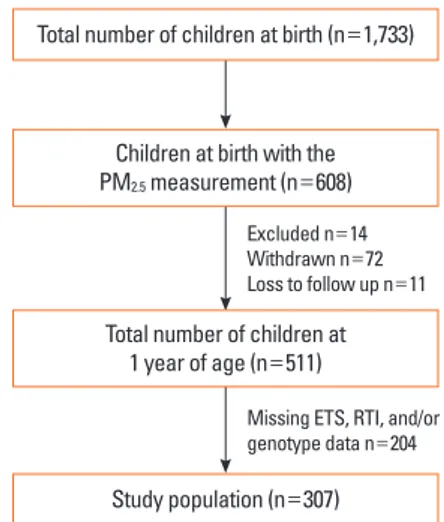
genotype data, 307 infants were finally included in the study (Fig. 1). Whether the 6- and 12-month-old infants had had any RTIs was determined by parental report of physician-diagnosed RTIs: “Has a doctor diagnosed RTIs in your child during the last 6 months?” Bronchiolitis, tracheobronchitis, and/or pneumo-nia were considered as LRTIs and common cold, sinusitis, otitis media, and/or croup as upper RTIs (URTIs).
Exposure assessment
Starting in May 2009, indoor PM2.5 samples were collected by
specialists during home visit between 26 and 36 weeks of preg-nancy. In addition, PM2.5 samples at 6 months after birth were
collected in the subgroup (n=75) for the applicants. PM2.5
con-centrations were measured 3 times in the parents’ bed room by using a particle discriminator (Model GT-331; SIBATA Co., Ja-pan) with a laser light-scattering optical particle counter for 5 minutes. The mean value of 3 measurements was used for eval-uation. The indoor PM2.5 values were log-transformed and
di-Fig. 1. Flow chart of the study population. Of infants with prenatal indoor PM2.5
measurements (n=608), 14 met the exclusion criteria, 72 were withdrawn from the study, and 11 were lost to follow-up. An additional 204 infants were then excluded because the RTIs, prenatal ETS exposure, or genotype data were missing. ETS, environmental tobacco smoke; PM2.5, fine particulate matter;
RTIs, respiratory tract infections.
Excluded n=14 Withdrawn n=72 Loss to follow up n=11
Missing ETS, RTI, and/or genotype data n=204
Total number of children at 1 year of age (n=511)
Study population (n=307) Total number of children at birth (n=1,733)
Children at birth with the
chotomized to high or low by using the median value before being entered into the regression models. Mothers were asked the following questions about their ETS exposure at home: “Have you been regularly exposed to passive smoking during your current pregnancy?”
The groups were stratified by exposure time. This led to 4 study population groups receiving the following combination of prenatal/postnatal exposures: prenatal ETS/prenatal PM2.5,
prenatal ETS/postnatal PM2.5, postnatal ETS/prenatal PM2.5,
and postnatal ETS/postnatal PM2.5. To assess whether the 2
in-door pollutants acted additively to increase RTI susceptibility in infancy, each group was divided into 4 groups according to their ETS exposure and whether the indoor PM2.5 levels were
high or low.
Genotyping
Genomic DNA was prepared from heparinized newborn um-bilical cord blood by using a G-DEX II kit (Intron, Seoul, Korea). ROS-related genes were analyzed as follows. The Nrf2 (rs6726395) and GSTP1 (rs1695) polymorphisms were geno-typed by using a TaqMan assay (ABI, Foster City, CA, USA). The
GSTM1 copy number was measured by real-time polymerase
chain reaction (PCR). The genotyping method is detailed in the Supplemental Material.
Bisulfite conversion and genome-wide methylation array
Nine subjects were selected from the study population to un-dergo genome-wide methylation analysis of cord blood ge-nomic DNA. Bisulfite conversion was performed by using the EZ DNA methylation kit (Zymo Research, Irvine, CA, USA) ac-cording to the manufacturer’s instructions. The bisulfite-con-verted genomic DNA was analyzed by using the Infinium Hu-man Methylation 450 Beadchip (Illumina, San Diego, CA, USA), with >450,000 probes covering 99% of reference se-quence genes, following the Illumina Infinium HD Methylation protocol. The 9 subjects and the methylation array are de-scribed in the Supplemental Material.
Statistical analysis
Chi-square and t tests were used to assess the significance of differences between the groups, as appropriate. The associa-tions between prenatal indoor PM2.5 and/or ETS exposure and
the incidence of RTIs at 12 months of age were analyzed by us-ing multiple logistic regression. Adjustments were made for po-tential confounding factors, namely, maternal age at delivery, maternal body mass index, maternal educational degree, gesta-tional age, delivery mode, infant sex, and family history of aller-gic diseases. The results are expressed as adjusted odds ratios (aORs) and 95% confidence intervals (CIs). All statistical analy-ses were performed by using SPSS 18.0 software (SPSS Inc., Chicago, IL, USA), with a P value <0.05 considered statistically significant.
Ethics statement
The study was approved by the Institutional Review Board of Asan Medical Center (IRB No. 2008-0616), Samsung Medical Center (IRB No. 2009-02-021), Yonsei University (IRB No. 4-2008-0588), and CHA Medical Center (IRB No. 2010-010). RESULTS
Study population characteristics
Table 1 summarizes the characteristics of the study popula-tion (n=307). The study populapopula-tion consisted of 171 boys and 136 girls. In total, 61.6% had been prenatally exposed to mater-nal ETS, and the mean indoor PM2.5 level during pregnancy was
6.08±7.64 μg/m3. The frequencies of the Nrf2 (rs6726395) GG,
GSTP1 (rs1695) AG or GG, and GSTM1 null genotypes were
40.1%, 36.9%, and 56.7%, respectively. The distribution of the 2 polymorphisms was in Hardy-Weinberg equilibrium. The inci-dences of LRTIs and URTIs by the age of 12 months were 16.3% and 76.2%, respectively.
Table 1 also shows the characteristics of the infants who were not included for reasons shown in Fig. 1. There were no signifi-cant differences between participants and non-participants, ex-cept in gestational age and the incidence of URTIs.
Risk factors for RTIs in infancy
High PM2.5 levels during pregnancy were independent risk
factors for LRTIs in infants (aOR=2.11; 95% CI: 1.12, 3.99) (Table 2). However, none of the early-life environmental factors in-creased the risk of URTIs. When we compared indoor PM2.5
lev-els according to RTIs and exposure time, prenatal indoor PM2.5
levels were higher in infants with LRTIs than in those without (mean=7.21 vs 5.71, respectively; 95% CI: 4.99, 9.44 vs 4.97, 6.45, respectively; P=0.119, data not shown). These differences were not distinct according to postnatal PM2.5 levels or the
pres-ence of URTIs.
Effects of prenatal exposure to both indoor PM2.5 and ETS on
RTIs susceptibility in infancy
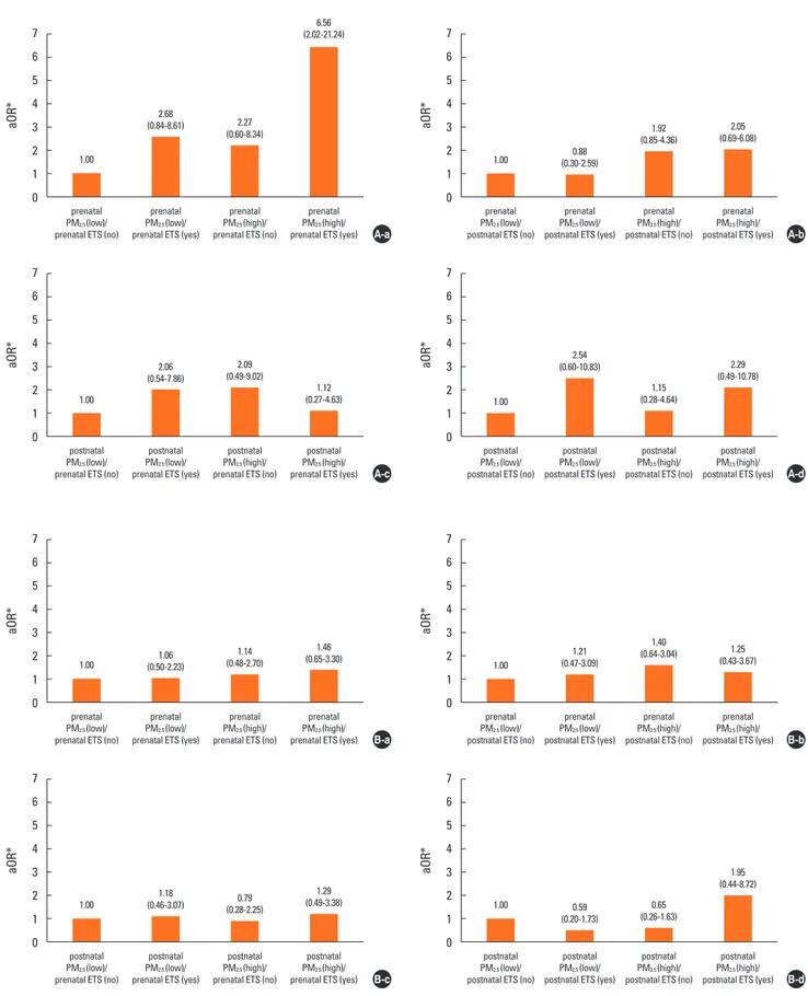
Prenatal high indoor PM2.5 and ETS exposure acted additively
to increase the risk of LRTIs (aOR=6.56; 95% CI: 2.02, 21.24) in infants (Fig. 2A and Table S1). Such an additive effect was not replicated when examining the effects of exposure to prenatal PM2.5/postnatal ETS, postnatal PM2.5/prenatal ETS, and
postna-tal PM2.5/postnatal ETS on any respiratory outcomes in infants.
In addition, such additive effects were not observed for URTIs risk, regardless of exposure time (Fig. 2B and Table S1).
Effect of GSTM1, GSTP1, and Nrf2 genotypes on the relationship between prenatal indoor PM2.5/ETS exposure and
RTIs in infancy
Prenatal exposure to both high indoor PM2.5 and ETS
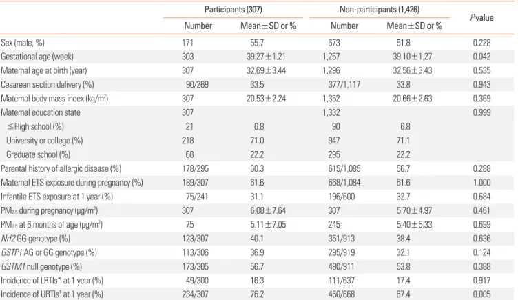
Table 1. Characteristics of infants who were included and excluded from the study
Participants (307) Non-participants (1,426)
P value
Number Mean±SD or % Number Mean±SD or %
Sex (male, %) 171 55.7 673 51.8 0.228
Gestational age (week) 303 39.27±1.21 1,257 39.10±1.27 0.042
Maternal age at birth (year) 307 32.69±3.44 1,296 32.56±3.43 0.535
Cesarean section delivery (%) 90/269 33.5 377/1,117 33.8 0.943
Maternal body mass index (kg/m2) 307 20.53±2.24 1,352 20.66±2.63 0.369
Maternal education state 307 1,332 0.999
≤High school (%) 21 6.8 90 6.8
University or college (%) 218 71.0 947 71.1
Graduate school (%) 68 22.2 295 22.2
Parental history of allergic disease (%) 178/295 60.3 615/1,085 56.7 0.288
Maternal ETS exposure during pregnancy (%) 189/307 61.6 668/1,084 61.6 1.000
Infantile ETS exposure at 1 year (%) 75/241 31.1 196/600 32.7 0.684
PM2.5 during pregnancy (μg/m3) 307 6.08±7.64 307 5.70±4.97 0.461
PM2.5 at 6 months of age (μg/m3) 75 5.11±7.05 245 5.40±5.33 0.699
Nrf2 GG genotype (%) 123/307 40.1 351/913 38.4 0.636
GSTP1 AG or GG genotype (%) 113/306 36.9 295/919 32.1 0.124
GSTM1 null genotype (%) 173/305 56.7 490/911 53.8 0.388
Incidence of LRTIs* at 1 year (%) 49/300 16.3 111/637 17.4 0.917
Incidence of URTIs† at 1 year (%) 234/307 76.2 450/668 67.4 0.005
*Lower respiratory tract infections: tracheobronchitis, pneumonia, and bronchiolitis; †Upper respiratory tract infections: common cold, sinusitis, otitis media, and
croup.
ETS, environmental tobacco smoke; GSTM1, glutathione S-transferase-mu 1; GSTP1, glutathione S-transferase-pi 1; Nrf2, nuclear factor erythroid 2-related factor; PM2.5, fine particulate matter; LRTIs, lower respiratory tract infections; URTIs, upper respiratory tract infections.
Table 2. Risk factors for respiratory tract infections in infancy
LRTIs URTIs
aOR* 95% CI P value aOR* 95% CI P value
Sex (male) 1.53 (1.01, 2.31) 0.046 1.04 (0.75, 1.45) 0.799
Parental history of allergic disease 0.87 (0.58, 1.30) 0.500 0.97 (0.70, 1.35) 0.863
Gestational age 0.94 (0.80, 1.10) 0.450 0.94 (0.83, 1.08) 0.393
Cesarean section delivery 1.14 (0.74, 1.75) 0.550 0.75 (0.53, 1.06) 0.098
Higher maternal education state 1.08 (0.74, 1.58) 0.683 1.12 (0.82, 1.54) 0.468
Maternal age at birth 0.99 (0.93, 1.05) 0.713 1.06 (1.01, 1.11) 0.019
Maternal body mass index 1.04 (0.96, 1.12) 0.363 0.94 (0.88, 1.01) 0.078
Higher PM2.5 during pregnancy 2.11 (1.12, 3.99) 0.021 1.23 (0.71, 2.15) 0.463
Higher PM2.5 at 6 months of age 0.89 (0.37, 2.18) 0.801 1.00 (0.52, 1.94) 0.999
Maternal ETS exposure during pregnancy 1.33 (0.87, 2.04) 0.193 1.15 (0.82, 1.62) 0.414
Infantile ETS exposure at 1 year 0.87 (0.53, 1.42) 0.576 0.79 (0.53, 1.17) 0.241
Nrf2 GG genotype 1.27 (0.82, 1.97) 0.290 0.55 (0.39, 0.78) 0.001
GSTP1 AG or GG genotype 1.53 (0.98, 2.38) 0.062 0.89 (0.62, 1.28) 0.523
GSTM1 null genotype 1.69 (1.07, 2.67) 0.024 0.91 (0.64, 1.30) 0.612
*Odds ratios were adjusted for maternal age, maternal body mass index, maternal educational state, infant sex, gestational age, delivery mode, and family history of allergy.
ETS, environmental tobacco smoke; GSTM1, glutathione S-transferase-mu 1; GSTP1, glutathione S-transferase-pi 1; Nrf2, nuclear factor erythroid 2-related factor; PM2.5, fine particulate matter; LRTIs, lower respiratory tract infections; URTIs, upper respiratory tract infections.
aOR* aOR* aOR* aOR* aOR* aOR* aOR* aOR* prenatal PM2.5 (low)/
prenatal ETS (no)
prenatal PM2.5 (low)/
prenatal ETS (no) postnatal PM2.5 (low)/
prenatal ETS (no)
postnatal PM2.5 (low)/
prenatal ETS (no)
prenatal PM2.5 (low)/
postnatal ETS (no)
prenatal PM2.5 (low)/
postnatal ETS (no) postnatal PM2.5 (low)/
postnatal ETS (no)
postnatal PM2.5 (low)/
postnatal ETS (no) prenatal
PM2.5 (low)/
prenatal ETS (yes)
prenatal PM2.5 (low)/
prenatal ETS (yes) postnatal PM2.5 (low)/
prenatal ETS (yes)
postnatal PM2.5 (low)/
prenatal ETS (yes)
prenatal PM2.5 (low)/
postnatal ETS (yes)
prenatal PM2.5 (low)/
postnatal ETS (yes) postnatal PM2.5 (low)/
postnatal ETS (yes)
postnatal PM2.5 (low)/
postnatal ETS (yes) prenatal
PM2.5 (high)/
prenatal ETS (no)
prenatal PM2.5 (high)/
prenatal ETS (no) postnatal PM2.5 (high)/
prenatal ETS (no)
postnatal PM2.5 (high)/
prenatal ETS (no)
prenatal PM2.5 (high)/
postnatal ETS (no)
prenatal PM2.5 (high)/
postnatal ETS (no) postnatal PM2.5 (high)/
postnatal ETS (no)
postnatal PM2.5 (high)/
postnatal ETS (no) prenatal
PM2.5 (high)/
prenatal ETS (yes)
prenatal PM2.5 (high)/
prenatal ETS (yes) postnatal PM2.5 (high)/
prenatal ETS (yes)
postnatal PM2.5 (high)/
prenatal ETS (yes)
prenatal PM2.5 (high)/
postnatal ETS (yes)
prenatal PM2.5 (high)/
postnatal ETS (yes) postnatal PM2.5 (high)/
postnatal ETS (yes)
postnatal PM2.5 (high)/
postnatal ETS (yes) 7 6 5 4 3 2 1 0 7 6 5 4 3 2 1 0 7 6 5 4 3 2 1 0 7 6 5 4 3 2 1 0 7 6 5 4 3 2 1 0 7 6 5 4 3 2 1 0 7 6 5 4 3 2 1 0 7 6 5 4 3 2 1 0 1.00 1.00 1.00 1.00 1.00 1.00 1.00 1.00 2.68 (0.84-8.61) 1.06 (0.50-2.23) 2.06 (0.54-7.86) 1.18 (0.46-3.07) 0.88 (0.30-2.59) 1.21 (0.47-3.09) 2.54 (0.60-10.83) 0.59 (0.20-1.73) 2.27 (0.60-8.34) 1.14 (0.48-2.70) 2.09 (0.49-9.02) 0.79 (0.28-2.25) 1.92 (0.85-4.36) 1.40 (0.64-3.04) 1.15 (0.28-4.64) 0.65 (0.26-1.63) 6.56 (2.02-21.24) 1.46 (0.65-3.30) 1.12 (0.27-4.63) 1.29 (0.49-3.38) 2.05 (0.69-6.08) 1.25 (0.43-3.67) 2.29 (0.49-10.78) 1.95 (0.44-8.72) A-c B-c A-a B-a A-d B-d A-b B-b
Fig. 2. Effect of combined exposure to both PM2.5 and ETS according to exposure time on susceptibility to (A) lower and (B) upper respiratory tract infections in
infan-cy. The groups were stratified by exposure time: (a) prenatal PM2.5/prenatal ETS; (b) prenatal PM2.5/postnatal ETS; (c) postnatal PM2.5/prenatal ETS; and (d) postnatal
PM2.5/postnatal ETS. Prenatal high indoor PM2.5 and ETS exposure acted additively to increase the risk of lower respiratory tract infections. *Adjustments for
mater-nal age at delivery, matermater-nal body mass index, matermater-nal educatiomater-nal degree, gestatiomater-nal age, delivery mode, infant sex, and family history of allergy. ETS, environ-mental tobacco smoke; PM2.5, fine particulate matter.
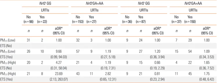
Nrf2 GG genotypes (aOR=8.18, 95% CI: 1.53, 43.72; aOR=7.37,
95% CI: 1.12, 48.66; and aOR=23.69, 95% CI: 2.13, 263.07, re-spectively). Such gene–environment interactions were not ob-served for URTIs (Tables 3-5).
Relationship between DNA methylation patterns and prenatal indoor PM2.5/ETS exposure
Prenatal indoor PM2.5/ETS exposure was associated
specifi-cally with 15 CpG sites, 6 of which were located in intergenic
re-gions. Of the remaining 9 CpG sites, 5 were hypomethylated and 4 were hypermethylated by PM2.5/ETS exposure. This
anal-ysis is described in the Supplemental Material. DISCUSSION
The present study showed that indoor PM2.5 and ETS
expo-sure may have an effect on RTIs in infants and revealed that the adverse effect may depend on the timing of the exposure. The
Table 3. Influence of the glutathione S-transferase-mu 1 (GSTM1) copy number variation on the results of prenatal exposure to both PM2.5 and ETS
GSTM1 present GSTM1 null GSTM1 present GSTM1 null
LRTIs LRTIs URTIs URTIs
No
(n=112) (n=17)Yes (n=137)No (n=32)Yes (n=36)No (n=87)Yes (n=37)No (n=150)Yes
n n (95% CI)aOR* n n (95% CI)aOR* n n (95% CI)aOR* n n (95% CI)aOR*
PM2.5 (Low) 29 2 1.00 34 3 1.00 6 26 1.00 10 27 1.00 ETS (No) PM2.5 (Low) 37 7 1.68 45 12 4.52 11 33 0.51 12 48 2.02 ETS (Yes) (0.24, 11.79) (0.87, 23.60) (0.14, 1.83) (0.68, 6.01) PM2.5 (High) 13 2 2.26 28 4 1.65 4 12 0.69 9 25 1.17 ETS (No) (0.26, 19.36) (0.24, 11.29) (0.13, 3.76) (0.38, 3.57) PM2.5 (High) 33 6 3.75 30 13 8.18 9 32 0.57 11 33 1.86 ETS (Yes) (0.52, 27.10) (1.53, 43.72) (0.14, 2.35) (0.60, 5.75)
*Odds ratios were adjusted for maternal age, maternal body mass index, maternal educational state, infant sex, gestational age, delivery mode, and family history of allergy.
aOR, adjusted odds ratio; CI, confidence interval; ETS, environmental tobacco smoke; PM2.5, fine particulate matter; GSTM1, glutathione S-transferase-mu 1; LRTIs,
lower respiratory tract infections; URTIs, upper respiratory tract infections.
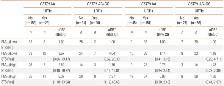
Table 4. Influence of the glutathione S-transferase-pi 1 (GSTP1) (rs1695) polymorphism on the results of prenatal exposure to both PM2.5 and ETS
GSTP1 AA GSTP1 AG+GG GSTP1 AA GSTP1 AG+GG
LRTIs LRTIs URTIs URTIs
No
(n=159) (n=29)Yes (n=91)No (n=20)Yes (n=45)No (n=151)Yes (n=27)No (n=86)Yes
n n (95% CI)aOR* n n (95% CI)aOR* n n (95% CI)aOR* n n (95% CI)aOR*
PM2.5 (Low) 38 3 1.00 25 2 1.00 9 33 1.00 7 20 1.00 ETS (No) PM2.5 (Low) 59 12 3.57 24 7 4.04 15 58 1.14 9 23 1.18 ETS (Yes) (0.66, 19.17) (0.62, 26.30) (0.41, 3.15) (0.29, 4.71) PM2.5 (High) 26 3 2.82 14 3 1.70 9 23 0.75 3 14 1.43 ETS (No) (0.40, 19.77) (0.19, 15.01) (0.24, 2.34) (0.28, 7.30) PM2.5 (High) 36 11 6.32 28 8 7.37 12 37 0.83 8 29 1.80 ETS (Yes) (1.18, 33.94) (1.12, 48.66) (0.28, 2.43) (0.41, 7.97)
*Odds ratios were adjusted for maternal age, maternal body mass index, maternal educational state, infant sex, gestational age, delivery mode, and family history of allergy.
aOR, adjusted odds ratio; CI, confidence interval; ETS, environmental tobacco smoke; PM2.5, fine particulate matter; GSTP1, glutathione S-transferase-pi 1; LRTIs,
ability of indoor PM2.5 and/or ETS to increase susceptibility to
LRTIs appeared to be stronger when the exposure occurred during the prenatal period rather than the postnatal period. This study also showed that the genetic polymorphisms
GSTM1, GSTP1 (rs1695), and/or Nrf2 (rs6726395) were further
associated with the increased susceptibility of LRTIs in indoor PM2.5/ETS-exposed infants. Thus, the susceptibility of LRTIs in
infancy may be shaped by gene-environment interactions be-tween ROS-related genes and prenatal indoor PM2.5/ETS
expo-sure. To our knowledge, this is the first study to evaluate the as-sociation between combined exposure to indoor PM2.5/ETS
and infants’ susceptibility to LRTIs that is associated with expo-sure time and genetic susceptibility.
Most people spend as much as 90% of their time indoors, espe-cially pregnant women and infants. Chronic exposure to indoor pollutants at home or school can increase air pollutant inhala-tion and significantly impact health.26,27 The interaction between
PM and ETS, the most important indoor air pollutants, modify their individual harmful effects on respiratory outcomes.7,8
How-ever, studies about the indoor PM concentration that would have an adverse health outcome and an interaction between PM and ETS, especially prenatal exposure, are scarce. Our study revealed the effect of indoor PM2.5 even in the low concentration
and the additive effect of indoor PM2.5/ETS exposure during the
prenatal period on the development of LRTIs in infancy. The fetal period is critical for lung and immune development. Although some epidemiologic studies showed that prenatal PM or ETS exposure increases the risk of wheezing, asthma, and respiratory infections,28-31 studies comparing the effect
ac-cording to exposure time are limited. A few studies revealed
that prenatal exposure has a stronger effect on respiratory out-comes than postnatal exposure.32-34 We also found its stronger
associations with combined exposures to indoor PM2.5/ETS
during the prenatal period than the postnatal period. However, this result must be interpreted with caution because it is diffi-cult to clearly separate exposure periods.
Air pollutants and tobacco smoke exert their harmful effects on health by inducing oxidative stress in exposed cells and tis-sues.18,19 Air pollutants can be directly absorbed to the fetal
cir-culation and produce ROS, ultimately inducing inflammatory and oxidative stress responses in the fetal lung.35 The fetus can
also be affected indirectly by the oxidative stress and inflamma-tory cytokine production induced in the placenta by the pollut-ants.36 Of particular interest in this regard are several
intracellu-lar antioxidant enzymes, including GSTM1 and GSTP1, which defend the airway epithelium from damage caused by oxidants and inflammation. These enzymes are regulated by the tran-scription factor Nrf2, which translocates to the nucleus after ox-idative stress induction.37 These enzymes in respiratory disease
pathogenesis after pollutant exposure is provided by results showing that children with the GSTM1 null genotype are more likely to develop asthma and wheezing after prenatal ETS expo-sure than children with the GSTM1 present genotype.20
Similar-ly, our study showed that while both prenatal indoor PM2.5 and
ETS exposure greatly increased the incidence of LRTIs in in-fants, this effect was particularly marked in the infants with the
GSTM1 null, GSTP1 (rs1695) AG or GG, or Nrf2 (rs6726395) GG
genotypes.
ROS reacts with lipids, proteins, and DNA, resulting in cell membrane damage, alteration of gene and protein expression,
Table 5. Influence of the nuclear factor erythroid 2-related factor (Nrf2) (rs6726395) on the results of prenatal exposure to both PM2.5 and ETS
Nrf2 GG Nrf2 GA+AA Nrf2 GG Nrf2 GA+AA
LRTIs LRTIs URTIs URTIs
No
(n=98) (n=22)Yes (n=153)No (n=27)Yes (n=36)No (n=87)Yes (n=37)No (n=150)Yes
n n (95% CI)aOR* n n (95% CI)aOR* n n (95% CI)aOR* n n (95% CI)aOR*
PM2.5 (Low) 31 2 1.00 32 3 1.00 9 24 1.00 7 29 1.00 ETS (No) PM2.5 (Low) 26 10 9.66 57 9 1.19 9 27 1.20 15 54 1.09 ETS (Yes) (0.99, 94.03) (0.27, 5.18) (0.36, 3.94) (0.34, 3.53) PM2.5 (High) 20 2 4.27 21 4 1.19 9 15 0.66 4 22 1.65 ETS (No) (0.31, 58.04) (0.19, 7.31) (0.19, 2.29) (0.36, 7.52) PM2.5 (High) 21 8 23.69 43 11 2.82 9 21 0.81 11 45 1.75 ETS (Yes) (2.13, 263.07) (0.65, 12.31) (0.23, 2.94) (0.48, 6.42)
*Odds ratios were adjusted for maternal age, maternal body mass index, maternal educational state, infant sex, gestational age, delivery mode, and family history of allergy.
aOR, adjusted odds ratio; CI, confidence interval; ETS, environmental tobacco smoke; PM2.5, fine particulate matter; Nrf2, nuclear factor erythroid 2-related factor;
and even cell death.18,37 Secondary mediators generated by
oxi-dant reactions with lipids, proteins, and other biomolecules contribute to the toxic effects of pollutants. Oxidative stress also induces MAP kinase and NF-κB activation, which may ulti-mately produce a variety of proinflammatory mediators. Proin-flammatory mediators from the airway epithelium play a criti-cal role in the pathogenesis of several pulmonary diseases. Pre-vious experimental studies supported the association between air pollutants and oxidative stress by demonstrating that anti-oxidant pretreatment attenuates oxidative stress and airway ep-ithelial cell injury induced by air pollutants.38-40
Prenatal exposure to environmental factors may affect disease susceptibility later in life by inducing epigenetic changes. A cross-sectional study of children under 18 years of age revealed that air pollutants and ETS both associate with significantly in-creased DNA methylation and dein-creased transcription of inter-feron gamma (IFN-γ) in T-effector cells and forkhead box tran-scription factor 3 (Foxp3) in T-regulatory cells.41 Interestingly,
GSTM1 and GSTP1 polymorphisms alter the ability of prenatal
tobacco smoke exposure to induce global DNA methylation.21
Although the sample size in our experiment was too small to make firm conclusions, our data suggest that the ability of pre-natal indoor PM2.5 and ETS exposure to promote LRTIs in
in-fancy may be due to DNA methylation alterations; the 9 CpG sites whose methylation was significantly altered by PM2.5/ETS
exposure were in subjects with LRTIs (Table S2 and Fig. S1). Further studies on this issue are required.
This study has several limitations. First, it was not possible to clearly distinguish between the effects of prenatal and postna-tal exposure or exposure that persisted during both the pre- and postnatal periods. Further studies on the effect of indoor PM and ETS exposure during specific prenatal and postnatal periods may help identify the mechanisms involved. Second, the RTIs and ETS data were derived from questionnaires, and the indoor PM2.5 levels were measured only 1 day between 26
and 36 weeks of pregnancy and at 6 months of age. Therefore, an information bias could not be excluded. Although question-naires may misclassify ETS exposure, previous studies have shown a fairly good correlation between self-reported ETS ex-posure and biomarkers of ETS exex-posure.5,42-44 Future studies
may gain greater sensitivity by using more objective and precise measures of RTIs, and smoke and indoor PM exposure. The third limitation is the relatively small study population, which is because the indoor PM2.5 measurements started later in the
COCOA study, and these data were thus only available for about one-third of the whole COCOA cohort. However, it is un-likely that the addition of indoor PM2.5 measurements to the
protocol introduced a selection bias because the study partici-pants and non-participartici-pants did not differ significantly in terms of their characteristics. An increase in the sample size would be likely to lead to more consistent and significant data. The fourth limitation is that we only selected 1 polymorphism from each
gene. However, these polymorphisms have been shown in sev-eral studies to contribute to asthma susceptibility.20,45-47
An important strength of our study is its prospective design: the indoor PM2.5 and ETS exposure data and the data on many
potential confounders were collected before the children were born. This is likely to have markedly reduced the study bias. An additional strength is that the PM2.5 measurement was
per-formed at home. This direct measurement of residential indoor PM2.5 probably estimates the actual exposure levels more
accu-rately than other indirect methods. Finally, we investigated ge-notypic data for GSTM1, GSTP1, and Nrf2 to determine gene– environment interactions between both PM2.5/ETS exposure
and LRTIs. These results imply that air pollutant exposure should be reduced, especially in genetically susceptible infants, and support a mechanism for oxidative stress in inducing ad-verse respiratory outcomes by air pollutants.
It should be noted that there was an important difference be-tween previous studies and ours, namely, that maternal ETS ex-posure was considered in our study. It was not possible to eval-uate the effect of maternal active smoking because the active smoking rate of Korean women is low: only 11.4% of the CO-COA cohort mothers had smoked before their pregnancy, of whom only 1 continued to smoke during pregnancy. Thus, ma-ternal ETS exposure was more likely to be an important source of pollutant exposure in our cohort than maternal smoking. CONCLUSIONS
Indoor PM2.5 and ETS exposure increases susceptibility to
LR-TIs in infants. This effect was particularly marked when the ex-posure occurred in the prenatal period. Moreover, the effect was modified by ROS-related gene polymorphisms. Along with studies suggesting that acute LRTIs in early life is associated with a long-standing susceptibility to all forms of lung disease, including asthma,15-17 our study highlights the importance of
health intervention strategies that focus on the indoor environ-ment in the prenatal period. Additional analyses of genetic and epigenetic variants may help individualize such strategies. Fur-ther studies of gene–environment interactions and epigenetic mechanisms that shape the effect of air pollutants on suscepti-bility to LRTIs and chronic lung diseases are warranted, along with studies assessing the association between LRTIs in infancy and the development of chronic lung diseases, such as asthma. ACKNOWLEDGMENTS
The authors thank Kyung-Shin Lee, Jin-Ah Park, and Hee-Suk Kim for organizing the data. We would also like to express our gratitude to Ja-Young Kwon, Suk-Joo Choi, Soo-Young Oh, Kyung-Ju Lee, and Hey-Sung Won for helping collect the ob-stetric data. We also thank Sung-Ok Kwon, Se-Young Oh, Kyung-Sook Lee, Yee-Jin Shin, Jong-Hwan Lim, Whan-Cheol
Kim, and Ho Kim for their participation in this study. REFERENCES
1. Hasheminassab S, Daher N, Shafer MM, Schauer JJ, Delfino RJ, Sioutas C. Chemical characterization and source apportionment of indoor and outdoor fine particulate matter (PM(2.5)) in retire-ment communities of the Los Angeles Basin. Sci Total Environ 2014;490:528-37.
2. Clougherty JE, Houseman EA, Levy JI. Source apportionment of indoor residential fine particulate matter using land use regression and constrained factor analysis. Indoor Air 2011;21:53-66. 3. Wallace LA, Mitchell H, O’Connor GT, Neas L, Lippmann M,
Kat-tan M, et al. Particle concentrations in inner-city homes of children with asthma: the effect of smoking, cooking, and outdoor pollu-tion. Environ Health Perspect 2003;111:1265-72.
4. Guarnieri M, Balmes JR. Outdoor air pollution and asthma. Lancet 2014;383:1581-92.
5. Rosa MJ, Jung KH, Perzanowski MS, Kelvin EA, Darling KW, Ca-mann DE, et al. Prenatal exposure to polycyclic aromatic hydrocar-bons, environmental tobacco smoke and asthma. Respir Med 2011; 105:869-76.
6. Miller RL, Garfinkel R, Horton M, Camann D, Perera FP, Whyatt RM, et al. Polycyclic aromatic hydrocarbons, environmental tobac-co smoke, and respiratory symptoms in an inner-city birth tobac-cohort. Chest 2004;126:1071-8.
7. Sonnenschein-van der Voort AM, de Kluizenaar Y, Jaddoe VW, Ga-briele C, Raat H, Moll HA, et al. Air pollution, fetal and infant to-bacco smoke exposure, and wheezing in preschool children: a population-based prospective birth cohort. Environ Health 2012; 11:91.
8. Rabinovitch N, Silveira L, Gelfand EW, Strand M. The response of children with asthma to ambient particulate is modified by tobac-co smoke exposure. Am J Respir Crit Care Med 2011;184:1350-7. 9. Nicolai T, Carr D, Weiland SK, Duhme H, von Ehrenstein O,
Wag-ner C, et al. Urban traffic and pollutant exposure related to respira-tory outcomes and atopy in a large sample of children. Eur Respir J 2003;21:956-63.
10. Jedrychowski W, Bendkowska I, Flak E, Penar A, Jacek R, Kaim I, et al. Estimated risk for altered fetal growth resulting from exposure to fine particles during pregnancy: an epidemiologic prospective co-hort study in Poland. Environ Health Perspect 2004;112:1398-402. 11. Hertz-Picciotto I, Park HY, Dostal M, Kocan A, Trnovec T, Sram R.
Prenatal exposures to persistent and non-persistent organic com-pounds and effects on immune system development. Basic Clin Pharmacol Toxicol 2008;102:146-54.
12. Herr CE, Ghosh R, Dostal M, Skokanova V, Ashwood P, Lipsett M, et al. Exposure to air pollution in critical prenatal time windows and IgE levels in newborns. Pediatr Allergy Immunol 2011;22:75-84. 13. Latzin P, Frey U, Armann J, Kieninger E, Fuchs O, Röösli M, et al.
Ex-posure to moderate air pollution during late pregnancy and cord blood cytokine secretion in healthy neonates. PLoS One 2011; 6:e23130.
14. Hertz-Picciotto I, Herr CE, Yap PS, Dostál M, Shumway RH, Ash-wood P, et al. Air pollution and lymphocyte phenotype proportions in cord blood. Environ Health Perspect 2005;113:1391-8.
15. Gern JE. Viral respiratory infection and the link to asthma. Pediatr Infect Dis J 2008;27:S97-103.
16. Sigurs N, Aljassim F, Kjellman B, Robinson PD, Sigurbergsson F, Bjarnason R, et al. Asthma and allergy patterns over 18 years after severe RSV bronchiolitis in the first year of life. Thorax 2010;65: 1045-52.
17. Holt PG, Sly PD. Viral infections and atopy in asthma pathogene-sis: new rationales for asthma prevention and treatment. Nat Med 2012;18:726-35.
18. Ciencewicki J, Trivedi S, Kleeberger SR. Oxidants and the patho-genesis of lung diseases. J Allergy Clin Immunol 2008;122:456-68. 19. Kim BJ, Lee SY, Kim HB, Lee E, Hong SJ. Environmental changes,
microbiota, and allergic diseases. Allergy Asthma Immunol Res 2014;6:389-400.
20. Gilliland FD, Li YF, Dubeau L, Berhane K, Avol E, McConnell R, et al. Effects of glutathione S-transferase M1, maternal smoking dur-ing pregnancy, and environmental tobacco smoke on asthma and wheezing in children. Am J Respir Crit Care Med 2002;166:457-63. 21. Breton CV, Byun HM, Wenten M, Pan F, Yang A, Gilliland FD.
Pre-natal tobacco smoke exposure affects global and gene-specific DNA methylation. Am J Respir Crit Care Med 2009;180:462-7. 22. Guerrero-Preston R, Goldman LR, Brebi-Mieville P, Ili-Gangas C,
Lebron C, Witter FR, et al. Global DNA hypomethylation is associ-ated with in utero exposure to cotinine and perfluorinassoci-ated alkyl compounds. Epigenetics 2010;5:539-46.
23. Flom JD, Ferris JS, Liao Y, Tehranifar P, Richards CB, Cho YH, et al. Prenatal smoke exposure and genomic DNA methylation in a mul-tiethnic birth cohort. Cancer Epidemiol Biomarkers Prev 2011;20: 2518-23.
24. Kim HB, Ahn KM, Kim KW, Shin YH, Yu J, Seo JH, et al. Cord blood cellular proliferative response as a predictive factor for atopic der-matitis at 12 months. J Korean Med Sci 2012;27:1320-6.
25. Yang HJ, Lee SY, Suh DI, Shin YH, Kim BJ, Seo JH, et al. The Cohort for Childhood Origin of Asthma and allergic diseases (COCOA) study: design, rationale and methods. BMC Pulm Med 2014;14:109. 26. Pawankar R, Canonica GW, Holgate ST, Lockey RF. World Allergy
Organization (WAO) white book on allergy. Milwaukee (WI): World Allergy Organization; 2011.
27. Yoon C, Ryu K, Kim J, Lee K, Park D. New approach for particulate exposure monitoring: determination of inhaled particulate mass by 24 h real-time personal exposure monitoring. J Expo Sci Envi-ron Epidemiol 2012;22:344-51.
28. Magnusson LL, Olesen AB, Wennborg H, Olsen J. Wheezing, asth-ma, hayfever, and atopic eczema in childhood following exposure to tobacco smoke in fetal life. Clin Exp Allergy 2005;35:1550-6. 29. Jedrychowski W, Perera FP, Maugeri U, Mrozek-Budzyn D, Mroz E,
Flak E, et al. Early wheezing phenotypes and severity of respiratory illness in very early childhood: study on intrauterine exposure to fine particle matter. Environ Int 2009;35:877-84.
30. Jedrychowski WA, Perera FP, Maugeri U, Mrozek-Budzyn D, Mroz E, Klimaszewska-Rembiasz M, et al. Intrauterine exposure to polycy-clic aromatic hydrocarbons, fine particulate matter and early wheeze. Prospective birth cohort study in 4-year olds. Pediatr Al-lergy Immunol 2010;21:e723-32.
31. Jedrychowski WA, Perera FP, Spengler JD, Mroz E, Stigter L, Flak E, et al. Intrauterine exposure to fine particulate matter as a risk factor for increased susceptibility to acute broncho-pulmonary infections in early childhood. Int J Hyg Environ Health 2013;216:395-401. 32. Raherison C, Pénard-Morand C, Moreau D, Caillaud D, Charpin D,
Kopfersmitt C, et al. In utero and childhood exposure to parental tobacco smoke, and allergies in schoolchildren. Respir Med 2007;
101:107-17.
33. Singh SP, Gundavarapu S, Peña-Philippides JC, Rir-Sima-ah J, Mishra NC, Wilder JA, et al. Prenatal secondhand cigarette smoke promotes Th2 polarization and impairs goblet cell differentiation and airway mucus formation. J Immunol 2011;187:4542-52. 34. Jedrychowski WA, Perera FP, Majewska R, Camman D, Spengler
JD, Mroz E, et al. Separate and joint effects of tranplacental and postnatal inhalatory exposure to polycyclic aromatic hydrocar-bons: prospective birth cohort study on wheezing events. Pediatr Pulmonol 2014;49:162-72.
35. Kelly FJ. Oxidative stress: its role in air pollution and adverse health effects. Occup Environ Med 2003;60:612-6.
36. Auten RL, Potts EN, Mason SN, Fischer B, Huang Y, Foster WM. Maternal exposure to particulate matter increases postnatal ozone-induced airway hyperreactivity in juvenile mice. Am J Respir Crit Care Med 2009;180:1218-26.
37. Auerbach A, Hernandez ML. The effect of environmental oxidative stress on airway inflammation. Curr Opin Allergy Clin Immunol 2012;12:133-9.
38. Wu W, Peden DB, McConnell R, Fruin S, Diaz-Sanchez D. Gluta-thione-S-transferase M1 regulation of diesel exhaust particle-in-duced pro-inflammatory mediator expression in normal human bronchial epithelial cells. Part Fibre Toxicol 2012;9:31.
39. Messier EM, Day BJ, Bahmed K, Kleeberger SR, Tuder RM, Bowler RP, et al. N-acetylcysteine protects murine alveolar type II cells from cigarette smoke injury in a nuclear erythroid 2-related factor-2-independent manner. Am J Respir Cell Mol Biol 2013;48:559-67. 40. Wu W, Muller R, Berhane K, Fruin S, Liu F, Jaspers I, et al.
Inflam-matory response of monocytes to ambient particles varies by high-way proximity. Am J Respir Cell Mol Biol 2014;51:802-9.
41. Kohli A, Garcia MA, Miller RL, Maher C, Humblet O, Hammond SK, et al. Secondhand smoke in combination with ambient air pol-lution exposure is associated with increasedx CpG methylation and decreased expression of IFN-gamma in T effector cells and Foxp3 in T regulatory cells in children. Clin Epigenetics 2012;4:17. 42. Wang IJ, Hsieh WS, Wu KY, Guo YL, Hwang YH, Jee SH, et al. Effect
of gestational smoke exposure on atopic dermatitis in the offspring. Pediatr Allergy Immunol 2008;19:580-6.
43. Carlsten C, Dimich-Ward H, DyBuncio A, Becker AB, Chan-Yeung M. Cotinine versus questionnaire: early-life environmental tobacco smoke exposure and incident asthma. BMC Pediatr 2012;12:187. 44. Yi O, Kwon HJ, Kim H, Ha M, Hong SJ, Hong YC, et al. Effect of
envi-ronmental tobacco smoke on atopic dermatitis among children in Korea. Environ Res 2012;113:40-5.
45. Masuko H, Sakamoto T, Kaneko Y, Iijima H, Naito T, Noguchi E, et al. An interaction between Nrf2 polymorphisms and smoking sta-tus affects annual decline in FEV1: a longitudinal retrospective co-hort study. BMC Med Genet 2011;12:97.
46. Lee E, Chang HY, Lee KS, Suh DI, Yu HS, Kang MJ, et al. The effect of perinatal anxiety on bronchiolitis is influenced by polymor-phisms in ROS-related genes. BMC Pulm Med 2014;14:154. 47. Kang SH, Jung YH, Kim HY, Seo JH, Lee JY, Kwon JW, et al. Effect of
paracetamol use on the modification of the development of asth-ma by reactive oxygen species genes. Ann Allergy Asthasth-ma Immu-nol 2013;110:364-369.e1.
48. Brasch-Andersen C, Christiansen L, Tan Q, Haagerup A, Vestbo J, Kruse TA. Possible gene dosage effect of glutathione-S-transferases on atopic asthma: using real-time PCR for quantification of GSTM1 and GSTT1 gene copy numbers. Hum Mutat 2004;24:208-14.