구순구개열환자의 상악 전방분절 골신장술식을 이용한
교정 치험례
유성훈, 최혜영, 유형석, 백형선, 차정열*
연세대학교 치과대학 교정학교실, 두개안면기형연구소ABSTRACT
Maxillary Anterior Segmental Distraction with Rigid External Device:
Case Report
Seong-Hun Yoo, Hye-Young Choi, Hyung-Seog Yu,
Hyoung-Seon Baik, Jung-Yul Cha*
Department of Orthodontics, Institute of Craniofacial Deformities,
Yonsei University, College of Dentistry, Seoul, KOREA
Maxillary anterior segmental distraction osteogenesis (DO) has been the alternative treatment option for patients with midfacial retrusion. The patient showed unilateral cleft lip and palate, and premaxillary distraction with rigid external device (RED) was planned to solve midface deficiency and to create alveolar space. Significant advancement of A point was observed, but relapse of A point was detected during consolidation period. The vertical position of the ANS was found to have moved downward. Axis of upper incisor decreased after DO. Maxillary anterior segmental DO is effective for treatment of patient with cleft lip and palate. The alveolar space is regained successfully, and the facial profile is improved without velopharyngeal problems.
Key words : Intraoral appliance, Maxillary anterior segmental distraction osteogenesis, RED
Introduction
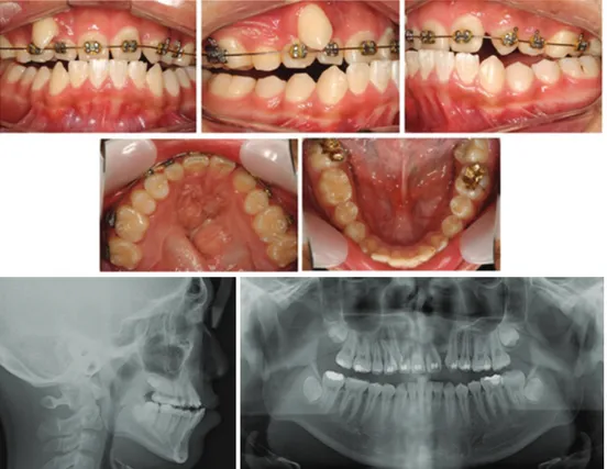
구순구개열 환자의 치료에서 중안모의 열성장 개선은 중요한 치료목표가 될 수 있다. 이러한 환 자들에게 심미적, 기능적 측면의 개선을 위하여 상악 분절골 수술을 포함하는 전통적인 수술치료 방법이 적용되어 왔다. 그러나 수술 범위가 넓어 지면서 골편의 이동방향에 따라 다양한 재발 경 향이 발생하는 것으로 보고되었다1,2). Le Fort osteotomy 술식의 대안으로 상악골 전방 신장술이 소개되었다. 그러나 이 술식의 결 과 과도하게 상악골이 전방 신장되면 인두간 폐 쇄부전(velopharyngeal incompetemcy)과 과비성 성(hypernasality)을 유발할 수 있다고 보고되었 다3-5). 또한 최근 보고에 따르면, 10 mm 이상의 상악골 전진술을 시행한 환자에서 발음상의 문제 를 경험할 가능성이 커진다고 보고되었다3,4). 대부분의 구순구개열 환자들은 상악 전치부와 소구치 부위에 선천적 치아 결손을 동반한 극심Figure 1. Pretreatment intraoral views and radiographs. 한 총생을 보인다. 그러므로 치아의 배열과 상악
열성장을 동시에 해결하기 위해서는 새로운 치조 골의 형성도 필수적이다.
구순구개열 환자에서 상악골 전방분절골 신장술 (Maxillary anterior segment distraction osteo-genesis)은 치조 공간의 형성과 반대 교합의 해소 에 적절한 술식이며 중안면 열성장 환자의 치료 대안으로 대두되고 있다2,6). 최근에 상악골 전방분절골 신장술에 다양한 종류 의 신장기를 이용하는 방법들이 보고되었다. Bengi 등과 Dolanmaz 등은 RPE스크류와 함께 사용할 것을 제안하였다. 하지만 치아 지지 장치는 전치부 에 가해지는 힘을 증가시켜 원하지 않는 치성 효과 가 발생될 수 있을 것으로 여겨진다5,7,8). 이러한 이 유로 Karakasis 와 Hadjipetrou는 두개의 원통형 신장기를 동반하는 골지지 장치를 이용한 신장술을 소개하였다1). 이러한 방법은 Rigid external device (RED)에 비하여 상대적으로 작은 두개부위의 손상 과 심미적으로 더 우수한 치료결과를 보인다9,10). 그러나 원통형 신장기는 신장 방향 유도가 어렵다 는 기술적인 이유로 널리 사용되지는 않는다. 반면 Iida등은 골 지지 유형의 신장기를 이용한 새로운 시스템을 소개하였다5). Iida는 효율적인 골지지 신 장기의 작용을 위하여 미니스크류를 사용하였다. 그러나 신장기의 종류에 따른 골격적 효과나 재발 양상에 대한 연구는 많지 않다. 이번 증례는 구순구개열 환자의 치료에 RED 장치를 이용하여 상악골 전방분절골 신장술(Ma-xillary anterior segmental DO)을 시행 후 골격 적, 치성 효과와 재발 양상을 보고하고자 하였다.
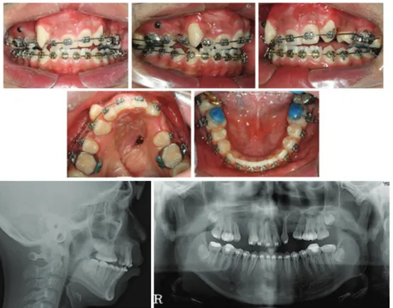
Figure 2. Intraoral views and radiographs after Distraction osteogenesis.
Diagnosis and Treatment planning
15세 남자 환자는 편측성 구순구개열을 보였다. 환자는 상악 좌측 측절치와 제2소구치의 결손, 상 악 우측 치열궁의 붕괴로 인한 high canine을 동 반한 7mm의 극심한 총생이 있었다. 상,하악은 모 두 열성장을 보였으며 상악 전치는 직립되었고 하 악 전치는 약간 돌출 되었다(SNA, 68.0゚; SNB, 68.8゚; U1 to SN, 93.6゚; IMPA, 97.5゚). 환자는 편측성 구순구개열을 동반한 골격성 III급 부정교 합으로 진단되었다(Figure 1). 환자의 안모와 교합을 개선하기 위해서 상악골 전방분절골 신장술을 계획하였다. 견치와 결손치 의 공간을 확보하기 위하여 골절단 선은 양측 소 구치와 견치 사이에 위치시켰으며 RED를 이용하 여 전방분절골을 신장하기로 하였다. 골절단의 방향과 외과용 플레이트의 위치는 측 모두부방사선 사진을 이용한 STO를 통하여 결정 하였으며, 신장력의 방향은 교합면과 평행하게 적 용하였다(Figure 2).
Surgical procedure
High Le Fort I osteotomy 와 함께 견치와 소구 치 사이에 vertical osteotomy가 시행되었다. 골절 단 후에 미니플레이트(Martin Medizin. Technik, Tuttlingen, Germany)에 miniscrew를 이용하여 전방분절을 고정하였다. 그리고 RED
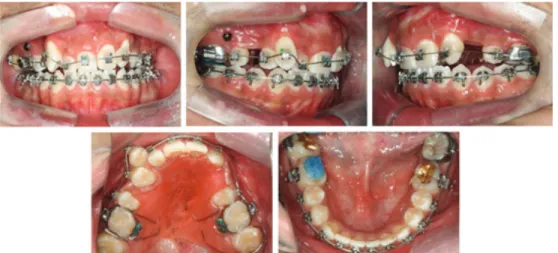
시스템(Mar-Figure 3. Intraoral views during treatment (9M after distraction osteogenesis). tin Medizin Technik, Tuttlingen, Germany)의
여섯 개의 두부고정용 스크류를 이용하여 두개관과 연결되었다11). 분절골의 전방이동을 위해 강선을 미 니플레이트와 연결하여 비강아래 부위를 관통하여 견인장치와 연결하였다.
Distraction protocol
골절단 후 7일 동안의 휴지기 이후에 골신장을 시작하였다. 하루에 1mm를 신장하였으며 오전과 오후에 0.5 mm 씩 나누어 신장하였다. 골신장 이후 경화기 기간은 1개월간 시행하였으며, 신장된 공간 은 가철성 유지 장치와 고정식 장치를 이용하여 유 지하였다(Figure 2-5).Results
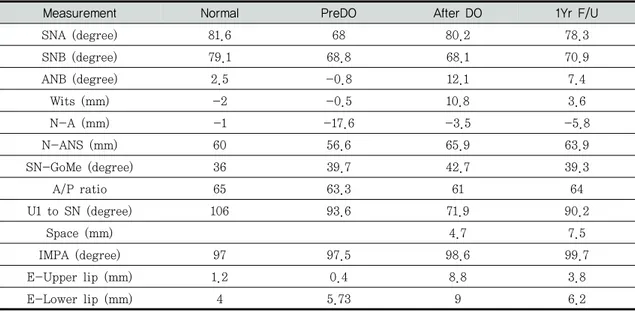
신장기간 동안 ANB값은 증가되었지만 유지 기 간 동안 감소되었다. 골 신장 후 A point는 전방 으로 14.1 mm 이동되었다(Table 1). RED 장치 사용 결과 ANS의 수직적인 위치는 하방으로 이 동되었다. 그리고 술 후 유지기간 동안 하방 이동 이 더 진행되었다(Figure 3). 골신장 후에 상악 전치부 각도(U1 to SN)는 21.7도 정도 감소되었다. 치조골 신장 공간은 4.7 mm가 생성되었다. 치료치조골의 신장으로 인하여 상악 견치의 배열을 위한 공간의 형성이 충분하게 형성되었으며 CT촬영을 통하여 신장된 결손부위 에 임플란트 식립을 위한 적절한 골질과 골양을 확인하였다(Figure 7).Discussion
A point의 위치는 신장 기간 동안 전방으로 14.1 mm 이동되었다. 이 결과는 이전의 상악골 신장술의 결과와 유사한 양이었다. Hashimoto등은 RED 장치 를 이용한 상악골 신장술을 시행한 결과 신장 기간 동안 ANS가 전방으로 7.5 mm 이동되었다고 보고하 였다12). 이번 증례에서는 골신장시 상악골의 전방분 절은 시계방향의 회전을 보였고, 그 결과 A point가 상악 전치보다 더욱 전방으로 이동하게 되었다.Figure 4. Intraoral views and radiographs after treatment.
Measurement Normal PreDO After DO 1Yr F/U
SNA (degree) 81.6 68 80.2 78.3 SNB (degree) 79.1 68.8 68.1 70.9 ANB (degree) 2.5 -0.8 12.1 7.4 Wits (mm) -2 -0.5 10.8 3.6 N-A (mm) -1 -17.6 -3.5 -5.8 N-ANS (mm) 60 56.6 65.9 63.9 SN-GoMe (degree) 36 39.7 42.7 39.3 A/P ratio 65 63.3 61 64 U1 to SN (degree) 106 93.6 71.9 90.2 Space (mm) 4.7 7.5 IMPA (degree) 97 97.5 98.6 99.7 E-Upper lip (mm) 1.2 0.4 8.8 3.8 E-Lower lip (mm) 4 5.73 9 6.2
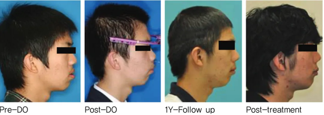
Pre-DO Post-DO 1Y-Follow up Post-treatment Figure 5. Comparison of extraoral views.
Figure 6. Superimposition of pre-Do, post-DO and 1Y-F/U. pre-Do post-DO 1Y-F/U 이번 증례에서 신장 기간 동안 전방부 분절골 의 수직적인 이동이 관찰되었다. 신장 기간 동안, 상악 전방분절골은 시계방향의 회전양상을 보였 다. 이것은 신장 방식에 따른 신장 방향과 분절골 의 무게 중심이 연관되어 있다. 상악의 무게 중심 은 상악 제1대구치의 상방 14.7 mm로 보고되었 다13). 최근 연구에서는 상악 전방부의 무게 중심 이 상악 무게 중심보다 전방에 위치하며, ANS 하 방의 견치 사이에 존재한다고 보고하였다14). 따라 서 RED를 이용한 경우, 상악 전방 분절골의 시계 방향 회전을 방지하기 위해서는 치조골 신장기가 RED와 결합될 필요가 있다. 상악전치부 경사도(U1 to SN)는 골신장 기간 동안 21.7 도가 감소되었다. RED 장치의 사용 시, 상악 전치가 구개 쪽으로 경사가 되며, 이를 해결 하기 위해서 신장 후에 고정식 교정장치를 이용하 여 전치부 토오크 조절을 시행하여야 한다. 그러 므로 상악골 전방분절의 회전 조절은 치료기간의 단축을 위해서 필수적이며 상악 전방분절의 저항 중심을 고려한 골신장기가 고안될 필요가 있다. 상악 전방분절골 신장술(Maxillary anterior segment DO)은 상악골 신장술에 비하여 신장 후에 입천장인두 기능부전(velopharyngeal incompetence) 가 발생될 수 있지만 반대교합 해결을 위하여 치조공 간의 확보가 필요한 환자에게 더욱 적합하다. 또한 상악 전방분절골 신장술은 상악골 신장시에 발생될 수 있는 개방교합이 흔히 발생하지 않는다. 최근의 연구에서 신생골의 생성과 함께 충분한 양의 치조골 공간 형성이 보고되었으며, 생성된 치조골질은 임플란트를 식립하기에 적합하였다15). Nosaka등은 개를 이용한 동물실험에서 신장된 치 조골의 골질과 골의 양은 방사선학과 조직학적으 로 우수한 결과를 나타내었다고 보고하였다14). 이 번 연구에서는 3D CT 상에서도 신장부위의 치조 골의 골질과 양이 우수한 것을 확인할 수 있었다 (Figure 7). 상악 전방분절골 신장술은 여러 장점이 있지만 치료 과정 중에 발생될 수 있는 전방 분절의 회
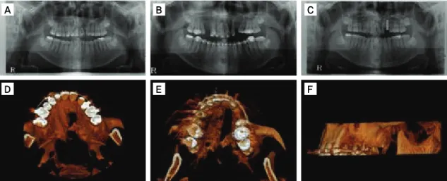
Figure 7. Investigation of alveolar bone using panoramic X-ray scanning and three-dimensional computed tomography. A, Pre-DO; B, post-DO; C, 1Y-F/U; D, axial view of pre-DO; E, axial view of post-DO; F, lateral view of post-DO.
B C A D E F 전을 고려해야 한다. 골신장시 원하지 않는 회전 을 방지하기 위해서는 구내장치와 RED 장치를 함께 사용하여 부작용을 최소화 할 수 있다. 하지 만 RED 장치는 비심미적이며 불편하기 때문에 임상적으로 충분한 경화기간을 유지하기 어렵다. 필요하다면 Face mask와 치조골 신장기를 이용 하여 장기적인 골경화가 가능하다. 골신장 기간 동안에 성공적으로 치조공간을 확 보할 수 있지만 골경화기 기간 동안에 유의할 만 한 공간 손실이 관찰되었다. 따라서 골신장 이후 에는 아크릴릭 레진의 공간유지장치가 필요하며, 성공적인 치료 결과의 유지를 위하여서는 장기간 의 유지가 필요하다(Figure 3).
Conclusions
이번 증례를 통하여 상악 전방분절골 신장술은 구순구개열 환자의 치조골 공간 확보, 신생골의 형성, 인두개 부전의 문제없는 측모 개선에 효과 적이라는 것을 알 수 있다. 상악 전방분절골 신장 술을 통하여도 많은 양의 신장이 가능하고 재발 이 적다는 장점이 있지만 신장 후에는 안정성 측 면에서 견고한 유지장치와 충분한 유지기간이 추 천된다.References
1. Karakasis D, Hadjipetrou L. Advancement of the anterior maxilla by distraction (case report). J Cranio Maxill Surg 2004; 32:150-154.
2. Tong ACK, Yan BSW, Chan TCK. Use of interdental distraction osteogenesis for orthodontic tooth alignment and correction of maxillary hypoplasia: a case report. Brit J Oral Max Surg 2003;41:185-187.
3. Ko EW, Figueroa AA, Guyette TW, Polley JW, Law WR. Velopharyngeal changes
after maxillary advancement in cleft pa-tients with distraction osteogenesis using a rigid external distraction device: A 1-year cephalometric follow-up. J Cra-niofac Surg 1999;10:312-320.
4. Harada K, Ishii Y, Ishii M, Imaizumi H, Mibu M, Omura K. Effect of maxillary distraction osteogenesis on velopharyn-geal function: A pilot study. Oral Surg Oral Med O 2002;93:538-543.
5. Iida S, Yagi T, Yamashiro T, Okura M, Takada K, Kogo M. Maxillary anterior segmental distraction osteogenesis with the Dynaform system for severe maxillary retrusion in cleft lip and palate. Plast Reconstr Surg 2007;120:508-516.
6. Liou EJW, Chen PKT, Huang CS, Chen YR. Interdental distraction osteogenesis and rapid orthodontic tooth movement: A novel approach to approximate a wide alveolar cleft or bony defect. Plast Reconstr Surg 2000;105:1262-1272.
7. Dolanmaz D, Karaman AI, Ozyesil AG. Maxillary anterior segmental advancement by using distraction osteogenesis: a case report. Angle Orthod 2003;73:201-205. 8. Bengi AO, Gurton AO, Okcu KM, Aydintug
YS. Premaxillary distraction osteogenesis with an individual tooth-borne appliance. Angle Orthodontist 2004;74:420-431. 9. Le BT, Eyre JM, Wehby MC, Wheatley MJ.
Intracranial migration of halo fixation pins: A complication of using an extraoral distraction device. Cleft Palate-Cran J 2001;38:401-404.
10. Rieger J, Jackson IT, Topf JS, Audet B. Traumatic cranial injury sustained from a fall on the rigid external distraction device. J Craniofac Surg 2001;12:237-241. 11. Minami K, Mori Y, Tae-Geon K, Shimizu
H, Ohtani M, Yura Y. Maxillary distrac-tion osteogenesis in cleft lip and palate patients with skeletal anchorage. Cleft Palate-Cran J 2007;44:137-141.
12. Hashimoto K, Otsuka R, Minato A, Sato- Wakabayashi M, Takada J, Inoue-Arai MS, et al. Short-term changes in tem-poromandibular joint function in subjects with cleft lip and palate treated with maxillary distraction osteogenesis. Orthod Craniofac Res 2008;11:74-81.
13. Ahn JG, Figueroa AA, Braun S, Polley JW. Biomechanical considerations in distraction of the osteotomized dento-maxillary complex. Am J Orthod Dento-facial Orthop 1999;116:264-270.
14. Melsen B, Fotis V, Burstone CJ. Vertical force considerations in differential space closure. Journal of clinical orthodontics : JCO 1990;24:678-683.
15. Van Sickels JE, Abadi B, Attisha R. Anterior segmental distraction for a Class III maxillary prosthetic defect in a cleft palate patient. The Journal of oral implantology. 2011;37:457-461.
16. Nosaka Y, Tsunokuma M, Hayashi H, Kakudo K. Placement of implants in distraction osteogenesis: a pilot study in dogs. The International journal of oral & maxillofacial implants. 2000;15:185-192.
교신 저자 Jung-Yul Cha
Room 726, Department of orthodontics, Yonsei University College of Dentistry, Seoul, South Korea, 250 Seongsanno, Seodaemun-gu, Seoul 120-752, South Korea
Tel : +82-2-2228-3103 / Fax : +82-2-363-3404 / E-mail : jungcha@yuhs.ac
Acknowledgement
This study was supported by a grant from the Korea Healthcare Technology R&D Project, Ministry for Health, Welfare & Family Affairs, The Republic of Korea (A090353).