Jin-Sun Kim, Jung-Sun Kim, Eui-Young Choi, Donghoon Choi and Yangsoo Jang
Catastrophic Thrombus Formation During Optical Coherence Tomography
Print ISSN: 0009-7322. Online ISSN: 1524-4539
Copyright © 2008 American Heart Association, Inc. All rights reserved.
is published by the American Heart Association, 7272 Greenville Avenue, Dallas, TX 75231
Circulation
doi: 10.1161/CIRCULATIONAHA.107.762096
2008;118:e101-e102
Circulation.
http://circ.ahajournals.org/content/118/6/e101
World Wide Web at:
The online version of this article, along with updated information and services, is located on the
http://circ.ahajournals.org/content/suppl/2008/08/12/118.6.e101.DC1.html
Data Supplement (unedited) at:
http://circ.ahajournals.org//subscriptions/
is online at:
Circulation
Information about subscribing to Subscriptions:
http://www.lww.com/reprints
Information about reprints can be found online at: Reprints:
document.
Permissions and Rights Question and Answer
this process is available in the
click Request Permissions in the middle column of the Web page under Services. Further information about Office. Once the online version of the published article for which permission is being requested is located,
can be obtained via RightsLink, a service of the Copyright Clearance Center, not the Editorial
Circulation
in
Requests for permissions to reproduce figures, tables, or portions of articles originally published Permissions:
at CONS KESLI on October 15, 2014 http://circ.ahajournals.org/
Downloaded from http://circ.ahajournals.org/ at CONS KESLI on October 15, 2014 Downloaded from
Catastrophic Thrombus Formation During Optical
Coherence Tomography
Jin-Sun Kim, MD; Jung-Sun Kim, MD, PhD; Eui-Young Choi, MD, PhD;
Donghoon Choi, MD, PhD; Yangsoo Jang, PhD
A
71-year-old patient underwent follow-up coronary an-giography to assess the patency of a left anterior descending (LAD) artery stent. Seven months previously, she had received percutaneous coronary intervention at the LAD because of anterior wall ST-elevation myocardial infarction. At that time, 2 drug-eluting stents (Cypher 3.0⫻28 mm and Cypher 2.75⫻28 mm) were inserted at the proximal and distal LAD. After percutaneous coronary intervention, she had taken 100 mg of aspirin, 75 mg of clopidogrel, and 200 mg of cilostazol daily. Coronary angiography at 7 months follow-up was performed as part of the protocol for the optical coherence tomography (OCT) registry. The follow-up coronary angiography revealed patent stents implanted in the LAD (Figure 1A). We also performed intravascular ultra-sound (IVUS) and OCT to evaluate neointimal coverage of the drug-eluting stents. IVUS revealed that the stents were partially covered with neointima without any evidence of thrombus during IVUS imaging (online-only Data Supple-ment Movie I). Subsequently, we performed OCT, which clearly illustrated partial neointimal coverage of stent struts. But during the OCT procedure, multiple thrombi formation were observed (Figure 2 and online-only Data Supplement Movie II). Coronary angiography immediately after OCT revealed thrombotic total occlusion of the distal LAD (Figure 1B and online-only Data Supplement Movie III). Therefore we immediately performed thrombi suction with a Throm-buster catheter (Kaneka Medics, Osaka, Japan). However, the thrombi were not completely cleared, with only partial recovery of the distal flow (online-only Data Supplement Movie IV). Fortunately, no significant deterioration occurred in the ECG or in the patient’s condition after the procedure. The patient recovered uneventfully.OCT is an in vivo light-based imaging modality for evaluating the coronary artery microstructure.1Because the resolution of
OCT imaging is higher than that of IVUS, its application in research is expanding.2,3In particular, attempts to detect
vulner-able plaques in vivo with OCT imaging and studies of the delicate interaction between vessel wall and stent strut, as well as of neointima growth, are actively ongoing.3,4
Unlike IVUS, however, OCT has a limitation in that it needs to create a blood-free zone. Therefore, balloon occlu-sion of the proximal vasculature for as long as 30 to 40 seconds is required.1This limitation can provoke myocardial
ischemia during the procedure and, in rare situations, intra-coronary thrombus formation as demonstrated in this case.
Disclosures
None.
References
1. Regar E, Schaar JA, Mont E, Virmani R, Serruys PW. Optical coherence tomography. Cardiovasc Radiat Med. 2003;4:198 –204.
2. Yabushita H, Bouma BE, Houser SL, Aretz HT, Jang IK, Jang IK, Schlendorf KH, Kauffman CR, Shishkov M, Kang DH, Halpern EF, Tearney GJ. Characterization of human atherosclerosis by optical coherence tomography. Circulation. 2002;106:1640 –1645.
3. Jang IK, Bouma BE, Kang DH, Park SJ, Park SW, Seung KB, Choi KB, Shishkov M, Schlendorf K, Pomerantsev E, Houser SL, Aretz HT, Tearney GJ. Visualization of coronary atherosclerotic plaques in patients using optical coherence tomography: comparison with intravascular ultrasound. J Am Coll Cardiol. 2002;39:604 – 609.
4. Matsumoto D, Shite J, Shinke T, Otake H, Tanino Y, Ogasawara D, Sawara T, Paredes OL, Hirata K, Yokoyama M. Neointimal coverage of sirolimus-eluting stents at 6-month follow-up: evaluated by optical coherence tomography. Eur Heart J. 2007;28:961–967.
From the Division of Cardiology, Yonsei Cardiovascular Center, Yonsei University College of Medicine, Seoul, South Korea.
The online-only Data supplement, which consists of movies, is available with this article at http://circ.ahajournals.org/cgi/content/full/118/6/e101/DC1.
Correspondence to Jung-Sun Kim, MD, Division of Cardiology, Yonsei Cardiovascular Center, Yonsei University College of Medicine, 120 –752, Shinchondong, Seodaemun-gu, Seoul, South Korea, E-mail [email protected]
(Circulation. 2008;118:e101-e102.)
© 2008 American Heart Association, Inc.
Circulation is available at http://circ.ahajournals.org DOI: 10.1161/CIRCULATIONAHA.107.762096
e101
Images in Cardiovascular Medicine
at CONS KESLI on October 15, 2014 http://circ.ahajournals.org/
Figure 1. A, Follow-up coronary angiography
revealed patent stents previously inserted at proxi-mal and distal LAD artery. B, After OCT imaging, total occlusion of distal LAD was observed on cor-onary angiography (arrow).
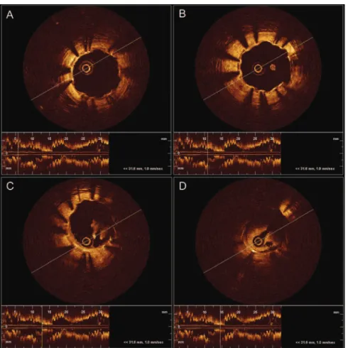
Figure 2. New thrombi were detected
during evaluation of the distal stent with OCT.
e102 Circulation August 5, 2008
at CONS KESLI on October 15, 2014 http://circ.ahajournals.org/