저작자표시-비영리-변경금지 2.0 대한민국 이용자는 아래의 조건을 따르는 경우에 한하여 자유롭게 l 이 저작물을 복제, 배포, 전송, 전시, 공연 및 방송할 수 있습니다. 다음과 같은 조건을 따라야 합니다: l 귀하는, 이 저작물의 재이용이나 배포의 경우, 이 저작물에 적용된 이용허락조건 을 명확하게 나타내어야 합니다. l 저작권자로부터 별도의 허가를 받으면 이러한 조건들은 적용되지 않습니다. 저작권법에 따른 이용자의 권리는 위의 내용에 의하여 영향을 받지 않습니다. 이것은 이용허락규약(Legal Code)을 이해하기 쉽게 요약한 것입니다. Disclaimer 저작자표시. 귀하는 원저작자를 표시하여야 합니다. 비영리. 귀하는 이 저작물을 영리 목적으로 이용할 수 없습니다. 변경금지. 귀하는 이 저작물을 개작, 변형 또는 가공할 수 없습니다.
Autophagy mechanisms in sputum and peripheral
blood cells of patients with severe asthma: a new
therapeutic target
By
Ga-Young Ban
Major in Medicine
Department of Medical Sciences
The Graduate School, Ajou University
Autophagy mechanisms in sputum and peripheral
blood cells of patients with severe asthma: a new
therapeutic target
By
Ga-Young Ban
A Dissertation Submitted to The Graduate School of
Ajou University in Partial Fulfillment of the
Requirements for
The Degree of PhD of Medicine
Supervised by
Hae Sim Park, M.D., Ph.D.
Major in Medicine
Department of Medical Sciences
The Graduate School, Ajou University
This certifies that the dissertation
ofGa Young Ban is approved.
SUPERVISORY COMMITTEE
Dong-Ho Nahm (Sign)
Hae-Sim Park (Sign)
Young-JoonChwae (Sign)
Young-Min Ye (Sign)
Gyu-Young Hur, (Sign)
The Graduate School, Ajou University
December, 18th, 2015
i
-ABSTRACT-
Autophagy mechanisms in sputum and peripheral blood
cells of patients with severe asthma: a new therapeutic
target
Background: Autophagy and genetic predisposition have been suggested to
potentially play roles in the development of asthma. However, little is known about the role of autophagy in the pathogenesis of severe asthma.
Objective:Wecompared autophagy in the sputum granulocytes, peripheral
blood cells (PBCs),and peripheral blood eosinophils (PBEs) betweenpatients with severe asthma and those with non-severe asthmaand investigatedthe functional effects of autophagy.
Methods:We enrolled 36patients with severe asthma, 14 with non-severe
asthma,and 23 normal healthy controls in this study. Sputum granulocytes, PBCs, and PBEswere isolated from each subject. Autophagy was evaluated based on the expression of microtubule-associated protein light chain 3 (LC3) by western blot, confocal microscopy, transmission electron microscopy, and flow cytometry. IL-8 levels were measured by ELISA. To induce autophagy, HL-60 cells, SAECs, and A549 cells were treated withIL-5, IL-1β, and TNF-α. To inhibit autophagy, PI3K inhibitors (LY29400 and 3-methyladenine [3-MA]) and hydroxychloroquine (HCQ) were used. Knockdown of ATG5 and Beclin-1 was performed in A549 cells, and thetherapeutic effects of dexamethasone were evaluated.
Results: Higherautophagy levels were noted in sputum granulocytes, PBCs,
and PBEs from patients with severe asthma than from patients with non-severe asthma and healthy controls (P<0.05 for all). IL-5 increased autophagy levels in both PBCs and PBEs (P<0.05). 3-MA attenuated the increased
expression of LC3-II and eosinophil cationic protein in HL-60 cells induced by IL-5 (P=0.034 for both). Dexamethasone did not affect autophagy levels in PBEs.IL-1β increased LC3-II expression and IL-8 production (P<0.01) in SAECs, and this was attenuated by LY294002, 3-MA, HCQ,and knockdown of ATG5 and Beclin-1 (in A549 cells) (P<0.01).
Conclusions and clinical relevance:Autophagy could play a role in the
pathogenesis of severe asthma. Autophagy modulation may be a novel therapeutic target for conventional therapy-resistant severe asthma.
iii
TABLE OF CONTENTS
ABSTRACT ••••••••••••••••••••••••••••••••••••••••••••••••••••••••••••••••••••••ⅰ TABLE OF CONTENTS•••••••••••••••••••••••••••••••••••••••••••••••••••••••ⅱ LISTS OF FIGURES•••••••••••••••••••••••••••••••••••••••••••••••••••••••••••ⅲ LISTS OF TABLES••••••••••••••••••••••••••••••••••••••••••••••••••••••••••••ⅳ ABBREVIATION••••••••••••••••••••••••••••••••••••••••••••••••••••••••••••••ⅴ Ⅰ. INTRODUCTION••••••••••••••••••••••••••••••••••••••••••••••••••••••••••1 II. MATERIALS AND METHODS•••••••••••••••••••••••••••••••••••••••••••3 A. MATERIALS••••••••••••••••••••••••••••••••••••••••••••••••••••••••••••••3 1. Study subjects•••••••••••••••••••••••••••••••••••••••••••••••••••••••••3 2.Antibodies and reagents
•••••••••••••••••••••••••••••••••••••••••••3 B. METHODS1. Cell culture••••••••••••••••••••••••••••••••••••••••••••••••••••••••••••3 2. Sputum induction and granulocyte isolation•••••••••••••••••••••••4 3. Human PBC isolation and autophagy induction•••••••••••••••••••4 III. RESULTS•••••••••••••••••••••••••••••••••••••••••••••••••••••••••••••••••••8 IV. DISCUSSION••••••••••••••••••••••••••••••••••••••••••••••••••••••••••••••16 V. CONCLUSION••••••••••••••••••••••••••••••••••••••••••••••••••••••••••••••20 REFERENCES••••••••••••••••••••••••••••••••••••••••••••••••••••••••••••••••••21 국문요약•••••••••••••••••••••••••••••••••••••••••••••••••••••••••••••••••••••••24
Fig. 1.Autophagy in sputum granulocytes and peripheral blood cells (PBCs) isolated from normal healthy controls (NC), patients with non-severe asthma (NSA), and patients with severe asthma (SA).
Fig. 2. Interleukin (IL)-5–induced autophagy in peripheral blood cells (PBCs) and HL-60 cells.
Fig. 3.The effects of anti-inflammatory drugs on autophagy in HL-60 cells. Fig. 4.Autophagy in human peripheral blood eosinophils (PBEs).
Fig. 5. Autophagy in human small primary airway epithelial cells (SAECs). Fig. 6.Induction of autophagy in A549 cells and the effects of
anti-inflammatory drugs.
Fig. 7.Functional effect of autophagy on interleukin (IL)-8 production from human small primary airway epithelial cells (SAECs) and human lung epithelial cells (A549).
v
ABBREVIATIONS 3-MA: 3-methyladenine
ECP: eosinophil cationic protein
ELISA: enzyme-linked immunosorbent assay E+P: E64D and pepstatin A
FBS:fetal bovine serum HCQ: hydroxychloroquine IL: interleukin
LAMP1: lysosomal-associated membrane protein 1 LC3: microtubule-associated protein light chain MBP: major basic protein
NC: normal healthy control NSA: non-severe asthma PBCs: peripheral bloodcells PBEs: peripheral blood eosinophils PBS: phosphate-saline buffer RBCs: red blood cells
TEM: transmission electron microscopy TNF-α: tumor necrosis factor-α
SA: severe asthma
1
I.
INTRODUCTION
Autophagy is widely involved in both pathophysiologicalprocesses (cancer, metabolic and neurodegenerative disorders, and cardiovascular and pulmonary diseases) and physiological processes such as aging. Autophagy affects many processes that have various effects on disease,andcan inhibit or promote disease progression(Choi et al., 2013). The adverse effects of autophagy are observed in lung cancer and acute lung injury induced by H5N1. In contrast, the beneficial roles of autophagy are seen in cystic fibrosis, tuberculosis, and sepsis. Autophagy was initially believed to be a cytoprotective response in the pathophysiological state of chronic obstructive pulmonary disease; however, accumulating data have revealedthat autophagy has both adverse and beneficial functions(Nakahira, 2013).
A few studies (Morris et al., 2011; Chan et al., 2012; Martin et al., 2012; Poon et al., 2012b) have suggested that autophagy may be involved in asthma. A recent study revealed an association between a genetic variant of ATG5, an autophagy-related gene, and the force expiratory volume in 1 second, implying that autophagy is relatedto reduced lung function in asthmatics. These studies also demonstrated that fibroblasts and epithelial cells isolated from bronchial biopsy tissue taken from patients with moderately severe asthma had agreater number of autophagosomesthan that taken fromhealthy controls(Poon et al., 2012b). The authors suggested that autophagy delays the apoptosis of fibroblasts and maintains chronic oxidative stress by promotingtransforming growth factor-β release from fibroblasts and airway remodeling; consequently, autophagy may contribute to the pathogenesis of severe asthma(Poon et al., 2012a).However, little is known about the role of autophagy in the pathogenesis of asthma, especially severe asthma.
controlled by the regular use of an inhaled corticosteroid with or without a long-acting β2 agonist. However, 5% to 10% of the asthmatic population is
poorly controlled or is only controlled by higher levels of recommended treatment such as systemic corticosteroids or biologics. This group of patients is defined as severe asthmatics (SA)(Bousquet et al., 2010). They suffer from frequent asthma exacerbations as well as greater morbidity and mortality, and consume a greater proportion of health care resources; this islargely attributed to medication, hospital admissions, and work loss (Macedo et al., 2009).
Airway inflammation and remodeling are key characteristics of asthma. Multicellular inflammatory processes involving eosinophils, neutrophils, T helper cells, monocytes, mast cells, basophils, and epithelial cells contribute to the pathogenesis of asthma (Holgate, 2011). Among these inflammatory cells, eosinophilic inflammation is particularly associated with more severe asthma in terms of airway remodeling, impaired lung function, and disease exacerbation (Fahy, 2009; Kim et al., 2013). Severe eosinophilic asthma is a major phenotype of severe asthma(Brusselle et al., 2013).However, the mechanism of how eosinophilia persists in severe eosinophilic asthma (despite the high dose of inhaled corticosteroid) is not well understood. Biologics (such as anti-IL-5)are anemerging class of drugs used to treat steroid-refractory severe asthma; however, the response to biologicsis diverse. One factor that affects the response to biologics is theeosinophilic phenotype(Reddy and Little, 2013); however, asthma is a heterogeneous disease, and the eosinophilic phenotype alone cannot fully explain the diverse responses to anti-IL-5 treatment.
We hypothesized that autophagyplaysa role in the activation and augmentation of airway inflammation in severe asthma, especially severe eosinophilic asthma. We evaluated autophagy in the sputum granulocytes, peripheral blood cells (PBCs), and peripheral blood eosinophils (PBEs) using immunoblot analysis, immunofluorescence microscopy (confocal microscopy),
3
and transmission electron microscopy (TEM) in patients withsevere asthma compared withthose with non-severe asthma and normal healthy controls. We investigated the changes in autophagy expression and its functional effects on human eosinophil-like (HL-60) cells, human lung epithelial (A549) cells, PBEs, and human primary small airway epithelial cells (SAECs) to elucidate the effects of IL-5,IL-1β, and tumor necrosis factor-α (TNF-α)on autophagy. We also used ex vivo and in vitroapproaches to investigate the effect of two representative anti-inflammatory drugs on autophagy and thus determine whetherour hypothesis, namely that autophagy contributes to the exacerbation of SA, can be applied to management of severe asthma.
II. MATERIALS AND METHODS
A. MATERIALS 1. Study subjects
We enrolled 36 patients with severe asthma (SA), 14 with non-severe asthma (NSA), and 33 normal healthy controls (NCs) in this study. All subjects were nonsmokers. All asthmatics were diagnosed by recurrent episodes of wheezing, dyspnea, cough, sputum, and evidence of either airway hyper-responsiveness to methacholine or reversible airway obstruction improved by a short-acting β2 agonist (Hwang et al., 2012). All patients in the SA group fit
the definition of severe asthma adopted by the American Thoracic Society Workshop (Wenzel et al., 2000). All subjects gave written informed consent at the time of enrollment, and the study was approved by the institutional review board of Ajou University Hospital (AJIRB-GEN-SMP-13-108). Peripheral blood and induced sputum were collected from the study subjects when the disease was stable and patients were taking their regular medication. Venous blood from the subjects was drawn into a BD Vacutainer® containing ACD
solution A (BD Biosciences, Franklin Lakes, NJ, USA) and processed within 2 h after collection.
2. Antibodies and reagents
Anti-microtubule-associated light chain 3 (LC3) rabbit monoclonal antibody and PE-conjugated anti-LC3 rabbit monoclonal antibody were purchased from Cell Signaling (Minneapolis, MN, USA). Anti-eosinophil cationic protein (ECP) rabbit polyclonal antibody was purchased from Santa Cruz (Dallas, TX, USA). Anti-major basic protein (MBP) mouse monoclonal antibody was purchased from GenWay Biotech Inc. (San Diego, CA, USA). Anti-lysosomal-associated membrane protein 1 (LAMP-1) mouse monoclonal antibody was purchased from Novus (Littleton, CO, USA). APC-conjugated anti-Siglec8 antibody was purchased from Biolegend (San Diego, CA, USA).
5
APC-conjugated anti-LAMP-1 mouse monoclonal antibody was purchased from BD Biosciences (San Jose, CA, USA).Anti-tubulin goat polyclonal antibody, anti-beta actin goat polyclonal antibody, dithiothreitol, trypan blue, rapamycin, human recombinant IL-5, human recombinant IL-1β, human recombinant TNF-α, dexamethasone, E64D, pepstatin A, hydroxychloroquine(HCQ), polyprene, puromycin, radioimmunoprecipitation buffer, 3-methyladenine(3-MA), and LY294002 were purchased from Sigma-Aldrich (St. Louis, Missouri, USA). RPMI-1640 medium, fetal bovine serum (FBS), penicillin, and streptomycin solution for cell culture were purchased from Gibco (Grand Island, NY, USA).
B. METHODS 1. Cell culture
A human lung airway epithelial cell line (A549), human eosinophil-like cells (HL-60),and primary human small airway epithelial cells (SAECs) were purchased from American Type Culture Collection (ATCC, Manassas, VA, USA). A549 cells were cultured in RPMI-1640 medium, and HL-60 cells were cultured in Iscove’s Modified Dulbecco’s Medium supplemented with 10% heat-inactivated FBS, penicillin (100 IU/ml), and streptomycin (50 μg/ml). SAECs were cultured in Airway Epithelial Cell Basal Medium (ATCC) supplemented with L-alanyl-L glutamine (2.4 mM), extract P (0.4%), plasma protein fraction (0.5%), epinephrine (1.0 μM), transferrin (5 μg/ml), T3 (10nM), hydrocortisone (5 μg/ml), human recombinant epidermal growth factor (5 ng/ml), human recombinant insulin (5 μg/ml) (all components were provided in a Small Airway Epithelial Cell Growth Kit, ATCC), penicillin (10 IU/ml), and streptomycin (10 μg/ml). SAECs cultured within three to eight passages were used for the experiments. All cells were grown at 37ºC in humidified air with 5% CO2.
2. Sputum induction and granulocyte isolation
described previously (Rutgers et al., 2000). Sputum samples were processed within 2 hafter induction using previously described methods(Pang et al., 1995; Urbanowicz et al., 2010). The total number of isolated sputum granulocytes and cell viability were determined by trypan blue staining and observed under an inverted microscope. To determine the fraction of granulocytes in cell mixtures, a cell smear of each sample was placed on a glass slide, air-dried, fixed in methanol, and stained with May-Grünwald-Giemsa (Sigma-Aldrich). Samples that contained at least 90% granulocytes were used for immunoblot and immunocytochemistry analysis.
3. Human PBC isolation and autophagy induction
Human PBCs were isolated from peripheral blood samples (5 ml) from the study subjects. Red blood cells (RBC) were lysed by adding 10 ml of RBC lysis buffer (NH4Cl, 0.15 M; Na2HCO3, 10 mM; EDTA, 0.1 M) into 5 ml of
blood followed by centrifugation at 1350 rpm for 10 min at 4ºC. The cell pellet was washed with 10 ml of Hanks’ balanced salt solution (Sigma-Aldrich) and cultured in RPMI-1640 medium supplemented with 10% heat-inactivated FBS.Cells (1 ´106) were seeded in each well of a six-well plate
(TPP) and treated with IL-5 (30 ng/ml) for 24 h. E64D (10 μM) and pepstatin A (15 μM) (E+P) were used to block the degradation of autophagolysosomes by inhibiting lysosomal proteases. When indicated, cells were co-treated with IL-5 and HCQ at different concentrations.
4. Human PBE isolation and autophagy induction
Human eosinophils were isolated from peripheral blood samples (30 ml) of patients with severe asthma. Blood was layered on a LymphoprepTM
(Axis-Shield, Oslo, Norway), followed by centrifugation at 2000 rpm, at 20ºC for 25 min without braking. Eosinophils were isolated from the RBC and granulocyte fraction by an Eosinophil Isolation Kit and MACS Column (MiltenylBiotec Inc., Auburn, CA, USA) according to the manufacturer’s instructions. Isolated eosinophils were labeled with APC-conjugated
anti-7
Siglec8 antibody and anti-ECP rabbit polyclonal IgG antibody to evaluate purity (>95%) by flow cytometry. Cell viability (>98%) was assessed under an inverted microscope by trypan blue staining. PBEs (5 ´105)were seeded in
each well of a 12-well plate (TPP, Trasadingen, Switzerland) in RPMI-1640 medium supplemented with 10% heat-inactivated FBS, penicillin (100 IU/ml), and streptomycin (50 μg/ml). Cells were treated with rapamycin (10 μM) or human recombinant IL-5 (100 ng/ml) for 6 h to induce autophagy. Dexamethasone (1 μM) was used to pretreat the cells for 1 h when indicated. To investigate autophagy flux, E+P was used to block the degradation of autophagolysosomes.
5. Autophagy induction in HL-60 cells and SAECs
HL-60 cells (1 ´106) or SAECs (5 ´105) were seeded in each well of a
six-well plate (TPP) and treated with rapamycin (10 μM), human recombinant IL-5 (10 ng/ml), IL-1β (100 ng/ml), or TNF-α (100 ng/ml) for 24 h. Cells were pretreated with dexamethasone (1 μM) for 1 h when indicated. To inhibit autophagy, cells were pretreated for 15 min with LY294002 (20 nM), a PI3K inhibitor. To inhibit the initial step of autophagy, another PI3K inhibitor, 3-MA (5 mM), was added during the last 6 h of the incubation period; HCQ (10 μM) was used to block autophagosome degradation by co-treatment with the autophagy inducers. Cell culture supernatants were collected for IL-8 concentration measurement by ELISA.
6. Knockdown of ATG5 and beclin-1 in A549 cells
Bacteria (Escherichia coli) stocks that contained MISSION short hairpin (sh) RNA against ATG5and Beclin-1 cloned into a pLKO.1 vector were purchased from Sigma-Aldrich. shRNAthat contained a nontarget (NT) sequence was used as a control. ATG5 andBeclin-1 shRNA were co-transfected with pMISSION-GAG-POL vector and pMISSION-VSV-G vector (Sigma) into HEK293TN cells (System Biosciences, Mountain View, CA, USA) using Lipofectamine®2000 Transfection Reagent (Invitrogen Life Technologies,
Carlsbad, CA, USA) according to the manufacturer’s protocol to produce pseudo lentiviral particles. Virus particles were collected 48 h after the transfection and infected into A549 cells with the presence of polyprene(8 μg/ml) in the culture medium. Lentivirus-infected A549 cells were selected by puromycin(2 μg/ml) to produce stable cell lines and used for experiments within 2 weeks after infection.
An equal number (1 ´106) of A549 cells of each group (ATG5, Beclin-1, and
NT knockdown) was seeded in each well of a six-well plate (TPP) and left untreated or treated with IL-1β (100 ng/ml) for 24 h. The culture supernatants were collected for IL-8 measurement by ELISA. Given that a knockdown ofATG5or Beclin-1 could affect cell proliferation (Wang et al., 2013), the cell proliferation rate at 24 h of each cell group was determined as described in the Methods section of the supplementary document. The measured IL-8 levels were adjusted for the cell proliferation rate of each cell group as follows: adjusted IL-8 level = measured IL-8 level/proliferation rate.
7. Measurement of IL-8 production by ELISA
IL-8 levels in cell culture supernatants were measured by a commercial enzyme-linked immunosorbent assay kit (ELISA, Endogen, Woburn, MA, USA) following the manufacturer’s protocol.
8. Western blot analysis for autophagy investigation
Autophagy was evaluated by using western blot to detect the autophagic markers – microtubule associated protein light chain 3 (LC3). In the elongation step of autophagosome formation, LC3-I is converted into LC3-II and anchors the autophagosome membrane; therefore, LC3-II has been considered a marker for autophagy (Choi et al., 2013). Cells were lysed in RIPA buffer that contained 1% of proteinase inhibitor cocktail solution (Roche Diagnostics, Indianapolis, IN, USA) to extract proteins. The protein concentration in cell lysis was measured by using a Bio-Rad Protein assay kit (Bio-Rad, Hercules, CA, USA). An equal amount of protein (30-50 μg) from
9
each cell lysis was loaded on 4-20% SDS-polyacrylamide gels and transferred to a polyvinylidene difluoride membrane (Bio-Rad). After blocking with 5% skim milk (Difco, Detroit, MI, USA) in PBS containing 0.5% of Tween 20 (PBS-T), the membrane was incubated with anti-LC3 rabbit polyclonal antibody (1:1000), anti-eosinophil cationic protein (ECP) rabbit polyclonal antibody (1:1000) overnight at 4℃. The blots were then washed with PBS-T and incubated with appropriate HRP-conjugated secondary antibodies (Santa Cruz) for 1 hr at room temperature (RT). The membrane was blotted for anti-beta actin or anti-tubulin as loading controls. Signals were detected using an ECL Plus Western Blotting Detection Reagents (GE Healthcare, Little Chalfont, UK). The intensity of bands was analyzed by gel doc system (Bio-Rad).
9. Detection of autophagy by confocal microscopy
After isolation, sputum granulocytes, PBCs, and PBEs were seeded onto poly-L-lysine–coated slides (1 ´104 cells/slide) and maintained at 37ºC for 1 h to
allow for cell attachment. Cells were then treated with the indicated drugs, followed by fixation with 4% paraformaldehyde in PBS for 20 min at room temperature (RT), permeabilization with 0.2% Triton-X100 in PBS for 10 min on ice, and blocking with PBS that contained 10% horse serum (Abcam, Cambridge, MA, USA) and 1% BSA (Amresco, Solon, OH, USA) for 1h at RT. Cells were then incubated for 1 h at 37ºC withanti-LC3 rabbit monoclonal antibody (1:300), anti-LAMP-1 mouse monoclonal antibody (1:500), and/oranti-MBP mouse monoclonal antibody (1:500) as an eosinophil-specific marker. Cells were then washed for 10 min six times with PBS containing 0.2% Tween-20, and then incubated with appropriate Alexa Fluor®-conjugated secondary antibodies (Invitrogen) for 50 min at 37ºC.
Nuclei were stained with 4´,6-diamidino-2-phenylindole (DAPI, Sigma-Aldrich). Coverslips were mounted onto glass slides using VECTASHIELD mounting medium (Vector, Burlingame, CA, USA), and the cells were
examined under a Zeiss LSM710 confocal microscope with a 63´oil objective lens (Carl Zeiss AG, Oberkochen, Germany). Images were analyzed using ZEN 2009 software (Carl Zeiss). Under a fluorescent microscope, autophagosomeswere observed as LC3 dots (puncta) in the cell cytoplasm due to the accumulation of LC3-II in autophagosomeswhen autophagy occurs(Klionsky et al., 2012). Additionally, lysosomes were observed as LAMP-1 puncta in the cell cytoplasm. Consequently, the fusion of autophagosomes and lysosomes in the final step of autophagy was detected by the co-localization of LC3 and LAMP-1 puncta. For quantification of autophagy in PBEs, the number of LC3/LAMP-1 puncta was counted in 50 cells randomly selected from each treatment condition in a blinded fashion.
10. Detection of autophagosomes by TEM
PBCs were isolated, fixed in 2.5% glutaraldehyde for 2 h at 4ºC, washed with PBS (0.1 M, pH 7.4), and post-fixed in 1% osmium tetroxide for 90 min. The specimens were then dehydrated through a graded ethanol series, exchanged through propylene oxide, and embedded in a mixture of embedding resin (Epon). Subsequently, ultrathin sections were obtained by an ultramicrotome (Leica Reichert Ultracut S, Wetzlar, Germany) with a diamond knife, double-stained with uranyl acetate and lead citrate, and examined by TEM at 80kV. The specimens were observed by TEM (Carl Zeiss 902A) to visualize the localization of double-membrane vesicles (which are considered to beautophagosomes)in the cell cytoplasm.
11. Evaluation of autophagy by flow cytometry
To detect autophagy in eosinophils among PBCs, we used the Cyto-ID® Green
autophagy detection kit (Enzo Life Sciences, Farmingdale, NY, USA) following the manufacturer’s protocol with some modifications. Briefly, after removal of RBCs, the white blood cells were incubated with an FC receptor blocker (eBioscience, San Diego, CA, USA) in PBS for 10 min, and then stained with APC-conjugated anti-Siglec8 antibody and Cyto-ID® green dye
11
in the 1X assay buffer provided with the kit.
To evaluate autophagy in purified PBEs, cells were fixed with 2% PFA for 10 min at RT followed by permeabilization with 0.1% Triton-X100 for 5 min on ice. Cells were then stained with PE-conjugated anti-LC3 rabbit monoclonal antibody and APC-conjugated anti-LAMP-1 mouse monoclonal antibody for 20 min at RT in the dark, followed by washing and resuspension in 100 μL of PBS and analyzed by flow cytometry using the FACSDiva software v6.0 (FACSCanto II, BD Biosciences). Cells were gated using the FSC-SSC, plot and eosinophils were gated with siglec8-positive cells.
12. Statistical analysis
Data for continuous variables were compared using the Mann-Whitney U test, and Pearson’s chi-squared or Fisher’s exact testwas used for categorical variables. All computations were performed using SPSS software, version 22.0 (IBM Corp., Armonk, NY, USA).GraphPad Prism 5.0 software
(GraphPad Inc., San Diego, CA, USA) was used for the production of graphs, and values are presented as the mean±standard deviation (SD) of at least three independent experiments.
III.RESULTS
1. Autophagy in sputum granulocytes and PBCs from patients in the SA and NSA groups
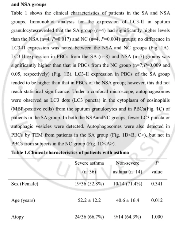
Table 1 shows the clinical characteristics of patients in the SA and NSA groups. Immunoblot analysis for the expression of LC3-II in sputum granulocytesrevealed that the SA group (n=4) had significantly higher levels than the NSA (n=4, P=0.017) and NC (n=4, P=0.004) groups; no difference in LC3-II expression was noted between the NSA and NC groups (Fig. 1A). LC3-II expression in PBCs from the SA (n=8) and NSA (n=7) groups was significantly higher than that in PBCs from the NC group (n=7;P=0.009 and 0.05, respectively) (Fig. 1B). LC3-II expression in PBCs of the SA group tended to be higher than that in PBCs of the NSA group; however, this did not reach statistical significance. Under a confocal microscope, autophagosomes were observed as LC3 dots (LC3 puncta) in the cytoplasm of eosinophils (MBP-positive cells) from the sputum granulocytes and in PBCs(Fig. 1C) of patients in the SA group. In both the NSAandNC groups, fewer LC3 puncta or autophagic vesicles were detected. Autophagosomes were also detected in PBCs by TEM from patients in the SA group (Fig. 1D<B, C>), but not in PBCs from subjects in the NC group (Fig. 1D<A>).
Table 1.Clinical characteristics of patients with asthma
Severe asthma (n=36) Non-severe asthma (n=14) P value Sex (Female) 19/36 (52.8%) 10/14 (71.4%) 0.341 Age (years) 52.2 ± 12.2 40.6 ± 16.4 0.012 Atopy 24/36 (66.7%) 9/14 (64.3%) 1.000
13
Total IgE (KU/L) 432.3 ± 612.5 708.5 ±1188.5 0.430
Peripheral eosinophil count (/µL) 412.0 ± 390.0 241.9 ± 129.7 0.340 Sputum eosinophils (%) 50.3 ± 37.8 13.2 ± 16.2 0.076 ECP (µg/L) 51.9 ± 45.9 39.2 ± 28.5 0.644 Baseline FEV1 (% predicted) 65.3 ± 20.1 96.8 ±12.8 <0.001
ECP, eosinophil cationic protein; FEV1, forced expiratory volume in 1 second
Fig. 1. Autophagy in sputum granulocytes and peripheral blood cells (PBCs) isolated from normal healthy controls (NC), patients with non-severe asthma (NSA), and patients with non-severe asthma (SA). LC3-II
blot. Data are shown as mean ± SD of relative LC3-II/tubulin expression; P values were obtained by the Mann-Whitney U test. (C) Autophagosomes (white arrows) depicted in sputum and peripheral blood eosinophils of patients with SA by confocal microscopy (scale bar, 10 μm). (D) Autophagosomes (black arrows) in PBCs observed under transmission electron microscopy (A, NC group; B and C, SA group; scale bar, 1100 nm).
DAPI, 4´,6-diamidino-2-phenylindole; LC3, microtubule-associated protein
light chain 3; MBP, major basic protein.
2. IL-5–induced autophagy in human PBCs and HL-60 cells
We compared the autophagy levels in PBCs between the SA and NC groups by LC3-II expression levels.E+P was added to investigate autophagy flux by blocking the degradation of autophagolysosomes in the PBCs. The baseline LC3-II expression in PBCs from patients with SA (n=3) tended to be higher than that in PBCs from NCs (n=3, P=0.059) and was further enhanced by E+P treatment (P=0.038) (Fig. 2A). After IL-5 treatment, LC3-II expression in PBCs fromSA patients was significantly higher than that in PBCs from NCs (P=0.014). Additionally, IL-5 significantly enhanced the LC3-II expression in PBCs in both the SA and NC groups (P=0.035 and 0.021, respectively). We next investigated the effect of HCQ, an anti-inflammatory drug, on autophagy in PBCs from patients with SA. HCQ is known to increase lysosomal pH and block autolysosomal fusion (Rubinsztein et al., 2007), which leads to the accumulation of autophagosomes. Consequently, HCQ is considered to be an inhibitor of autophagy flux (Harhaji-Trajkovic et al., 2012). As shown in Figure 2B, HCQ at a 5-μM concentration significantly increased LC3-II expression in the PBCs as a result ofautophagosome accumulation (P=0.021).
To evaluate the functional effect of autophagy on human eosinophils, HL-60 cells were treated with IL-5 (a key eosinophil-activating cytokine) or
15
rapamycin (a well-known autophagy inducer that works by blocking the mammalian target of rapamycin protein) with or without 3-MA treatment (a PI3K inhibitor that can inhibit autophagy induction). Figure 2C shows that IL-5 significantly increased LC3-II expression (P=0.034), which was attenuated by 3-MA treatment (P=0.034). In confocal microscopy analysis, IL-5 increased the number of LC3 puncta in the cytoplasm of HL-60 cells (Fig.3A). However, IL-5, but not rapamycin, increased the expression of ECP (P=0.034), which was also decreased significantly by 3-MA treatment (P=0.034) (Fig. 2C).Additionally, HCQ enhanced LC3-II expression in HL-60 cells in a dose-dependent manner (Fig. 3C), while dexamethasone had no effect (Fig. 3B).
Fig. 2. Interleukin (IL)-5–induced autophagy in peripheral blood cells (PBCs) and HL-60 cells. (A) Expression of LC3-II in PBCs from normal
healthy controls (NC) and patients with severe asthma (SA). Cells were treated with IL-5 (30 ng/ml) for 24 h, and E64D (10 μM) and pepstatin A (15 μM) (E+P) were used to block the degradation of autophagosomes. (B) The effect of hydroxychloroquine (HCQ) on IL-5–induced LC3-II expression in PBCs isolated from patients with SA. Cells were co-treated with IL-5 (30 ng/ml) and HCQ (1 and 5 μM) for 24 h. (C) The expression of LC3-II and eosinophil cationic protein (ECP) in HL-60 cells treated with rapamycin (10
μM) or IL-5 (10 ng/ml). 3-methyladenine (3-MA, 5 mM) was added during the last 6 h of the treatment period to inhibit autophagy induction. Data are shown as mean ± SD of relative LC3-II/tubulin or ECP/tubulin expression from at least three independent experiments. P values were obtained by the Mann-Whitney U test. LC3, microtubule-associated protein light chain 3.
Fig.3.The effects of anti-inflammatory drugs on autophagy in HL-60 cells.To induce autophagy, HL-60 cells were treated with 10 μM of rapamycin
or 10 ng/mL of human IL-5 recombinant for 24 hr.(A) LC3 puncta in the cytosol of HL-60 cells treated with IL-5 observed under a confocal microscope at 400X. (B) Effects of dexamethasone and (C) HCQ treatment on rapamycin- and IL-5-induced autophagy in HL-60 cells. DAPI, 4´,6-diamidino-2-phenylindole; LC3, microtubule-associated protein light chain 3.
3. Autophagy induction in human PBEs
We investigated the expression of autophagyin PBEs among the PBCs isolated from subjects in the SA (n=23), NSA (n=14), and NC groups (n=23) using flow cytometry with an anti-Siglec8 antibody as an eosinophil marker and a Cyto-ID® Autophagy detection kit (Fig. 4A). The level of autophagy in
17
NC group (P=0.013). The autophagy levels in PBEs of the SA group tended to be higher than that in PBEs of the NSA group, but this did not reach statistical significance (P=0.204).
Next, we evaluated the effect of IL-5 on autophagy induction in PBEs purified from three patients with SA. Considering that autophagosomes fuse with lysosomes after induction to form autophagolysosomes, we used anti-LC3 antibody as a marker for autophagosomesand LAMP-1 antibody as a marker for lysosomes to detect the presence ofautophagolysosomes. To investigate autophagy flux, E+P was used to inhibit the degradation of autophagolysosomes. We observed that both IL-5 and rapamycin significantly increased the percentage of LC3+/LAMP-1+ double-positive cells (P=0.001 and P<0.001, respectively) as determined by flow cytometry analysis (Fig. 4B), which indicated increased autophagy flux. In confocal microscopy analysis, autophagolysosomes were defined as LC3 puncta that co-localized with LAMP-1 puncta in the cell cytoplasm (Fig. 4C). IL-5 and rapamycin consistently and significantly increased the number of LC3+/LAMP-1+ puncta
in PBEs (P<0.001 for both) (Fig. 4D), indicating increased autophagy flux. Dexamethasone did not significantly affect the autophagy flux induced by IL-5 in PBEs isolated from patients in the SA group (Fig. 4B–D), but significantly reduced IL-5–induced autophagy in PBEs isolated from two NSA patients (data not shown).
Fig. 4.Autophagy in human peripheral blood eosinophils (PBEs). (A)
Autophagy levels in PBEs isolated from normal healthy controls (NC), patients with non-severe asthma (NSA), and patients with severe asthma (SA) as measured by flow cytometry using a Cyto-ID® autophagy detection kit.
Effect of interleukin (IL)-5 and dexamethasone (Dexa) on autophagy in PBEs isolated from patients in the SA group investigated by flow cytometry (B) and confocal microscopy (C, D). PBEs were treated with IL-5 (100 ng/ml) or rapamycin (Rapa, 10 μM) for 6 h. Dexa (1 μM) was added to pretreat the cells for 1 h when indicated. E64D (10 mM) and pepstatin A (15 mM) (E+P) were used to inhibit autophagosome degradation. (B) PBEs were labeled with PE-conjugated anti-LC3 antibody and APC-PE-conjugated anti-LAMP-1 antibody. Autophagy levels were evaluated by the percentage of LC3+/LAMP-1+cells.
19
Data are shown as the fold increase of autophagy levels (mean ± SD). (C, D) Cells were stained with anti-LC3 and anti-LAMP-1 antibodies, and autophagosomes were defined as LC3 puncta co-localized with LAMP-1 puncta in the cell cytosol. Shown are (C) a representative figure and (D) the mean numbers of LC3+/LAMP-1+ puncta in each cell evaluated from at least
three independent experiments (scale bar, 10 μm). All P values were obtained by the Mann-Whitney U test. LC3, microtubule-associated protein light chain 3; LAMP-1, lysosomal-associated membrane protein 1.
4. Autophagy induction inhuman primary SAECs
SAECs cells were treated with rapamycin as well as two proinflammatory cytokines (IL-1β and TNF-α) to induce autophagy. To confirm that the drugs induced autophagy rather than inhibited autophagosome degradation, HCQ was used to block the fusion of autophagosomes and lysosomes. Rapamycin and IL-1β significantly increased LC3-II expression both in the presence and absence of HCQ (P<0.001 for all), while TNF-α did not (Fig. 5A). Consequently, IL-1β was used as an inducer of autophagy in the following experiments. We observed that dexamethasone significantly attenuated IL-1β–induced autophagy in SAECs in the presence or absence of HCQ (P=0.016 and 0.004, respectively) (Fig. 5B). As shown in Figure5C, the two PI3K inhibitors, LY294002 and 3-MA, significantly attenuated the effect of IL-1β on LC3-II expression (P=0.004 for both), while HCQ increased the LC3-II expression induced by IL-1β (P=0.037).
Fig. 5. Autophagy in human small primary airway epithelial cells (SAECs). (A) Expression of LC3-II inSAECs treated with interleukin (IL) 1β
(100 ng/ml), tumor necrosis factor-α (TNF-α) (100 ng/ml), or rapamycin (Rapa, 10 μM) for 24 h. (B) Effect of dexamethasone on IL-1β–induced LC3-II expression in SAECs. Cells were pretreated with dexamethasone (1 μM) for 1 h and then treated with IL-1β (100 ng/ml) for 24 h. (A, B) Cells were co-treated with hydroxychloroquine (HCQ, 10 μM) to block autophagosome degradation when indicated. (C) Effect of autophagy inhibitors on IL-1β– induced LC3-II expression in SAECs. Cells were co-treated with IL-1β (100 ng/ml) and several autophagy inhibitors, including LY294002 (20 nM) and HCQ (10 μM). 3-methyladenine (3-MA, 5 mM) was added in the last 6 h of IL-1β treatment to block autophagy induction. Representative immunblot data from at least three independent experiments are shown. Autophagy levels were quantified by relative LC3-II/actin expression (mean ± SD). All P values were obtained by the Mann-Whitney U test. LC3, microtubule-associated protein light chain 3.
5.Autophagic induction inhuman lung epithelial cells (A549 cells) and the effects of anti-inflammatory drugs
A549 cells were treated with rapamycin or two proinflammatory cytokines (TNF-α and IL-1β) to induce autophagy in human lung epithelial cells. Rapamycin could induce LC3-II expression. TNF-α and IL-1β could induce LC3-II expression; however, higher expression was noted after IL-1β treatment compared to TNF-α treatment (Fig.6A). Therefore, IL-1β was treated in the following experiments. Increased LC3-II expressions by TNF-α and IL-1 decreased after 3MA treatment (Fig.6B). Dexamethasone did not affect the LC3-II expression in rapamycin or IL-1β treated A549 cells; however, HCQ could increase the LC3-II expression in either rapamycin or IL-1β treated cells in dose dependent manners (Fig. 6C-D).LY294002 and 3-MA, but not HCQ, significantly attenuated IL-1β induced IL-8 production
21
from A549 cells (P=0.01 and P=0.004; respectively, Fig. 6E).
Fig.6.Induction of autophagy in A549 cells and the effects of anti-inflammatory drugs.
To induce autophagy, A549 cells were treated with rapamycin (10 µM), human TNF-α recombinant (10 ng/mL) or human IL-1β recombinant (10 ng/mL) for 24 hours. (A) LC3-II expression in A549 cells treated with rapamycin, TNF-α or IL-1β. Effects of 3-methyladenine (3-MA, 5 nM) (B), dexamethasone (1 µM) (C) or HCQ (10 µM) (D) treatment on LC3-II expression in A549 cells treated with rapamycin, TNF-α and IL-1β. 3-MA was added to the culture medium in the last 6 hr of incubation time. (E) IL-8 levels in cell culture supernatants (measured by ELISA) produced by A549 cells treated with IL-1β in the presence or absence of different autophagy inhibitors. LC3, microtubule-associated protein light chain 3.
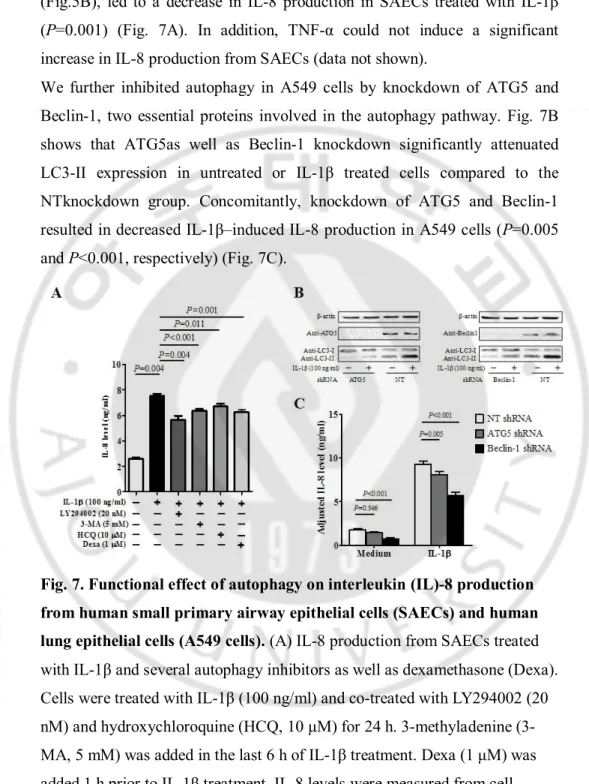
6. Functional effect of autophagy on IL-8 production from human SAECs and A549 cells
To determine the role of autophagy in airway inflammation, we investigated the functional effects of autophagy on IL-8 production from SAECs. As shown in Fig. 7A, IL-1β significantly induced IL-8 production from SAECs (P=0.004). The autophagy inhibitors, including LY294002, 3-MA, and HCQ, slightly but significantly attenuated the effect of IL-1β on IL-8 production from SAECs (P=0.004, P<0.001 and P=0.011, respectively). Treatment with dexamethasone, which was shown to inhibit IL-1β–induced autophagy
(Fig.5B), led to a decrease in IL-8 production in SAECs treated with IL-1β (P=0.001) (Fig. 7A). In addition, TNF-α could not induce a significant increase in IL-8 production from SAECs (data not shown).
We further inhibited autophagy in A549 cells by knockdown of ATG5 and Beclin-1, two essential proteins involved in the autophagy pathway. Fig. 7B shows that ATG5as well as Beclin-1 knockdown significantly attenuated LC3-II expression in untreated or IL-1β treated cells compared to the NTknockdown group. Concomitantly, knockdown of ATG5 and Beclin-1 resulted in decreased IL-1β–induced IL-8 production in A549 cells (P=0.005 and P<0.001, respectively) (Fig. 7C).
Fig. 7. Functional effect of autophagy on interleukin (IL)-8 production from human small primary airway epithelial cells (SAECs) and human lung epithelial cells (A549 cells). (A) IL-8 production from SAECs treated
with IL-1β and several autophagy inhibitors as well as dexamethasone (Dexa). Cells were treated with IL-1β (100 ng/ml) and co-treated with LY294002 (20 nM) and hydroxychloroquine (HCQ, 10 μM) for 24 h. 3-methyladenine (3-MA, 5 mM) was added in the last 6 h of IL-1β treatment. Dexa (1 μM) was added 1 h prior to IL-1β treatment. IL-8 levels were measured from cell culture supernatants by ELISA. (B, C) Effect of genetic inhibition of
23
autophagy on IL-8 production from A549 cells. To inhibit autophagy, A549 cells were infected with pseudo lentiviral particles that contained ATG5 or Beclin-1 short hairpin (sh) RNA. A nontarget (NT) shRNA sequence was used as a knockdown control. A549 cells that expressed ATG5, Beclin-1, or NT shRNA were treated with IL-1β (100 ng/ml) for 24 h to induce autophagy. (B) The expression of LC3-II was determined by western blot analysis, and (C) IL-8 levels in the cell culture supernatants were measured by ELISA. All data are shown as the mean ± SD of at least three independent experiments. P values were obtained by the Mann-Whitney U test. LC3, microtubule-associated protein light chain 3.
IV.DISCUSSION
This is the first study to showthateosinophil and epithelial cell autophagy plays arole in the activation and augmentation of airway inflammation in patients with SA. This was demonstrated by our novel autophagy findings in the sputum granulocytes, PBCs, and PBEsof patients with SA,supporting ex
vivo and in vitrofindings using SAECs, eosinophil-like and epithelial cell
lineswith functional studies. Several previousstudies on the role of autophagy in the pathogenesis of asthma have revealedthat the major determinants of autophagy in asthma are respiratory viral infection and genetic predisposition(Jyothula and Eissa, 2013).However, these studies cannot explain the relationship between autophagy and the pathogenesis ofSA. Moreover, few functional studies have been performed. In the present study, we conducted ex vivoexperiments to investigatemechanisms of autophagyusingairway and peripheral inflammatory cells from patients with SAand compared them with cells from patients with NSA. Our hypothesis was that SA and autophagy are positively associated, as has been previously suggested (Poon et al., 2012b; Jyothula and Eissa, 2013). Our results are the first to demonstratethat the expression ofautophagyin both sputum granulocytes and PBCs from patients with SA was mostly from eosinophils. Through multiple methods, including immunoblot analysis, TEM, and confocal microscopy, we also determined that study participants in the NSA and NC groups had minimal expression of autophagy. These findingsagree with a previous study involving a cohort of adult patients with asthma, which demonstrated that a greater number of double-membrane autophagosomes were detected inthe fibroblasts and epithelial cells isolated from bronchial biopsy tissuesfrom patients withmoderately SAthan fromnormal controls(Poon et al., 2012b).Autophagosome formation was found in tissue from a patient with SA; however, it wasnotquantified. The present study
25
qualitatively and quantitatively confirmed autophagy in sputum, PBCs,and PBEs from patients with SAusing immunoblot analysis, confocal microscopy,and flow cytometry with aCyto-ID® autophagy detection kit.
Cyto-ID® is a novel fluorescent probe that allows fortheinvestigation
ofautophagy using flow cytometry as a noninvasive method. Additionally, in contrast to immunoblot analysis, which is a relatively complicated and time-consuming method, the Cyto-ID® autophagy detection kit can be conducted
with ease and represents a peripheral diagnostic tool for autophagy detection. In the present study, we demonstrated autophagy expression comparable with that of conventional immunoblot analysis, and the specificity of the dye has been validated using flow cytometry(Chan et al., 2012); however, further studies involving a larger cohort are required to validate its use as a diagnostic tool.
The detection of autophagy using a diverse set of quantitative methods is not direct evidence for the role of autophagyin thepathogenesis ofSA. Therefore, we extendedfunctional studies using autophagy-induced cell line models with the treatment of 3-MA and anti-inflammatory drugs. We hypothesized that autophagy could contributes to the pathogenesis ofSAthrough the activation of eosinophils and epithelial cells, which areknown to play a role in SA.First, the autophagy findings were confirmed in both ex vivo and in vitro experiments based on our identification ofautophagy in sputum granulocytes, PBCs,and PBEs from patients with SA (not from patients with NSA). Autophagy was induced by IL-5 as well as rapamycin in PBCs and PBEsisolated from patients with SAas well as in HL-60 cells.However, rapamycin did not increase ECP, while IL-5 increased ECP expression in HL-60 cells. To clarify whether the increase in ECP was due to the synergistic effect of autophagy rather than the effect of IL-5 on eosinophils, we compared the expressionlevels of ECP before and after 3-MA treatment and found that autophagy blockade attenuated ECP expression. These results collectively
suggest that IL-5 (a key Th2 cytokine) can induce autophagy in asthmatic airways.In addition, IL-5 itself,as well as IL-5-induced autophagy, can increase ECP productionfrom eosinophils,which impliesthat autophagy can contribute to the pathogenesis ofSAby facilitating eosinophil activation. Moreover, anti-IL5 antibody may be a potential biologic treatmentwith which to suppress the autophagy-mediated activation pathway in eosinophils. Eosinophilic activation is a prominent feature of the airway inflammatory response in asthma; eosinophils are recruited to the lungs and airways by cytokines, particularly IL-5, and by chemokines, mostly from theeotaxin family(Uhm et al., 2012),in both allergic(Rosenberg et al., 2013) and non-allergic asthma(Brusselle et al., 2013). Eosinophil infiltration contributes to mucous hypersecretion and results in airway hyper-reactivity(Brusselle et al., 2013).ECP is one of the most widely used clinical biomarkers of eosinophil activation and airway inflammation in asthma, and production correlates withthe severity of asthma(Koh et al., 2007).ECP stimulates mucous secretion in the airways and histamine release from basophils and mast cells. Our data provide new evidence thatautophagyin eosinophils can contribute to airway inflammation in SAand may lead to more severe airwayinflammation by enhancing eosinophil activation.
Second, we confirmed our autophagy findings in airway epithelial cells to investigate the additionaleffects of autophagy in the pathogenesis of SA. IL-1β and TNF-α are known prototypical, pro-inflammatory, microbe-induced stimuli that act on respiratory epithelial cells(O’Gorman et al., 2005; Thomas et al., 2007). We found that IL-1β inducedautophagy,while TNF-α did not. There was also an increase in IL-8 production in A549cells and SAECstreated with IL-1β,while this was suppressed by treatment with several autophagy inhibitors and knockdowns of autophagy-related proteins. Asthmatic epithelium usually has chronic wounds leading to infiltration of the epithelium by diverse pro-inflammatory stimuli including allergens, microorganisms, and
27
pollutants,which facilitate inflammation(Holgate, 2013). Epithelial cells contribute to the inflammatory effect on the airway by producing various inflammatory mediators such as transforming growth factor-β, vascular endothelial growth factor, and IL-8. Transforming growth factor-β activates subepithelial mesenchymal cells to release from the matrix and proliferate along with proinflammatory cells. Subepithelial cell proliferation produces a thick subepithelial layer, which is believed to be a distinct feature of SA. In addition, IL-8,an important neutrophil chemoattractant,stimulates neutrophils to increase mucus secretion in fatal asthma(Brightling et al., 2012),facilitates the transient arrest of eosinophils,and increases the contact time between eosinophils and endothelial cells,potentiating theimmune response(Ulfman et al., 2001). IL-8 is also a crucial chemokine that contributes to epithelial– eosinophil interaction in the pathogenesis of asthma (Sexton and Walsh, 2002).Therefore, we speculate that there might be a novel autophagy-associated pathway besides the conventional pathway through the epithelial cell-IL-8-neutrophil/eosinophil axis in patients with SA.
Third, we evaluated the therapeutic effects of anti-inflammatory agents (including steroids) on autophagy in vitroand determined whether they act on autophagy pathways. Steroids act to inhibit inflammation by preventing the transcription of several cytokines or receptors that are relevant in asthma and by reducing the survival of certain inflammatory cells such as eosinophils(Barnes, 1996). In the present study, we found no change in autophagy levels before and after dexamethasonetreatment in PBEs from patients in the SA group. Steroids are a major anti-inflammatory drug usedto control asthma; however, a substantial group of patients with SA are resistant to steroids.Our results indicate that autophagy occurring in eosinophils may be a possible mechanism of steroid-resistant asthma.These findings indicate that autophagy modulation might be a novel therapeutic approach to manage SA. In contrast to the effect of dexamethasone in PBEs from patients with
SA,autophagicprotein expression was decreased by dexamethasone treatment in SAECs. The SAECs used in the present study were isolated from healthy airway epithelial cells, which clearlycontributed to the goodresponse to dexamethasone.Additional studies evaluating the effect of dexamethasone on primary airway epithelial cells collected from patients with SA are needed.
HCQ, another anti-inflammatory agent, decreased autophagy in rapamycin-induced and IL-5-rapamycin-induced autophagy models in HL-60 cells and PBCs. These data suggestthat HCQ might be used as anautophagy-inhibiting, anti-inflammatory agent for the management ofSA. However,one study(Banh, 2011) failed to show the effectiveness of HCQ in patients with SA. Further studies are required to investigate the potential usefulness of HCQ in the management of SAconsidering the viral and genetic susceptibility factors that are reportedlyassociated with autophagy in asthma(Martin et al., 2012; Poon et al., 2012b; Jyothula and Eissa, 2013).
Although the majority of asthma patients can be effectively treated with currently available medications, a substantial subset of patients remains uncontrolled. SA is defined as “asthma which requires treatment with high dose inhaled corticosteroid plus a second controller to prevent it from becoming ‘uncontrolled’ or which remains ‘uncontrolled’ despite this
therapy”(Chung et al., 2014). The prevalence of SAhas been estimated to be 5% to 10% and this group of patients account for a relatively large proportion of resource expenditure.Considering there is anunmet need of new therapy for SA, we expect our data providea significant impact on the noveltherapeutic approach for SA.
Our study did not elucidate how autophagy enhances eosinophil/epithelial cell activation to promote airway inflammation inSA. Autophagy is one of the major mechanisms ofcell death; however, it is not an inevitable cell death process and often protects against cell death(Madeo et al., 2010). In stressful conditions, or throughmTOR inhibition (e.g., with rapamycin), autophagy can
29
remove damaged organelles and help extend life(Levine et al., 2008).We speculate that autophagy may facilitate eosinophil/epithelial cell activation and survival and ultimately increase the level of airway inflammation in patients with SA. Further studies are required to determine whetheran autophagy-associated pathway can prolong the survival of eosinophils and epithelial cells and whether this pathway might be a potential target for new treatments for SA.
V. CONCLUSION
In conclusion, autophagy that occurs in eosinophils and airway epithelial cells could play a role in the pathogenesis of SA, andautophagy modulationmay be a novel therapeutic approach to conventional therapy-resistant SA.
31
REFERENCES
1. Banh HL: Unconventional treatment options in severe asthma: an overview. J Pharm Pharm Sci 14: 387-399, 2011
2. Barnes PJ: Molecular mechanisms of steroid action in asthma. J
Allergy Clin Immunol 97: 159-168, 1996
3. Bousquet J, Mantzouranis E, Cruz AA, Ait-Khaled N, Baena-Cagnani CE, Bleecker ER, Brightling CE, Burney P, Bush A, Busse WW, Casale TB, Chan-Yeung M, Chen R, Chowdhury B, Chung KF, Dahl R, Drazen JM, Fabbri LM, Holgate ST, Kauffmann F, Haahtela T, Khaltaev N, Kiley JP, Masjedi MR, Mohammad Y, O'Byrne P, Partridge MR, Rabe KF, Togias A, van Weel C, Wenzel S, Zhong N, Zuberbier T: Uniform definition of asthma severity, control, and exacerbations: document presented for the World Health Organization Consultation on Severe Asthma. J Allergy Clin Immunol 126: 926-938, 2010
4. Brightling CE, Gupta S, Gonem S, Siddiqui S: Lung damage and airway remodelling in severe asthma. Clin Exp Allergy 42: 638-649, 2012
5. Brusselle GG, Maes T, Bracke KR: Eosinophils in the spotlight: Eosinophilic airway inflammation in nonallergic asthma. Nat Med 19: 977-979, 2013
6. Chan LL, Shen D, Wilkinson AR, Patton W, Lai N, Chan E, Kuksin D, Lin B, Qiu J: A novel image-based cytometry method for autophagy detection in living cells. Autophagy 8: 1371-1382, 2012 7. Choi AM, Ryter SW, Levine B: Autophagy in human health and
disease. N Engl J Med 368: 651-662, 2013
8. Chung KF, Wenzel SE, Brozek JL, Bush A, Castro M, Sterk PJ, Adcock IM, Bateman ED, Bel EH, Bleecker ER, Boulet LP, Brightling C, Chanez P, Dahlen SE, Djukanovic R, Frey U, Gaga M,
Gibson P, Hamid Q, Jajour NN, Mauad T, Sorkness RL, Teague WG: International ERS/ATS guidelines on definition, evaluation and treatment of severe asthma. Eur Respir J 43: 343-373, 2014
9. Fahy JV: Eosinophilic and neutrophilic inflammation in asthma: insights from clinical studies. Proc Am Thorac Soc 6: 256-259, 2009 10. Harhaji-Trajkovic L, Arsikin K, Kravic-Stevovic T, Petricevic S,
Tovilovic G, Pantovic A, Zogovic N, Ristic B, Janjetovic K, Bumbasirevic V, Trajkovic V: Chloroquine-mediated lysosomal dysfunction enhances the anticancer effect of nutrient deprivation.
Pharm Res 29: 2249-2263, 2012
11. Holgate ST: The sentinel role of the airway epithelium in asthma pathogenesis. Immunol Rev 242: 205-219, 2011
12. Holgate ST: Mechanisms of asthma and implications for its prevention and treatment: A personal journey. Allergy Asthma
Immunol Res 5: 343-347, 2013
13. Hwang EK, Jin HJ, Nam YH, Shin YS, Ye YM, Nahm DH, Park HS: The predictors of poorly controlled asthma in elderly. Allergy Asthma
Immunol Res 4: 270-276, 2012
14. Jyothula SSSK, Eissa NT: Autophagy and role in asthma. Curr Opin
Pulm Med 19: 30-35, 2013
15. Kim YM, Kim YS, Jeon SG, Kim YK: Immunopathogenesis of allergic asthma: more than the th2 hypothesis. Allergy Asthma
Immunol Res 5: 189-196, 2013
16. Klionsky DJ, Abdalla FC, Abeliovich H, Abraham RT, Acevedo-Arozena A, Adeli K, Agholme L, Agnello M, Agostinis P, Aguirre-Ghiso JA: Guidelines for the use and interpretation of assays for monitoring autophagy. Autophagy 8: 445-544, 2012
17. Koh GC, Shek LP, Goh DY, Van Bever H, Koh DS: Eosinophil cationic protein: is it useful in asthma? A systematic review. Respir
33
Med 101: 696-705, 2007
18. Levine B, Sinha SC, Kroemer G: Bcl-2 family members: dual regulators of apoptosis and autophagy. Autophagy 4: 600-606, 2008 19. Macedo P, Hew M, Torrego A, Jouneau S, Oates T, Durham A,
Chung KF: Inflammatory biomarkers in airways of patients with severe asthma compared with non-severe asthma. Clin Exp Allergy 39: 1668-1676, 2009
20. Madeo F, Tavernarakis N, Kroemer G: Can autophagy promote longevity? Nat Cell Biol 12: 842-846, 2010
21. Martin LJ, Gupta J, Jyothula SS, Kovacic MB, Myers JMB, Patterson TL, Ericksen MB, He H, Gibson AM, Baye TM: Functional variant in the autophagy-related 5 gene promotor is associated with childhood asthma. PLoS One 7: e33454, 2012
22. Morris S, Swanson MS, Lieberman A, Reed M, Yue Z, Lindell DM, Lukacs NW: Autophagy-Mediated Dendritic Cell Activation Is Essential for Innate Cytokine Production and APC Function with Respiratory Syncytial Virus Responses. J Immunol 187: 3953-3961, 2011
23. Nakahira K: Autophagy: a potential therapeutic target in lung diseases.
Am J Physiol Lung Cell Mol Physiol 15: L93-107, 2013
24. O’Gorman MT, Jatoi NA, Lane SJ, Mahon BP: IL-1β and TNF-α induce increased expression of CCL28 by airway epithelial cells via an NFκB-dependent pathway. Cell Immunol 238: 87-96, 2005
25. Pang G, Clancy R, Reeves G: Isolation and functional characterization of T cells from human sputum. Clin Exp Immunol 102: 642-648, 1995
26. Poon A, Eidelman D, Laprise C, Hamid Q: ATG5, autophagy and lung function in asthma. Autophagy 8: 694-695, 2012a
CJ, Eidelman DH, Olivenstein R, Martin JG, Weiss ST, Hamid Q, Laprise C: Genetic and histologic evidence for autophagy in asthma pathogenesis. J Allergy Clin Immunol 129: 569-571, 2012b
28. Reddy D, Little FF: Glucocorticoid-resistant asthma: more than meets the eye. J Asthma 50: 1036-1044, 2013
29. Rosenberg HF, Dyer KD, Foster PS: Eosinophils: changing perspectives in health and disease. Nat Rev Immunol 13: 9-22, 2013 30. Rubinsztein DC, Gestwicki JE, Murphy LO, Klionsky DJ: Potential
therapeutic applications of autophagy. Nat Rev Drug Discov 6: 304-312, 2007
31. Rutgers S, Timens W, Kaufmann H, van der Mark TW, Koeter G, Postma D: Comparison of induced sputum with bronchial wash, bronchoalveolar lavage and bronchial biopsies in COPD. Eur Respir J 15: 109-115, 2000
32. Sexton DW, Walsh GM: Eosinophil–epithelial cell interactions: an important facet of asthmatic inflammation. Clin Exp Allergy 32: 811-813, 2002
33. Thomas LH, Wickremasinghe MIY, Friedland JS: IL-1β stimulates divergent upper and lower airway epithelial cell CCL5 secretion. Clin
Immunol 122: 229-238, 2007
34. Uhm TG, Kim BS, Chung IY: Eosinophil development, regulation of eosinophil-specific genes, and role of eosinophils in the pathogenesis of asthma. Allergy Asthma Immunol Res 4: 68-79, 2012
35. Ulfman LH, Joosten DP, van der Linden JA, Lammers J-WJ, Zwaginga JJ, Koenderman L: IL-8 induces a transient arrest of rolling eosinophils on human endothelial cells. J Immunol 166: 588-595, 2001
36. Urbanowicz RA, Lamb JR, Todd I, Corne JM, Fairclough LC: Enhanced effector function of cytotoxic cells in the induced sputum
35
of COPD patients. Respir Res 11: 76-84, 2010
37. Wang W, Fan H, Zhou Y, Duan P, Zhao G, Wu G: Knockdown of autophagy-related gene BECLIN1 promotes cell growth and inhibits apoptosis in the A549 human lung cancer cell line. Mol Med Rep 7: 1501-1505, 2013
38. Wenzel S, Fahy J, Irvin C, Peters S, Spector S, Szefler S: Proceedings of the ATS workshop on refractory asthma: current understanding, recommendations, and unanswered questions. Am J Respir Crit Care
- 국문요약-
중증 천식환자의 가래와 말초혈액에서 자가소화작용의
역할
: 새로운 치료의 표적
아주대학교대학원 의학과/의학전공 반 가 영 (지도교수: 박해심) 배경: 자가소화작용은 염증세포의 사멸과 생존에 영향을 미치는 것 으로 알려져 있다. 하지만 만성 기도 염증이 주된 기전인 중증 천식 의 병인기전에 자가소화작용이 어떠한 역할을 하는지에 대한 연구 는 적다. 목적: 중증 천식 환자와 일반 천식 환자의 객담 과립구, 말초혈액 세포와 말초혈액 호산구에서 자가소화작용의 표현 정도를 비교하였 으며 그 역할을 연구하였다. 방법: 중증 천식 환자 33명, 일반 천식 환자 14명과 건강 대조군 23명을 등록하였다. 각 대상자들로부터 객담 과립구, 말초혈액 세포 와 말초혈액 호산구를 획득하였다. 자가소화작용은 웨스턴블롯, 공 초점 현미경, 전자현미경과흐름세포측정기로 측정한 미소관 관련 단 백질 경쇄 3(LC3)으로 평가하였다. 인터루킨 (IL)-8은 ELISA 로 측정하였다. 자가소화작용을 유도하기 위해 HL-60세포, 단일 소기 도 상피세포, A549 세포를 각각 IL-5, IL-1β와 TNF-α로 처리 하였다. 자가소화작용을 억제하기 위해 PI3K 억제제를 사용하였다.37 A549 세포에서는 ATG5와Beclin-1 넉다운을 하였으며 덱사메타 손의 치료 효과를 평가하였다. 결과: 일반 천식 환자나 건강 대조군 보다 중증 천식 환자에서 자가 소화작용이 유의하게 증가되어 있었다 (P<0.05). IL-5는 말초혈액 세포와 말초혈액 호산구에서 자가소화작용을 증가시켰다(P<0.05). HL-60 세포에서 PI3K 억제제는 IL-5에 의해 증가된 LC3 와 호 산구양이온단백을 감소시켰다 (P=0.034). 덱사메타손은 말초혈액 호산구의 자가소화작용에 영향을 미치지 못했다. IL-1β는 단일 소 기도 상피세포에서 LC-3 발현과 IL-8 생성을 증가시켰고
(P<0.01) 이는PI3K 억제제와 ATG5, Beclin-1 넉다운에 의해 감 소되었다(P<0.01).
결론: 자가소화작용은 중증천식의 병인기전에 역할을 할 것이다. 자 가소화작용의 조절은 일반적인 치료에 불응하는 중증천식의 새로은 치료 표적이 될것이다.