저작자표시-비영리-변경금지 2.0 대한민국 이용자는 아래의 조건을 따르는 경우에 한하여 자유롭게 l 이 저작물을 복제, 배포, 전송, 전시, 공연 및 방송할 수 있습니다. 다음과 같은 조건을 따라야 합니다: l 귀하는, 이 저작물의 재이용이나 배포의 경우, 이 저작물에 적용된 이용허락조건 을 명확하게 나타내어야 합니다. l 저작권자로부터 별도의 허가를 받으면 이러한 조건들은 적용되지 않습니다. 저작권법에 따른 이용자의 권리는 위의 내용에 의하여 영향을 받지 않습니다. 이것은 이용허락규약(Legal Code)을 이해하기 쉽게 요약한 것입니다. Disclaimer 저작자표시. 귀하는 원저작자를 표시하여야 합니다. 비영리. 귀하는 이 저작물을 영리 목적으로 이용할 수 없습니다. 변경금지. 귀하는 이 저작물을 개작, 변형 또는 가공할 수 없습니다.
Outcome Analysis of Helmet Therapy
and Counter Positioning for
Deformational Plagiocephaly
by
Se Yon Kim
Major in Medicine
Department of Medical Sciences
The Graduate School, Ajou University
Outcome Analysis of Helmet Therapy
and Counter Positioning for
Deformational Plagiocephaly
by
Se Yon Kim
A Dissertation Submitted to The Graduate School of
Ajou University in Partial Fulfillment of the Requirements for the
Degree of Master of Medicine
Supervised by
Shin-Young Yim, M.D., Ph.D.
Major in Medicine
Department of Medical Sciences
This certifies that the dissertation
of SeYon Kim is approved.
SUPERVISORY COMMITTEE
Shin-Young Yim
Moon-Sung Park
Jeong-In Yang
The Graduate School, Ajou University
May, 10th, 2013
i - ABSTRACT -
Outcome Analysis of Helmet Therapy and Counter Positioning for
Deformational Plagiocephaly
Objective: To compare effectiveness between helmet therapy and counter positioning for deformational plagiocephaly (DP).
Method: Retrospective data of children diagnosed with DP who visited our clinic from November 2010 to October 2012 were reviewed. The subjects who were 10 months of age or younger, and showed 10 mm or more of diagonal difference were included for analysis. For DP treatment, information on both helmet therapy and counter-positioning was given and either of two was chosen by each family. Head circumference, cranial asymmetry measures such as diagonal difference, cranial vault asymmetry index, radial symmetry index and ear shift were obtained by 3 dimensional head-surface laser scan at the time of initiation and termination of therapy.
Results: Total 27 subjects were included in this study, where 21 had helmet therapy and 6 underwent counter positioning. There was no significant difference of baseline characteristics, head circumference and cranial asymmetry measurements at the initiation of therapy. The mean duration of therapy was 4.30±1.27 months in the helmet group and 4.08±0.95 months in the counter positioning group (p=0.770). While cranial asymmetry measurements improved in both groups, significantly more improvement was observed with helmet therapy. There was no significant difference of the head circumference growth between two groups at the end of therapy.
Conclusion: This study showed that helmet therapy resulted in more favorable outcomes in correcting cranial and ear asymmetry than counter positioning on moderated to severe DP without compromising head growth. Further study with more subjects and longer follow-up period is warranted.
ii
TABLE OF CONTENTS
ABSTRACT………..……. i
TABLE OF CONTENTS ...……….………... ii
LIST OF FIGURES ….……….………... iii
LIST OF TABLES ………... iv
I. INTRODUCTION ……..………...………..………. 1
II. MATERIALS AND METHODS ……….... 3
A. Subjects ……….………...……….………..…… 3 B. Helmet therapy ……...………....………. 3 C. Counter positioning .………...………..………... 4 D. Anthropometric measurements ………..…. 5 E. Statistical analysis ………..………...…….………….… 6 III. RESULTS ……….………...………..… 8
A. Baseline patient characteristics ……..………...……….….………… 8
B. Comparison of anthropometric measurements at the conclusion of therapy .….….... 9
C. Head growth …...…...……….…….………..……… 10
IV. DISCUSSION ……..……….….………..…… 11
V. CONCLUSION ….………..……….……….……… 16
REFERENCES ………...……….……….……….. 17
iii
LIST OF FIGURES
Figure 1. Counter positioning instruction ………..…………..……… 4 Figure 2. Pictures showing anatomical landmarks (A) and reference plane (B)…....……... 5 Figure 3. (A) Diagonal difference and Cranial vault asymmetry index (B) Radial symmetry index. (C) Ear shift …...….... 6
iv
LIST OF TABLES
Table 1. Baseline patient characteristics ….………..…..……….…… 8 Table 2. Comparison of anthropometric measurements at the initiation of therapy ... 8 Table 3. Comparison of anthropometric measurements at the conclusion of therapy ...… 10
- 1 -
I. INTRODUCTION
Plagiocephaly literally means “oblique head,” combining the Greek words plagios, meaning oblique, and kephalē, meaning head (Kalra and Walker, 2012). The term can be used to describe asymmetric head shapes resulting from both craniosynostotic and nonsynostotic causes. Deformational plagiocephaly (DP) arises from repeated external pressure to the same area of the flexible cranium during the fetal or neonatal periods (Mortenson et al., 2012). When this repeated pressure is applied to one side of the occiput, it results in ipsilateral occipital flattening, frontal bone protrusion, and anterior shifting of the ipsilateral ear (Looman and Flannery, 2012).
The incidence of DP has risen since 1992, when the American Academy of Pediatrics started its Back to Sleep campaign, encouraging parents to place infants on their backs to decrease the incidence of Sudden Infant Death Syndrome (1992). The incidence of DP was only 0.3% in the 1970s (Dunn, 1974), but the current reported estimates since 1992 ranges from 2% (Roby et al., 2012) to 47% (Boere-Boonekamp and van der Linden-Kuiper, 2001), depending on the diagnostic criteria used and the age of the children involved (Boere-Boonekamp and van der Linden-Kuiper, 2001; Hutchison et al., 2004; Bialocerkowski et al., 2008; Saeed et al., 2008; Thompson et al., 2009; Roby et al., 2012). Despite the fact that the American Academy of Pediatrics released recommendations to prevent DP in 2003 (Persing et al., 2003), the incidence of DP remains high (Bialocerkowski et al., 2008; Saeed et al., 2008; Thompson et al., 2009; Roby et al., 2012).
Clarren et al (Clarren et al., 1979) introduced helmet therapy for children with DP in 1979. The first helmet was made of fiberglass and modeled on a plaster cast of the subject’s head. Four children wore these helmets, and their cranial asymmetry improved without any serious complications. Helmets used for DP treatment are currently custom-made for each individual. They are constructed to apply full-contact external pressure to the protruding area and to allow space where bony expansion is desired (Schaaf et al., 2010; Mortenson et al., 2012). In 1998, the Food and Drug Administration of the United States approved the cranial orthosis as a class II medical device, intended for infants between 3 and 18 months of age with moderate to severe DP.
- 2 -
The current treatment for patients with moderate to severe DP consists of either helmet therapy or counter positioning, with reports supporting the efficacy of both techniques (Moss, 1997; O'Broin et al., 1999; Thompson et al., 2009), although with varying results on comparing the efficacy between them (Pollack et al., 1997; Loveday and de Chalain, 2001; Bialocerkowski et al., 2005; Graham et al., 2005; Katzel et al., 2010; Lipira et al., 2010). A study conducted in 70 DP patients reported a greater improvement in the diagonal difference (DD) measurement with helmet therapy than with counter positioning, but the same improvement was not seen in the cranial vault asymmetry index (CVAI) (Lipira et al., 2010). A longitudinal study of 298 DP patients revealed that helmet therapy has a more favorable correction outcome than counter positioning; however, helmet therapy has a longer treatment time (Graham et al., 2005). There are no definable standard criteria for starting helmet therapy and treatment decision is influenced more strongly by factors other than medical evidence. One study reported that physician’s preference exists between specialties suggesting neurosurgeons are less likely to prescribe helmet therapy for DP than plastic surgeons (Lee et al., 2010).
The therapeutic effect of both helmet therapy and counter positioning on ear shift remains inconclusive, with few results reported. Kluba et al (Kluba et al., 2012) reported the effects of helmet therapy on ear shift in patients with DP, implying that helmet therapy has a corrective effect on pre-existing ear shift. However, the same study showed that ear shift can arise de novo during helmet therapy in patients who did not have ear shift at the beginning of treatment.
After reviewing both the English and the Korean literature, we found no studies comparing the efficacy of helmet therapy and counter positioning on ear shift in patients with DP. Also, to the best of our knowledge, there are no Korean studies comparing the general efficacy of these 2 therapeutic options for patients with DP. The aim of this study was to compare the effectiveness of helmet therapy and counter positioning for correcting cranial asymmetry and ear shift in Korean patients with DP.
- 3 -
II. MATERIAL AND METHODS
In this retrospective study, we analyzed the medical records of patients diagnosed and treated for DP at our clinic, part of a tertiary medical center in Korea, between November 2010 and October 2012.
A. Subjects
All children with a diagnosis of DP were evaluated for study inclusion; DP was defined as nonsynostotic unilateral occipital flattening. The diagnosis of DP was confirmed by 3-dimensional (3D) computed tomography, performed to exclude the diagnosis of
craniosynostotic plagiocephaly. Children with known neurodevelopmental disorders such as cerebral palsy, autistic spectrum disorders, genetic aberrations, and metabolic diseases were excluded.
Included patients had a DD greater than 10 mm and were 10 months of age or younger. According to the protocol at our clinic, the caregivers of patients meeting these criteria were given information about both helmet therapy and counter positioning; the treatment decision was made by the caregivers. Patients treated with helmet therapy were designated the helmet therapy group, and patients treated with counter positioning were designated the counter positioning group.
B. Helmet therapy
In the helmet therapy group, a custom-molded helmet was made for each child based on their initial 3D head-surface laser scan (STARscanner laser data acquisition system; Orthomerica Products Inc., Orlando, FL, USA). The helmet (Orthokorea, Seoul, Korea) consisted of a polyethylene foam liner and a copolymer outer shell. Caregivers were told to keep the child in the helmet as much as possible, with greater than 23 hours of daily use recommended. After the initial helmet fitting, follow-up laser scans were performed every 3– 4 weeks to adjust for both head growth and skull-shape change. Helmet therapy was terminated when subjects met 1 of the following criteria: 1) DD was less than 6 mm and CVAI was less than 3.5%; 2) no further helmet adjustment was possible due to cranial
- 4 - growth.
C. Counter positioning therapy
In the counter positioning group, the following program was recommended:
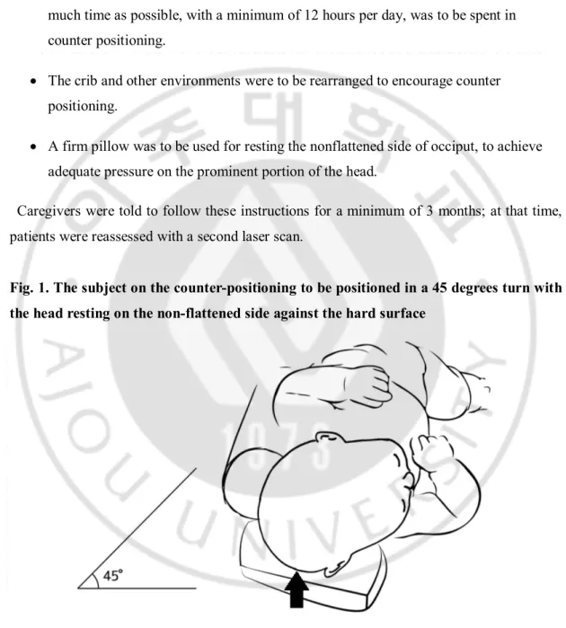
· When the child was lying on its back, the head was to be positioned in a 45-degree turn, resting against a hard surface on the nonflattened side of occiput (Fig. 1). As much time as possible, with a minimum of 12 hours per day, was to be spent in counter positioning.
· The crib and other environments were to be rearranged to encourage counter positioning.
· A firm pillow was to be used for resting the nonflattened side of occiput, to achieve adequate pressure on the prominent portion of the head.
Caregivers were told to follow these instructions for a minimum of 3 months; at that time, patients were reassessed with a second laser scan.
Fig. 1. The subject on the counter-positioning to be positioned in a 45 degrees turn with the head resting on the non-flattened side against the hard surface
- 5 - D. Anthropometric measurements
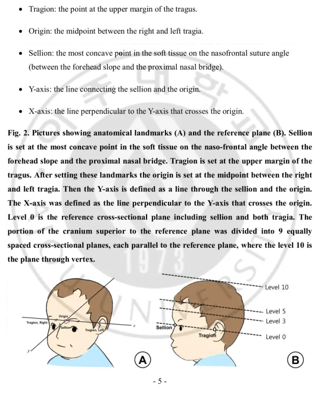
Anthropometric measurements were obtained at the initiation and termination timepoints of each therapy, using the 3D head-surface laser scanning device. The following landmarks were set for anthropometric measurement (Fig. 2A) and reference plane and planes levels were set as shown in Figure 2B.
· Tragion: the point at the upper margin of the tragus. · Origin: the midpoint between the right and left tragia.
· Sellion: the most concave point in the soft tissue on the nasofrontal suture angle (between the forehead slope and the proximal nasal bridge).
· Y-axis: the line connecting the sellion and the origin.
· X-axis: the line perpendicular to the Y-axis that crosses the origin.
Fig. 2. Pictures showing anatomical landmarks (A) and the reference plane (B). Sellion is set at the most concave point in the soft tissue on the naso-frontal angle between the forehead slope and the proximal nasal bridge. Tragion is set at the upper margin of the tragus. After setting these landmarks the origin is set at the midpoint between the right and left tragia. Then the Y-axis is defined as a line through the sellion and the origin. The X-axis was defined as the line perpendicular to the Y-axis that crosses the origin. Level 0 is the reference cross-sectional plane including sellion and both tragia. The portion of the cranium superior to the reference plane was divided into 9 equally spaced cross-sectional planes, each parallel to the reference plane, where the level 10 is the plane through vertex.
- 6 -
The following anthropometric measurements were obtained at levels 3 and 5 (Fig. 3): · DD (mm): the difference between 2 diagonal cranial diameters, 30 degrees from the
Y-axis (Meyer-Marcotty et al., 2012); DD = longer diagonal -shorter diagonal. · CVAI (%): DD/short cranial diagonal × 100 (Loveday and de Chalain, 2001). · Radial symmetry index (RSI) (mm): Starting at the front of the head, vector lengths
were measured at 15-degree intervals and summed for each side of the head. The absolute value of the difference between the right-sided sum and the left-sided sum was the RSI. An RSI of 0 mm indicated perfect symmetry (Plank et al., 2006). · Ear shift (mm): the anteroposterior distance between the right and left tragia,
perpendicular to the X-axis at the level 0 plane (Meyer-Marcotty et al., 2012). Ear shift was measured twice by a single blinded observer, using Adobe Acrobat Pro (Adobe Systems Inc., San Jose, CA, USA). No significant difference was found between the 2 measurements; the mean value was used for this study.
Figure. 3. (A) Diagonal difference (mm) = longer diagonal (AB)–shorter diagonal (CD). Cranial vault asymmetry index (%) = DD/CD x 100. (B) Radial symmetry index (mm) = |(A1 + A2 + … + A11) – (B1 + B2 + … + B11)|. (C) The ear shift (mm)
=|a-b|
E. Statistical analysis
- 7 -
USA), was used for statistical analysis. The statistical significance of the results was tested using the nonparametric Mann-Whitney U-test. The chi-square test was used for comparing dichotomous variables such as patient sex and the occipital flattening side. Statistical significance was set at a p-value of < 0.05. Post hoc statistical power analysis was done and statistical power was set at >0.8.
- 8 -
III. RESULTS
A. Baseline patient characteristics
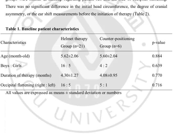
The helmet therapy group consisted of 21 patients, with 6 patients in the counter positioning group. The baseline patient characteristics are presented in Table 1. The mean patient age in the helmet therapy group was 5.62 months, compared with 5.60 months in the counter positioning group (p = 0.884). The mean duration of therapy was 4.30 months in the helmet therapy group and 4.08 months in the counter positioning group (p = 0.770). The groups were similar in makeup regarding patient sex and the side of occipital flattening. There was no significant difference in the initial head circumference, the degree of cranial asymmetry, or the ear shift measurements before the initiation of therapy (Table 2).
Table 1. Baseline patient characteristics
Characteristics Helmet therapy
Group (n=21)
Counter-positioning
Group (n=6) p-value
Age (month-old) 5.62±2.06 5.60±2.04 0.884
Boys : Girls 16 : 5 4 : 2 0.639
Duration of therapy (months) 4.30±1.27 4.08±0.95 0.770
Occipital flattening (right : left) 16 : 5 5 : 1 0.716
- 9 -
Table 2. Comparison of anthropometric measurements at the initiation of therapy
Measures Helmet therapy
group (n=21) Counter-positioning group (n=6) p-value Head circumference (mm) Level 3 431.39±17.34 433.03±20.49 0.816 Level 5 417.85±17.94 421.05±18.82 0.484 Diagonal difference (mm) Level 3 13.28±3.57 11.38±3.30 0.220 Level 5 14.94±3.60 13.75±4.28 0.560
Cranial vault asymmetry index (%)
Level 3 9.95±2.68 8.54±2.90 0.180
Level 5 11.76±2.89 10.86±3.89 0.600
Radial asymmetry index (mm)
Level 3 56.29±12.72 51.93±15.27 0.448
Level 5 66.20±17.01 66.00±20.83 0.953
Ear shift (mm) 5.67±2.39 6.09±3.53 0.899
*p<0.05
All values are expressed as means ± standard deviation
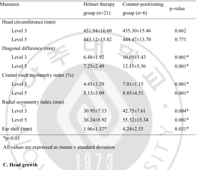
B. Comparison of anthropometric measurements at the conclusion of therapy
Comparison of the anthropometric measurements (DD, CVAI, RSI) at the level 3 and level 5 planes revealed a significant improvement in cranial asymmetry in the helmet therapy group at the end of the treatment period (Table 2). The mean DD at levels 3 and 5 at the end of treatment was 6.48 mm and 7.25 mm, respectively, for the helmet therapy group and 10.05 mm and 12.15 mm, respectively, in the counter positioning group (p = 0.01). The CVAI at levels 3 and 5 was 4.45% and 5.13%, respectively, in the helmet therapy group and 7.01% and 8.85%, respectively, in the counter positioning group (p = 0.01). The RSI showed significant improvement in the helmet therapy group (30.95 mm at level 3 and 36.24 mm at level 5) compared with the counter positioning group (42.75 mm at level 3 and 55.2 mm at level 5). Ear shift improved significantly in the helmet therapy group showing mean ear shift
- 10 -
of 1.96 mm compared with 4.24 mm in the counter positioning group at the end of treatment period(p = 0.031). Post hoc statistical power analysis show effective power in our statistical analysis.
Table 3. Comparison of anthropometric measurements at the conclusion of therapy
Measures Helmet therapy
group (n=21) Counter-positioning group (n=6) p-value Head circumference (mm) Level 3 451.94±16.60 455.30±15.46 0.662 Level 5 443.12±15.82 444.47±13.70 0.771 Diagonal difference (mm) Level 3 6.48±1.92 10.05±1.43 0.001* Level 5 7.25±2.49 12.15±5.56 0.001*
Cranial vault asymmetry index (%)
Level 3 4.45±1.29 7.01±1.15 0.001*
Level 5 5.13±3.09 8.85±4.53 0.001*
Radial asymmetry index (mm)
Level 3 30.95±7.13 42.75±7.61 0.004*
Level 5 36.24±8.92 55.52±15.34 0.001*
Ear shift (mm) 1.96±1.37* 4.24±2.53 0.031*
*p<0.05
All values are expressed as means ± standard deviation C. Head growth
The head circumference enlarged in all patients during treatment. There was no significant difference in head circumference before therapy, with a mean value at level 3 of 431.39 mm in the helmet therapy group and 433.03 mm in the counter positioning group, with level 5 measuring 417.85 mm and 421.05 mm, respectively. The lack of significant difference persisted after treatment, with a mean head circumference of 451.94 mm vs. 455.30 mm at level 3, and 443.12 mm vs. 444.47 mm at level 5. These results suggest that neither helmet therapy nor the counter positioning used in this study interfered with head growth.
- 11 -
IV. DISCUSSION
Our study compared helmet therapy with counter positioning in patients with moderate to severe DP and found better efficacy for helmet therapy in terms of correcting cranial
asymmetry; ear shift measurements also showed a larger degree of correction in the helmet-therapy group. After reviewing both the English and the Korean literature, we believe that ours is the first report to compare the effectiveness of these 2 techniques on ear shift in patients with DP, and we believe this is the first Korean report to compare the general efficacy of these 2 therapeutic options in children with DP.
Conservative management, whether with helmet therapy or counter positioning, is the major therapeutic option for the treatment of DP. The superiority of helmet therapy is not yet conclusively established, due to potential biases in prior studies such as inconsistent
diagnostic criteria, varying clinical severity of DP, varying age of therapy onset, different durations of therapy, and different measurement techniques (Bialocerkowski et al., 2005). Loveday and de Chalain (Loveday and de Chalain, 2001) proposed a definition of DP as a CVAI > 3.5%. The clinical severity classification of DP is determined by skull asymmetry, most commonly expressed as the DD. Hutchison et al (Hutchison et al., 2005) proposed a classification system, defining mild DP as a DD between 3 mm and 10 mm, moderate as a DD between 10 mm and 12 mm, and severe as a DD greater than 12 mm. Another study reported that a normal value for DD, determined by evaluating 36 healthy children without DP between the ages of 4 and 12 months, is 3 mm ± 1 mm (Graham et al., 2005). A study by Yoo et al (Yoo et al., 2012) of 108 Korean children classified DP into mild (DD between 6 mm and 10 mm), moderate (11 mm to 15 mm), and severe (at least 16 mm).
A recent systematic review focusing on the conservative management of DP reported that favorable outcomes are 1.3 times more likely with helmet therapy than with counter
positioning; however, this result is not conclusive as study biases exist regarding the clinical severity of the condition, patient age, and duration of therapy (Xia et al., 2008). In a study by Clarren et al (Clarren, 1981), all patients were offered helmet therapy but 10 caregivers refused this treatment; most of these caregivers had children with mild DP, resulting in more
- 12 -
severe DP in the helmet-therapy group. In another study by Graham et al (Graham et al., 2005), comparing helmet therapy with counter positioning, patients were treated with helmet therapy only after they failed to respond to counter positioning. Counterpositioning has a role in correcting mild DP, but its use seems less effective in correcting moderate to severe DP. In moderate to severe DP, helmet therapy applies more pressure on protruded area of occiput than counter positioning. Thus, greater correction can be expected. Our study analyzed only those subjects with moderate to severe DP; the results suggest that helmet therapy is superior to counter positioning in correcting cranial asymmetry in these patients.
In the present study, both ear shift and cranial asymmetry showed greater improvement in the helmet therapy group than in the counter positioning group. Ear shift is one of the major cosmetic concerns in moderate to severe DP (Argenta et al., 2004). However, there are only a couple of reports in the literature investigating this phenomenon, and these provide conflicting results. Kluba et al (Kluba et al., 2012) reported that helmet therapy improves ear shift in patients who demonstrate this problem prior to treatment, but not in those who did not have this problem prior to treatment. They even reported that ear shift develops during treatment in patients who were without this problem at the beginning of treatment. Katzel et al (Katzel et al., 2011) reported that the parents’ perception of ear shift improves after helmet therapy in their children, even though the actual measurements show no statistical difference after treatment. However, other cranial asymmetry measurements such as CVAI and RSI do improve significantly, along with the parents’ perception of the child’s appearance. A prospective study by Meyer-Marcotty, comparing the effects of helmet therapy in 20 patients with DP and 20 control subjects without the condition, showed that a comparison of
measurements before and after helmet therapy showed no significant difference in ear shift, even though other cranial asymmetry measurements such as DD improved significantly. The helmet provides external force on the prominent areas of the skull, resulting in a volume shift to the flat areas in the relatively soft skull of a young child. Thus, the parallelogram-shaped cranium seen in DP can be corrected to a symmetric, oval-shaped cranium, and ear shift should also be corrected (Schaaf et al., 2010; Mortenson et al., 2012). The results of the present study indicate that ear shift does indeed show significant improvement with helmet therapy.
- 13 -
10months of age or younger. Mean initiation age of subjects in this study was 5.62 months ranging from 3.13 to 10 months in the helmet therapy group and 5.60 months ranging from 3.23 to 8.46 months in the counter positioning group. Before 3months of age, children cannot control head. Therefore, most of caregivers cannot notice DP so most of DP patients come to the clinic after 3 months of age. Spontaneous correction of DP is reported to be possible according to brain growth up to 4 months of age (Clarren, 1981; Littlefield, 2001). The criteria of 10 months of age in our clinic’s protocol was according to previous study reporting decreased rated of successful correction of cranial asymmetry in patients with the initiation age of helmet therapy older than 9.1 months (Yoo et al., 2012).
In this study, the head circumference in both groups of patients appeared to grow during therapy. This implies that neither helmet therapy nor counter positioning interfere with head growth in children. However, we did not evaluate development in terms of cognition, language, motor function, or social functioning, all of which are very important aspects in the treatment of DP. Shamji et al (Shamji et al., 2012) conducted a 9-year study of 80 children with DP, using a questionnaire administered to parents concerning the cosmetic and cognitive outcomes of the condition. Twenty-one percent of parents reported language difficulties in their children, 28% reported motor difficulties, and 15% of children required special education; these values exceed the average rate of delayed development in the general population, reportedly 5–6%.In subgroup analysis, left-sided DP was related to language difficulties and the need for special education.
In a longitudinal study of 227 children with DP and 232 children without DP, beginning at infancy and lasting 18 months, children with DP showed lower cognition and language scores on the Bayley Scales of Infant and Toddler Development, third edition (BSID-III) (Collett et al., 2011). Subjects with known risk factors for delayed development were not included in this longitudinal study; these risk factors included a history of delivery at less than 35 weeks gestational age, known neurodevelopmental disorders, brain injury, significant hearing or vision problems, a single major malformation or 3 or more minor malformations, craniofacial microsomia, a non-English-speaking mother, and a history of adoption. Looking closely at the results, 8.3% of children with DP and 0.6% of children without DP scored in the delayed cognition range (less than 85% of the standard score), making it 13.8 times more likely that children with DP would experience this delay. Children
- 14 -
with DP were also 1.8 to 3.9 times more likely to score in the delayed range for language, motor skills, and adaptive behavior. Despite this observed difference between DP and control children, most study subjects scored within the normal range on BSID-III testing, regardless of their DP status. Although we cannot rule out the possibility, it should not be assumed from these results that DP causes delayed development; subtle neuromuscular problems related to delayed development may precede DP, resulting in restriction of movement and subsequent skull deformation. In a follow-up study, evaluating the same subjects through 36 months of age, the difference in BSID-III scores between subjects with and without DP persisted (Collett et al., 2013).
DP is often associated with prematurity, or maternal multiparity, some of which are also known risk factors for developmental delay (Kalra and Walker, 2012). The fact that these may be confounding factors should be considered when comparing developmental scores between children with and without DP.In this study, we excluded children with known neurodevelopmental disorders such as cerebral palsy, autistic spectrum disorders, and genetic or metabolic diseases. Further studies should be done to examine the adverse effects of helmet therapy or counter positioning on development.
The natural course of DP and the long-term effects of helmet therapy and counter positioning are not fully understood. A large study by Boere-Boonekamp et al (Boere-Boonekamp and van der Linden-Kuiper, 2001) in 7609 Dutch infants revealed that 9.9% had occipital asymmetry on physical examination, and 45% of these infants had persistent asymmetry after 2 years of follow-up. A 2-year prospective cohort study of 200 infants born between 2001 and 2002 in a single hospital in New Zealand (Hutchison et al., 2004) reported that the prevalence of plagiocephaly and/or brachycephaly is highest at 4 months, but
diminishes as infants grow older. This normal progression can solve the cosmetic issues in children with mild DP, but those with moderate to severe DP may have persistent cosmetic problems despite some naturally occurring improvement. Unfortunately, there is a lack of precise data on the extent of natural improvement and the long-term effectiveness of either helmet therapy or counter positioning. To the best of our knowledge, no study has yet followed groups of children who underwent either helmet therapy or counter positioning through adulthood. A report of 28 patients, treated with helmet therapy for DP and then followed for 5 years, revealed that the effect of treatment appears to regress after its
- 15 -
termination, although this regression was not statistically significant (Lee et al., 2008). In the present study, patients were assessed immediately after the conclusion of therapy; further study is therefore needed to validate the long-term efficacy of helmet therapy versus counter positioning.
We used measurements provided by a 3D laser head-surface scanner to analyze patients’ head shape. Plank et al (Plank et al., 2006) report that the device we used in our study provides accurate, reproducible measurements. They determined that the CVAI and RSI are the most valuable measurements for assessing head shape in children with DP. However, our study has some limitations that should be considered. First, we had a small sample size, related to the strict selection criteria for study subjects. Children had to be 10 months of age or younger, with moderate to severe DP (DD of 10 mm or greater). Second, the therapy group was determined by parental preference, which may have introduced bias. As we included only children with moderate to severe DP, the helmet therapy group had more subjects. Parents whose children have severe symptoms are quite possibly more likely to choose a more aggressive therapeutic option such as helmet therapy. Such bias could not be controlled because of the retrospective nature of this study; a prospective, randomized controlled trial with a large sample of patients could help to minimize this bias.
- 16 -
V. CONCLUSION
We showed that helmet therapy in children with moderate to severe DP results in
significantly more favorable outcomes in the correction of cranial and ear asymmetry than counter positioning therapy. The treatment does not appear to compromise growth of the head circumference. To the best of our knowledge, ours is the first ever report to compare the efficacy of helmet therapy and counter positioning on ear shift, and the first Korean report to compare the efficacy of these 2 therapeutic options in patients with DP.
- 17 -
REFERENCE
1. American Academy of Pediatrics AAP Task Force on Infant Positioning and SIDS: Positioning and SIDS. Pediatrics 89: 1120-1126, 1992
2. Argenta L, David L, Thompson J: Clinical classification of positional plagiocephaly.
J Craniofac Surg 15: 368-372, 2004
3. Bialocerkowski AE, Vladusic SL, Howell SM: Conservative interventions for positional plagiocephaly: a systematic review. Dev Med Child Neurol 47: 563-570, 2005
4. Bialocerkowski AE, Vladusic SL, Wei Ng C: Prevalence, risk factors, and natural history of positional plagiocephaly: a systematic review. Dev Med Child Neurol 50: 577-586, 2008
5. Boere-Boonekamp MM, van der Linden-Kuiper LL: Positional preference: prevalence in infants and follow-up after two years. Pediatrics 107: 339-343, 2001 6. Clarren SK: Plagiocephaly and torticollis: etiology, natural history, and helmet
treatment. J Pediatr 98: 92-95, 1981
7. Clarren SK, Smith DW, Hanson JW: Helmet treatment for plagiocephaly and congenital muscular torticollis. J Pediatr 94: 43-46, 1979
8. Collett BR, Gray KE, Starr JR, Heike CL, Cunningham ML, Speltz ML: Development at age 36 months in children with deformational plagiocephaly.
Pediatrics 131: e109-115, 2013
9. Collett BR, Starr JR, Kartin D, Heike CL, Berg J, Cunningham ML, Speltz ML: Development in toddlers with and without deformational plagiocephaly. Arch
Pediatr Adolesc Med 165: 653-658, 2011
10. Dunn PM: Congenital sternomastoid torticollis: An intrauterine postural deformity.
Arch Dis Child 49: 824-825, 1974
11. Graham JM, Jr., Gomez M, Halberg A, Earl DL, Kreutzman JT, Cui J, Guo X: Management of deformational plagiocephaly: repositioning versus orthotic therapy.
- 18 -
12. Hutchison BL, Hutchison LA, Thompson JM, Mitchell EA: Plagiocephaly and brachycephaly in the first two years of life: a prospective cohort study. Pediatrics 114: 970-980, 2004
13. Hutchison BL, Hutchison LA, Thompson JM, Mitchell EA: Quantification of plagiocephaly and brachycephaly in infants using a digital photographic technique.
Cleft Palate Craniofac J 42: 539-547, 2005
14. Kalra R, Walker ML: Posterior plagiocephaly. Childs Nerv Syst 28: 1389-1393, 2012 15. Katzel EB, Koltz PF, Sbitany H, Emerson C, Girotto JA: Real versus perceived
improvements of helmet molding therapy for the treatment of plagiocephaly. Plast
Reconstr Surg 126: 19e-21e, 2010
16. Katzel EB, Koltz PF, Sbitany H, Girotto JA: Treatment of plagiocephaly with helmet molding therapy: do actual results mimic perception? Cleft Palate Craniofac J 48: 205-209, 2011
17. Kluba S, Schreiber R, Kraut W, Meisner C, Reinert S, Krimmel M: Does helmet therapy influence the ear shift in positional plagiocephaly? J Craniofac Surg 23: 1301-1305, 2012
18. Lee A, Van Pelt AE, Kane AA, Pilgram TK, Govier DP, Woo AS, Smyth MD: Comparison of perceptions and treatment practices between neurosurgeons and plastic surgeons for infants with deformational plagiocephaly. J Neurosurg Pediatr 5: 368-374, 2010
19. Lee RP, Teichgraeber JF, Baumgartner JE, Waller AL, English JD, Lasky RE, Miller CC, Gateno J, Xia JJ: Long-term treatment effectiveness of molding helmet therapy in the correction of posterior deformational plagiocephaly: a five-year follow-up.
Cleft Palate Craniofac J 45: 240-245, 2008
20. Lipira AB, Gordon S, Darvann TA, Hermann NV, Van Pelt AE, Naidoo SD, Govier D, Kane AA: Helmet versus active repositioning for plagiocephaly: a three-dimensional analysis. Pediatrics 126: e936-945, 2010
21. Littlefield TR: Food and Drug Administration regulation of orthotic cranioplasty.
- 19 -
22. Looman WS, Flannery AB: Evidence-based care of the child with deformational plagiocephaly, Part I: assessment and diagnosis. J Pediatr Health Care 26: 242-250; quiz 251-243, 2012
23. Loveday BP, de Chalain TB: Active counterpositioning or orthotic device to treat positional plagiocephaly? J Craniofac Surg 12: 308-313, 2001
24. Meyer-Marcotty P, Bohm H, Linz C, Kunz F, Keil N, Stellzig-Eisenhauer A, Schweitzer T: Head orthesis therapy in infants with unilateral positional plagiocephaly: an interdisciplinary approach to broadening the range of orthodontic treatment. J Orofac Orthop 73: 151-165, 2012
25. Mortenson P, Steinbok P, Smith D: Deformational plagiocephaly and orthotic treatment: indications and limitations. Childs Nerv Syst 28: 1407-1412, 2012
26. Moss SD: Nonsurgical, nonorthotic treatment of occipital plagiocephaly: what is the natural history of the misshapen neonatal head? J Neurosurg 87: 667-670, 1997 27. O'Broin ES, Allcutt D, Earley MJ: Posterior plagiocephaly: proactive conservative
management. Br J Plast Surg 52: 18-23, 1999
28. Persing J, James H, Swanson J, Kattwinkel J, American Academy of Pediatrics Committee on P, Ambulatory Medicine SoPS, Section on Neurological S: Prevention and management of positional skull deformities in infants. American Academy of Pediatrics Committee on Practice and Ambulatory Medicine, Section on Plastic Surgery and Section on Neurological Surgery. Pediatrics 112: 199-202, 2003
29. Plank LH, Giavedoni B, Lombardo JR, Geil MD, Reisner A: Comparison of infant head shape changes in deformational plagiocephaly following treatment with a cranial remolding orthosis using a noninvasive laser shape digitizer. J Craniofac
Surg 17: 1084-1091, 2006
30. Pollack IF, Losken HW, Fasick P: Diagnosis and management of posterior plagiocephaly. Pediatrics 99: 180-185, 1997
31. Roby BB, Finkelstein M, Tibesar RJ, Sidman JD: Prevalence of positional plagiocephaly in teens born after the "Back to Sleep" campaign. Otolaryngol Head
Neck Surg 146: 823-828, 2012
- 20 -
Dis Child 93: 82-84, 2008
33. Schaaf H, Malik CY, Streckbein P, Pons-Kuehnemann J, Howaldt HP, Wilbrand JF: Three-dimensional photographic analysis of outcome after helmet treatment of a nonsynostotic cranial deformity. J Craniofac Surg 21: 1677-1682, 2010
34. Shamji MF, Fric-Shamji EC, Merchant P, Vassilyadi M: Cosmetic and cognitive outcomes of positional plagiocephaly treatment. Clin Invest Med 35: E266, 2012 35. Thompson JT, David LR, Wood B, Argenta A, Simpson J, Argenta LC: Outcome
analysis of helmet therapy for positional plagiocephaly using a three-dimensional surface scanning laser. J Craniofac Surg 20: 362-365, 2009
36. Xia JJ, Kennedy KA, Teichgraeber JF, Wu KQ, Baumgartner JB, Gateno J: Nonsurgical treatment of deformational plagiocephaly: a systematic review. Arch
Pediatr Adolesc Med 162: 719-727, 2008
37. Yoo HS, Rah DK, Kim YO: Outcome analysis of cranial molding therapy in nonsynostotic plagiocephaly. Arch Plast Surg 39: 338-344, 2012
- 21 - - 국문 요약 -
변형 사두증 환아에서 헬멧치료와 능동적 두위변경 치료의
두상 교정 효과 비교
아주대학교 대학원 의학과 김 세 온 (지도교수: 임 신 영) 목적: 변형 사두증은 지속적으로 반복되는 외부 압력에 의해 두상이 변형되는 영유아기의 흔한 근골격계 질환으로 본 연구에서는 변형 사두증 환아에서 헬멧 치료와 능동적 두위변경 치료의 두상 교정 효과를 비교하고자 하였다. 방법: 2010년 11월부터 2012년 10월까지 아주대학교병원 사경치료클리닉에서 변형사두증으로 진단 후 치료받은 환아의 의무기록을 검토하여 연령이 10개월 이하이며 치료 시작전 머리의 좌우 대각선의 차이가 10mm 이상인 환아의 치료 전과 치료 후의 두상의 레이저 스캔 기록을 분석하였다. 결과: 21명의 헬멧치료를 받은 환아와, 6명의 능동적 두위변경 치료를 받은 환 아의 기록이 분석되었다. 두 환자군 간의 성별, 연령, 치료기간, 변형사두증 방향 등의 기본 임상적 특징에는 서로 유의한 차이가 없었으며 치료 전의 두상형태의 측정값에서도 서로 유의한 차이가 없는 것으로 나타났다. 하지만 치료 후의 두상 형태 측정값에서는 헬멧치료를 받은 군에서 더 우수한 교정효과가 나타났으며 이는 통계적으로 유의하였다. 결론: 본 연구에서는 헬멧치료가 능동적 두위변경 치료보다 더 우수한 치료효과 를 보였다. 추후 대규모의 전향적 연구가 더 필요할 것으로 생각된다.- 22 -