ABSTRACT
Hematopoietic stem cells (HSCs) in bone marrow are pluripotent cells that can constitute the hematopoiesis system through self-renewal and differentiation into immune cells and red blood cells. To ensure a competent hematopoietic system for life, the maintenance of HSCs is tightly regulated. Although autophagy, a self-degradation pathway for cell homeostasis, is essential for hematopoiesis, the role of autophagy key protein Atg5 in HSCs has not been thoroughly investigated. In this study, we found that Atg5 deficiency in hematopoietic cells causes survival defects, resulting in severe lymphopenia and anemia in mice. In addition, the absolute numbers of HSCs and multiple-lineage progenitor cells were significantly decreased, and abnormal erythroid development resulted in reduced erythrocytes in blood of Vav_Atg5−/−
mice. The proliferation of Lin−Sca-1+c-Kit+ HSCs was aberrant in bone marrow of Vav_Atg5−/−
mice, and mature progenitors and terminally differentiated cells were also significantly altered. Furthermore, the reconstitution ability of HSCs in bone marrow chimeric mice was significantly decreased in the presence of Atg5 deficiency in HSCs. Mechanistically, impairment of autophagy-mediated clearance of damaged mitochondria was the underlying cause of the HSC functional defects. Taken together, these results define the crucial role of Atg5 in the maintenance and the reconstitution ability of HSCs.
Keywords: Autophagy; Atg5 protein; Hematopoiesis; Hematopoietic stem cells
INTRODUCTION
Hematopoietic stem cells (HSCs) are pluripotent cells that reside in the bone marrow and can differentiate into all blood cell lineages. The maintenance of HSCs is essential to ensure hematopoiesis is viable for the life of the organism. The HSC niche, a specific microenvironment where these cells form and reside, plays a pivotal role in maintaining HSCs via cell–cell contact and/or production of chemokines and cytokines (1,2). Furthermore, cell-intrinsic factors, such as GATA-2 (3) and Bim-1 (4,5) regulate HSC
Original Article
Received: Dec 16, 2018 Revised: Jan 17, 2019 Accepted: Jan 21, 2019 *Correspondence to Heung Kyu Lee
Laboratory of Host Defenses, Graduate School of Medical Science and Engineering, Korea Advanced Institute of Science and Technology, 291 Daehak-ro, Yuseong-gu, Daejeon 34141, Korea.
E-mail: [email protected]
Copyright © 2019. The Korean Association of Immunologists
This is an Open Access article distributed under the terms of the Creative Commons Attribution Non-Commercial License (https:// creativecommons.org/licenses/by-nc/4.0/) which permits unrestricted non-commercial use, distribution, and reproduction in any medium, provided the original work is properly cited. ORCID iDs Hi Eun Jung https://orcid.org/0000-0001-6501-6810 Ye Ri Shim https://orcid.org/0000-0003-3967-9471 Ji Eun Oh https://orcid.org/0000-0003-2511-7064 Dong Sun Oh https://orcid.org/0000-0002-9566-180X Heung Kyu Lee
https://orcid.org/0000-0002-3977-1510
Hi Eun Jung 1, Ye Ri Shim 1, Ji Eun Oh 2, Dong Sun Oh 1,2,
Heung Kyu Lee 1,2,3,*
1 Biomedical Science and Engineering Interdisciplinary Program, Korea Advanced Institute of Science and Technology, Daejeon 34141, Korea
2 Graduate School of Medical Science and Engineering, Korea Advanced Institute of Science and Technology, Daejeon 34141, Korea
3 KAIST Institute for Health Science and Technology, Korea Advanced Institute of Science and Technology, Daejeon 34141, Korea
The autophagy Protein Atg5 Plays
a Crucial Role in the Maintenance
and Reconstitution Ability of
Conflicts of Interest
The authors declare that the research was conducted in the absence of any commercial or financial relationships that could be construed as a potential conflict of interest. Abbreviations
CLP, common lymphoid progenitor; GMP, granulocyte-macrophage progenitor; HSC, hematopoietic stem cell; KAIST, Korea Advanced Institute of Science and Technology; LK, Lin−c-Kit+; LMPP, lymphoid-primed multipotent progenitor; LSK, Lin−Sca-1+c-Kit+; MkP, megakaryocyte progenitor; NKP, NK cell progenitor
Author Contributions
Conceptualization: Jung HE, Shim YR, Oh JE, Oh DS, Lee HK; Data curation: Jung HE, Shim YR, Lee HK; Formal analysis: Jung HE, Shim YR; Investigation: Jung HE, Shim YR, Oh JE, Oh DS; Methodology: Jung HE, Shim YR; Writing - original draft preparation: Jung HE, Shim YR; Writing - review and editing: Lee HK.
self-renewal and quiescence, and the anti-apoptotic Bcl-2 family proteins are required for survival of HSCs under stress conditions (6,7). Recently, several studies revealed that autophagy is related to the maintenance of HSCs attributing to its pivotal role in cellular homeostasis and cell survival. Autophagy is a highly conserved lysosome-dependent degradation pathway in eukaryotes and is used to maintain homeostasis by degrading old or damaged cellular proteins and organelles (8-10). mTOR complex 1, which is negative regulator of autophagy, is essential for regulation of HSC quiescence (11,12), and autophagy-regulating transcription factor Forkhead box O3a induces autophagy to protects HSCs from metabolic stress (13). Previously, it has been suggested that the essential autophagy machinery component Atg7 is required for the maintenance of HSC integrity, production of both lymphoid and myeloid progenitors, and for suppression of myeloproliferation (14). Also, hematopoietic cell specific Atg7 deficiency lead to altered erythroid developmental stages and lethal anemia (15). Furthermore, another essential autophagy molecule Atg5 plays an important role in B cell development (16), plasma cell differentiation (17), development of innate lymphocytes (18) and is associated with acute myeloid leukemia (19); however, the role of Atg5 in the self-renewal and differentiation of HSCs has not been investigated thoroughly.
Here, we demonstrated the role of Atg5 as a regulator for maintaining the number and proliferation of HSCs. Atg5 deficiency in HSCs resulted in a survival defect with severe lymphopenia and anemia. The absence of Atg5 results in aberrant proliferation of Lin− Sca-1+c-Kit+ (LSKs) and significant reduction of HSCs, mature progenitors, and terminally differentiated cells. Furthermore, the reconstitution ability of HSCs was significantly decreased following hematopoietic cell-specific Atg5 deficiency. Our findings suggest that Atg5 plays a crucial role in the maintenance and reconstitution ability of HSCs.
MATERIALS AND METHODS
Mice
Mice were housed in a specific pathogen-free facility at Korea Advanced Institute of Science and Technology (KAIST). Vav-iCre mice were purchased from Jackson Laboratories (Bar Harbor, ME, USA), and Atg5flox/flox mice (20) were gifted from Akiko Iwasaki (Yale University,
New Haven, CT, USA). Vav_Atg5-/- mice were obtained by crossing the Vav-iCre mice and
Atg5flox/flox mice, and their genotypes were confirmed using tail genomic DNA. Littermate
Atg5flox/flox mice were used as control mice. In all experiments, sex- and age-matched
mice between 7–14 weeks of age were used. All animal procedures were approved by and performed according to the standards of the Institutional Animal Care and Use Committee of KAIST (KA2016-18).
Bone marrow, spleen, lymph node, and blood isolation
Mice were euthanized with carbon dioxide gas, and bone marrow cells were isolated from tibias and femurs of hind legs from WT and Vav_Atg5−/− mice using a syringe with DMEM
containing 1% FBS (Welgene, Daegu, Korea). The lymph nodes were removed, minced using a razor, and incubated in PBS containing 1% FBS with 2 mg/ml of collagenase IV (Worthington Biochemical Corporation, Lakewood, NJ, USA) and 30 μg/ml of DNase I (Roche, Basel, Switzerland) for 30 min at 37°C. Then cells were centrifuged at 1,500 rpm for 5 min at 4ºC and treated with HBSS buffer containing 5% FBS and 5 mM EDTA for 5 min at 37°C. Lymph nodes and spleens were disrupted through a 70-μm cell strainer and plunger
using a syringe. After isolation, all cells were treated with ammonium-chloride-potassium lysis buffer for 5 min at room temperature to remove the RBCs. Whole blood cell samples were obtained from hearts using a syringe in 4% trisodium citrate buffer to prevent blood solidification. The isolated cells were counted using a hematocytometer following staining with 1% trypan blue (SPL Life Sciences, Pocheon, Korea) for exclusion of the dead cells.
Flow cytometry
For flow cytometry analysis, cells were stained with the indicated antibodies. FITC anti-mouse CD3 (17A2; BD Bioscience, Franklin Lakes, NJ, USA), CD11b (M1/70; BD Bioscience), B220 (RA3-6B2; BD Bioscience), Ly6C (AL-21; BD Bioscience), and Ly6G (1A8; BD
Bioscience) antibodies were used as lineage markers. PE anti-mouse CD48 (HM48-1; BD Bioscience), PerCPCy5.5 mouse Sca-1 (D7; BioLegend, San Diego, CA, USA), PECy7 anti-mouse c-Kit (CD117, 2B8; eBioscience, San Diego, CA, USA), APC anti-anti-mouse CD150 (TC15-12F12.2; BioLegend), biotin anti-mouse Flt3 (A2F10; BioLegend), APC anti-mouse CD127 (IL-7Rα, A7R34; BioLegend), Alexa Fluor 647 anti-mouse CCR9 (CW-1.2; BioLegend), PE anti-mouse CD122 (TM-β1; BioLegend), PECy7 anti-mouse NK1.1 (PK136; eBioscience), APC anti-mouse DX5 (CD49b, DX5; BD Bioscience), PE anti-mouse CD105 (MJ7/18; BioLegend), PECy7 anti-mouse CD41 (MWReg30; BioLegend), APCCy7 anti-mouse CD16/CD32 (93; BioLegend), biotin anti-mouse c-Kit (2B8; BD Bioscience), streptavidin-PE (BD Bioscience), and streptavidin-Alexa Fluor 700 (Life Technologies, Carlsbad, CA, USA) antibodies as well as DAPI were used to characterize the hematopoietic progenitor cells. PerCPCy5.5 anti-mouse F4/80 (BM8; eBioscience), APCCy7 anti-mouse CD11b (M1/70; BD Bioscience), Alexa Fluor 647 anti-mouse MHC class II (M5/114.15.2; BD Bioscience), APC anti-mouse CD11b (M1/70; BD bioscience), PE anti-mouse Siglec-F (E50-2440; BD bioscience), PerCPCy5.5 anti-mouse Ly6G (1A8; BD bioscience), PECy7 mouse CD11c (N418; BioLegend), APCCy7 anti-mouse Ly6C (AL-21; BD Bioscience), FITC anti-anti-mouse CD3, PE anti-anti-mouse CD8a (53-6.7; BioLegend), PerCPCy5.5 anti-mouse B220 (RA3-6B2; BD Bioscience), and APC anti-mouse CD4 (GK1.5; BioLegend) antibodies as well as DAPI were used to analyze immune cell populations according to previously described methods (21).
For intracellular staining, cells were stained with biotin anti-mouse lineage markers (CD3e, CD11b, Gr-1, B220 (BD Bioscience), FITC or PerCPCy5.5 anti-mouse Sca-1 (BioLegend), PECy7 anti-mouse c-Kit, and streptavidin-APCCy7 (BD Bioscience) antibodies. After washing by FACS buffer, the cells were stained with FITC labeled Annexin V (BD Bioscience), or cells were fixed and permeabilized with Fixation/Permeabilization Solution Kit (BD Biosciences) and stained with PE anti-mouse 53BP1 (Novus Biologicals, Centennial, CO, USA) or Ki-67 (eBioscience) antibodies. For mitochondrial staining, MitoTracker Green, MitoTracker Red, and MitoSox (Thermo Fisher, Waltham, MA, USA) were used according to manufacturer's instructions. Flow cytometry experiments were performed on a FACS Fortessa (BD Biosciences), and data were analyzed using FlowJo software (Tree Star, Inc., San Diego, CA, USA).
BrdU incorporation assay
Mice were injected intraperitoneally with 1 mg/ml of BrdU (BD Biosciences) in 200 μl DPBS. After 24 h, the mice were sacrificed, and bone marrow cells were isolated. BrdU staining was performed according to the manufacturer's instructions. For surface staining, 2×10⁶ bone marrow cells were stained with PE anti-mouse CD11b, CD3, B220, Ly6C, and Ly6G antibodies (BD bioscience), PerCPCy5.5 anti-mouse Sca-1 antibodies, and PECy7 anti-mouse c-Kit antibodies for 30 min on ice. Cells were examined using FACS Fortessa, and data were analyzed with FlowJo software (Tree Star, Inc.).
Bone marrow reconstitution
A total of 2×10⁶ bone marrow cells were transplanted intravenously into lethally irradiated CD45.1+ mice. Eight wk after transplantation, reconstitution of recipient bone marrow cells was confirmed in peripheral blood using PE anti-mouse CD45.1 (A20; BD Bioscience) and APC anti-mouse CD45.2 (104; BD Bioscience) antibodies.
Statistics
All data are presented as the mean ± SEM. Statistical significance was determined by the Student's t-test using GraphPad Prism (GraphPad Inc., La Jolla, CA, USA).
RESULTS
Atg5 deficiency in hematopoietic cells causes survival defects in mice
Because Atg5 deficiency in mice is neonatally lethal, we generated hematopoietic cell-specific Atg5 knockout mice to elucidate the role of Atg5 during hematopoiesis. As Vav, one of the guanine nucleotide exchange factors, is expressed in hematopoietic cells regardless of lineage or developmental stage, we crossed Atg5flox/flox mice with Vav-iCre animals to generate
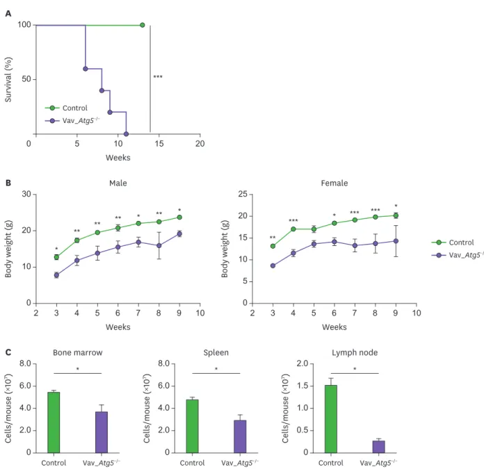
Vav_Atg5−/− mice and monitored the survival of newborn pups. Vav_Atg5−/− mice died within
11 wk (Fig. 1A) and exhibited body weights that were lower than control mice (Fig. 1B). These results indicated that deficiency in Atg5 in hematopoietic systems leads to a survival defect. We hypothesized that the decreased survival of Vav_Atg5−/− mice is attributable to the
impairment of hematopoiesis, because hematopoietic cell-specific Atg7 deficiency developed severe anemia and lymphopenia (14). To determine the effect of Atg5 on hematopoiesis, we assessed total cell numbers in bone marrow, spleen, and lymph nodes. As predicted, Vav_Atg5−/− mice displayed significantly diminished total cell numbers in these compartments
compared to control mice (Fig. 1C).
Hematopoietic cell-specific Atg5-deficient mice suffer from severe
lymphopenia
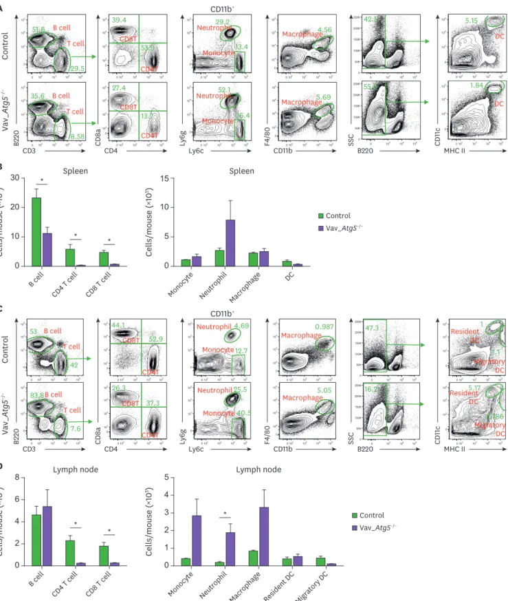
Next, we analyzed the immune cell populations in Vav_Atg5−/− mice. T cells were significantly
reduced in spleens (Fig. 2A and B) and lymph nodes (Fig. 2C and D), while B cells were diminished only in spleens (Fig. 2A and B). Myeloid cells were slightly increased in spleens (Fig. 2A and B) and lymph nodes (Fig. 2C and D) compared to control mice, but these differences failed to reach statistical significance except for the neutrophils in lymph nodes (Fig. 2C and D). These results indicated that Atg5 is crucial for the development of lymphoid cells and that hematopoietic cell-specific Atg5 gene deletion causes severe lymphopenia.
Atg5 deficiency results in defective erythropoiesis
Next, we examined erythropoiesis in Vav_Atg5−/− mice, because the overall hematopoiesis
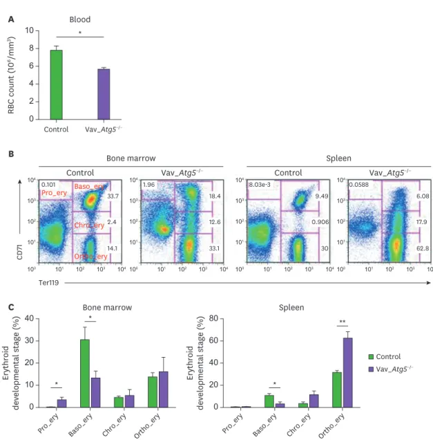
includes lymphopoiesis, myelopoiesis, and erythropoiesis. Previous study has reported that Atg5 was highly expressed in the pro-erythroblast and the expression levels were gradually reduced depending on the stage of erythropoiesis (15), suggesting that Atg5 is potentially associated with erythroid maturation. RBCs in peripheral blood were counted using a hemocytometer, and bone marrow and spleen samples were stained with CD71 and Ter119 to assess the developmental stage of the erythrocytes. Vav_Atg5−/− mice showed a reduced
number of RBCs in peripheral blood compared with control animals (Fig. 3A), indicating that Vav_Atg5−/− mice suffer from anemia. The distribution of erythrocyte developmental stages
erythroblasts were decreased concomitant with an increase in the proportions of other stages (Fig. 3C). These results indicate that Atg5 deficiency in hematopoietic cells leads to altered erythroid differentiation and causes RBC reduction.
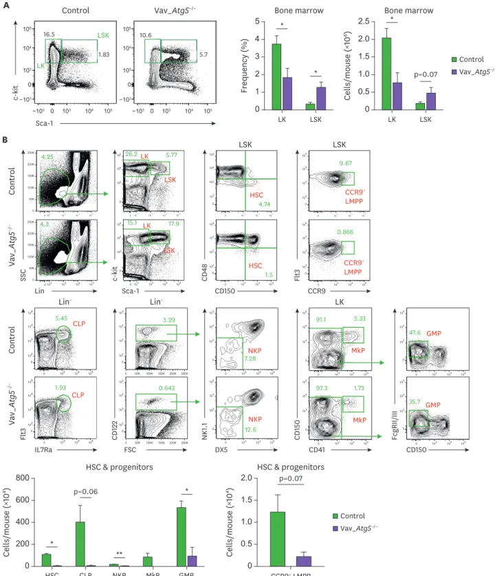
Atg5 is essential for the HSCs maintenance and reconstitution ability
In bone marrow, hematopoietic cells are divided into Lin−c-Kit+ (LK) cells enriched for myeloid progenitors and megakaryocyte-erythroid progenitors and LSK cells comprising long-term HSCs, short-term HSCs, and multipotent progenitors (22,23). Therefore, we analyzed LK and LSK populations to clarify the developmental impairment of HSCs and hematopoietic progenitor cells in Vav_Atg5−/− mice. LSKs in bone marrow were increased * 0 6.0 8.0 Cells/mouse ( ×10 7) Control Bone marrow Vav_Atg5−/− C 4.0 2.0 * 0 6.0 8.0 Cells/mouse ( ×10 7) Control Spleen Vav_Atg5−/− 4.0 2.0 * 0 1.5 2.0 Cells/mouse ( ×10 7) Control Lymph node Vav_Atg5−/− 1.0 0.5 0 30 10 Body weight (g) 2 Male Weeks B 3 4 5 6 7 8 9 10 20 0 25 5 Body weight (g) 2 Female Weeks 3 4 5 6 7 8 9 10 15 20 10 * * ** ** ** ** * Control Vav_Atg5−/− ** *** * *** *** * *** 100 Survival (%) 0 Weeks A 5 10 15 20 50 Control Vav_Atg5−/−
Figure 1. Vav_Atg5−/− mice display defects in survival, body weights, and total hematopoietic cell number. Starting 3 wk after birth, survival (A) and body weights
(B) of newborn control and Vav_Atg5−/− pups were monitored every wk. (C) The number of bone marrow, spleen, and lymph node cells from these animals were
counted (n=3). Data are representative of 3 independent experiments and presented as mean ± SEM.
Control Vav_ Atg 5 −/− A CD3 B220 CD4 CD11b+ CD 8a Ly6c Ly 6g CD11b F4/80 B220 SSC MHC II CD 11 c 0 102 103 104 105 0 103 104 105 35.6 8.58 0 103 104 105 0 50K 100K 150K 200K 250K55.5 0 102 103 104 105 0 103 104 105 5.15 0 103 104 105 0 50K 100K 150K 200K 250K42.5 0 102 103 104 105 0 102 103 104 105 4.56 0 102 103 104 105 0 103 104 105 13.4 29.2 0 102 103 104 105 0 102 103 104 105 39.4 53.9 0 102 103 104 105 0 102 103 104 105 27.4 13.7 0 102 103 104 105 0 103 104 105 51.6 29.5 B cell T cell CD8T CD4T Macrophage Neutrophil Monocyte DC 0 102 103 104 105 0 103 104 105 16.4 52.1 0 102 103 104 105 0 102 103 104 105 5.69 0 102 103 104 105 0 103 104 105 1.84 B cell T cell CD8T CD4T Macrophage Neutrophil Monocyte DC 0 102 103 104 105 0 103 104 105 83.8 7.6 0 102 103 104 105 0 103 104 105 53 42 B cell T cell B cell T cell CD11b+ Control Vav_ Atg 5 −/− C CD3 B220 CD4 CD 8a Ly6c Ly 6g CD11b F4/80 B220 SSC MHC II CD 11 c 0 102 103 104 105 0 102 103 104 10526.3 37.3 0 102 103 104 105 0 103 104 105 40.5 25.5 0 102 103 104 105 0 103 104 105 12.7 4.69 0 102 103 104 105 0 102 103 104 105 5.05 0 102 103 104 105 0 102 103 104 105 44.1 52.9 CD8T CD4T 0 102 103 104 105 0 102 103 104 105 0.987 Macrophage 0 103 104 105 0 50K 100K 150K 200K 250K 47.3 0 102 103 104 105 0 103 104 105 1.1 1 0 103 104 105 0 50K 100K 150K 200K 250K 16.2 0 102 103 104 105 0 103 104 105 0.786 5.17 Neutrophil Monocyte Migratory DC Resident DC Neutrophil Monocyte Migratory DC Resident DC CD8T CD4T Macrophage * 0 20 30 Cells/mouse ( ×10 6) B cell CD4 T cell CD8 T cell Spleen B * * 10 0 10 15 Cells/mouse ( ×10 5)
Monocyte Neutrophil Macrophage DC Spleen 5 Control Vav_Atg5−/− * * * 0 4 8 Cells/mouse ( ×10 6) B cell CD4 T cell CD8 T cell Lymph node D 6 2 0 3 5 Cells/mouse ( ×10 5)
Monocyte Neutrophil Macrophage Resident DC Migratory DC
Lymph node 2 4 1 Control Vav_Atg5−/−
Figure 2. Vav_Atg5−/− mice suffer from severe lymphopenia. Spleen (A, B) and lymph nodes (C, D) were isolated from control and Vav_Atg5−/− mice. B cells
(CD3−B220+), CD4 (CD3+CD4+) T cells, CD8 (CD3+CD8a+) T cells, dendritic cells (DC; B220−CD11C+MHC class II+) in spleen, resident DCs (B220−CD11c+MHC class IIInt) and migratory DCs (B220−CD11c+MHC class IIHigh) in lymph nodes, neutrophils (CD11b+Ly6G+), monocytes (CD11b+Ly6CHigh), and macrophage (CD11b+F4/80+) were classified by flow cytometry. Representative FACS plots (A, C) and absolute cell numbers (B, D) were calculated (n=3). Data are representative of three independent experiments and presented as mean ± SEM.
in Vav_Atg5−/− mice, while LKs were significantly decreased in these mice compared with
controls (Fig. 4A). We then analyzed HSCs and other hematopoietic progenitors in bone marrow. HSCs were characterized as LSK CD48−CD150+ cells. Interestingly, HSCs in bone marrow of Vav_Atg5−/− mice were significantly reduced compared with controls, indicating
that reduced HSC numbers were not attributable to the LSK expansion (Fig. 4B). For the lymphoid progenitor compartment, lymphoid-primed multipotent progenitors (LMPPs), common lymphoid progenitors (CLPs), and NK cell progenitors (NKPs) were analyzed, while megakaryocyte progenitors (MkPs) and granulocyte-macrophage progenitors (GMPs) were analyzed for the myeloid progenitor compartment. In Vav_Atg5−/− mice, NKPs,
and GMP were significantly reduced (Fig. 4B), while CLPs, CCR9+ LMPPs, and MkPs were diminished but without statistical significance. These results indicate that Atg5 is crucial for
* 0 8 10 RBC count ( 10 6/mm 3) Control Blood Vav_Atg5−/− A 6 4 2 0 20 40 Erythroid developmental stage (%)
Pro_ery Baso_ery Chro_ery Ortho_ery
Bone marrow 30 10 0 40 80 Erythroid developmental stage (%)
Pro_ery Baso_ery Chro_ery Ortho_ery
Spleen 60 20 * * * ** C Control Vav_Atg5−/− Pro_ery Baso_ery Chro_ery Ortho_ery B Ter119 CD 71 101 101 102 103 104 100 102 103 104 101 101 102 103 104 100 102 103 104 101 101 102 103 104 100 102 103 104 101 101 102 103 104 100 102 103 104 0.101 33.7 2.4 14.1 1.96 18.4 12.6 33.1 8.03e-3 0.0588 9.49 0.906 30 6.08 17.9 62.8 Control Bone marrow
Vav_Atg5−/− Control Vav_Atg5−/−
Spleen
Figure 3. Erythropoiesis is altered following Atg5 deficiency. (A) The number of RBCs in peripheral blood were counted in control and Vav_Atg5−/− mice.
(B) Erythroid developmental stages in bone marrow and spleen were assessed by flow cytometry. Pro-erythroblasts (Pro_ery; Ter119−CD71High), basophilic erythroblasts (Baso_ery; Ter119+CD71High), chromatophilic erythroblasts (Chro_ery; Ter119+CD71Med), and orthochromatophilic erythroblasts (Ortho_ery; Ter119+CD71−) were characterized (n=4). Data are representative of three independent experiments and presented as mean ± SEM.
A Control Vav_Atg5−/− LK LSK Control Vav_ Atg 5 −/− Lin SSC Sca-1 c-kit CD150 CD 48 CCR9 Flt 3 Control Vav_ Atg 5 −/− IL7Ra Flt 3 FSC CD 122 DX5 NK 1.1 CD41 CD 150 CD150 FcgRII/III p=0.06 * ** * B 0 400 800 Cells/mouse ( ×10 4) HSC CLP NKP MkP GMP HSC & progenitors 200 600 Control Vav_Atg5−/− 0 103 104 105 0 102 103 104 105 4.74 0 102 103 104 105 0 50K 100K 150K 200K 250K 4.25 0 102 103 104 105 0 50K 100K 150K 200K 250K 4.3 0 102 103 104 105 0 3 4 5 15.1 17.9 0 103 104 105 0 102 103 104 10 10 10 10 10 10 10 5 1.5 0 102 103 104 105 0 3 4 5 26.2LK 5.77 LSK HSC LSK LK LSK HSC LSK LK Lin− Lin− 0 103 104 105 0 102 103 104 105 0.866 0 103 104 105 0 102 103 104 105 9.67 CCR9+ LMPP CCR9+ LMPP 10 10 10 10 10 10 0 103 104 105 0 102 103 104 105 1.93 0 103 104 105 0 102 103 104 105 5.45 CLP 0 103 104 105 0 50K 100K 150K 200K 250K 0 102 103 104 105 0.643 0 103 104 105 0 3 4 5 12.6 0 50K 100K 150K 200K 250K 0 102 103 104 105 3.29 0 3 4 5 7.28NKP CLP NKP 0 103 104 105 0 103 104 105 97.3 1.73 0 103 104 105 0 103 104 105 91.1 5.33 0 103 104 105 0 103 104 105 35.7 0 103 104 105 0 103 104 105 47.6 MkP GMP MkP GMP p=0.07 0 1.5 2.0 Cells/mouse ( ×10 4) CCR9+ LMPP HSC & progenitors 1.0 0.5 Sca-1 c-kit−103 −103 103 103 0 0 104 104 105 105 −103 −103 103 103 0 0 104 104 105 105 16.5 1.83 10.6 5.7 Control Vav_Atg5−/− 0 2 5 Frequency ( % ) LK LSK Bone marrow 4 3 1 * * 0 1.5 2.5 Cells/mouse ( ×10 6) LK LSK Bone marrow 2.0 1.0 0.5 * p=0.07
Figure 4. Atg5 deficiency leads to defective maintenance of HSCs and developmental impairment of hematopoietic progenitor cells. (A, B) Bone marrow cells isolated from 11-wk-old from control and Vav_Atg5−/− mice were analyzed. (A) Frequency and absolute number of LK and LSK cells were assessed by flow
cytometry (n=4–5). (B) HSCs were characterized as LSK CD48−CD150+, and multiple progenitor cells were characterized in CLPs (Lin−IL-7Ra+Flt3+), CCR9+ LMPPs (LSK Flt3+), NKPs (Lin−CD122+NK1.1−DX5−), MkPs (LK CD41+CD150+), and GMPs (LK CD41−FcgRII/III+) (n=3). Data are representative of three independent experiments and presented as mean ± SEM.
the maintenance of HSCs and that a defect in HSCs results in developmental impairment of hematopoietic progenitor cells in Vav_Atg5 mice.
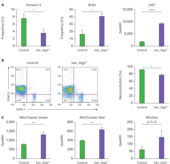
Next, we assessed proliferation and apoptosis in Atg5-deficient hematopoietic cells. Bone marrow cells were stained with anti-Ki67, BrdU antibodies and Annexin V. As shown in Fig. 5A, the LSK compartment in Vav_Atg5−/− mice exhibited enhanced resistance to apoptosis and higher
proliferation rates compared with controls. Specifically, the Annexin V+ populations in LSK cells were reduced in Vav_Atg5−/− mice. As Annexin V binds phosphatidylserine on the apoptotic cell
surface, the reduction of Annexin V+ populations suggests that LSK apoptosis was decreased in Vav_Atg5−/− mice. Furthermore, BrdU+ populations and Ki67 levels were enhanced in LSKs of Vav_Atg5−/− mice, suggesting that hematopoietic cell-specific Atg5 deficiency results in aberrant
proliferation of LSKs. ** 0 1,500 2,000 GeoMFI Control MitoTracker Green Vav_Atg5−/− C 1,000 500 ** 0 600 800 GeoMFI Control MitoTracker Red Vav_Atg5−/− 400 200 p=0.14 0 200 250 GeoMFI Control MitoSox Vav_Atg5−/− 150 100 50 * 0 101 101 102 103 104 100 102 103 104 101 101 102 103 104 100 102 103 104 8 10 Frequency ( % ) Control Control Annexin V Vav_Atg5−/− Vav_Atg5−/− A 6 4 2 * 0 40 50 Frequency ( % ) Control BrdU Vav_Atg5−/− 30 20 10 **** 0 10,000 15,000 GeoMFI Control Ki67 Vav_Atg5−/− 5,000 B * 0 80 100 Reconstitution ( % ) Control CD45.1 CD 45.2 Vav_Atg5−/− 60 40 20 1.67 90.3 3.61 4.41 4.01 73.7 9.54 12.7
Figure 5. Hematopoietic cell specific Atg5 deficiency results in aberrant proliferation and mitochondrial dysfunction in hematopoietic cells. (A) Apoptosis and cell proliferation assays of LSKs from control and Vav_Atg5−/− mice were performed using Annexin V, BrdU, and Ki67 staining. For the BrdU assay, proliferation
of LSK cells was analyzed 24 h after BrdU injection (n=4–5). (B) Approximately 2×10⁶ of total bone marrow cells from control and Vav_Atg5−/− mice were injected
intravenously into lethally irradiated CD45.1+ mice. After 8 weeks, percentage of reconstitution of transplanted bone marrow in peripheral blood was confirmed by flow cytometry (n=3). (C) Mitochondrial functions in bone marrow LSK cells from control and Vav_Atg5−/− mice were assessed using MitoTracker Green,
MitoTracker Red, and MitoSox (n=4–5). Data are representative of 3 independent experiments and presented as mean ± SEM.
In addition, we assessed the reconstitution ability of HSCs with Atg5 deficiency.
Approximately 2×10⁶ bone marrow cells from control and Vav_Atg5−/− (CD45.2+) mice were injected via tail vein into lethally irradiated CD45.1+ congenic mice. After 8 wk, more than 90% of cells were reconstituted in peripheral blood from the recipients transplanted with control bone marrow cells, while the Vav_Atg5−/− bone marrow cells led to reconstitution
of about 70% of the cells in the hematopoietic system of recipients (Fig. 5B). These results indicate that Atg5 affects not only the maintenance but also the reconstitution ability of HSCs. Autophagy contributes to the maintenance of intracellular homeostasis via removal of
damaged mitochondria, and the absence of autophagy leads to accumulation of dysfunctional mitochondria and ROS. Therefore, we hypothesized that Atg5 deficiency in hematopoietic cells results in mitochondrial disturbance. We analyzed LSK populations in bone marrow to determine whether Atg5 deficiency mediates mitochondrial dysfunction in hematopoietic cells. We assessed the mitochondrial functions in bone marrow LSK cells using MitoTracker Green, MitoTracker Red, and MitoSox. As expected, LSK cells showed enhanced mitochondrial mass and membrane potentials in Vav_Atg5−/− mice, while mitochondrial superoxide levels were
slightly, but not significantly, increased (Fig. 5C). These results indicate that Atg5 deficiency causes unregulated accumulation of mitochondria in LSKs.
DISCUSSION
Autophagy is crucial for the maintenance of cellular homeostasis via elimination of impaired cytosolic components, such as damaged mitochondria and misfolded proteins, and this process therefore plays an essential role in cell survival (24). During the early neonatal starvation period, autophagy promotes survival of newborn pups by degrading self-proteins to supply amino acids (25). In mesenchymal stromal/stem cells, autophagy is induced under hypoxic condition and promotes proangiogenic activity via enhancing angiogenin and VEGF production (26). Furthermore, autophagy and autophagy-related proteins have been reported as crucial regulators for hematopoiesis. For example, Atg7 is known to play an important role in the maintenance of HSCs, and Atg7 deficiency in hematopoietic cells causes severe anemia and lymphopenia in vivo (14,15). Also, Atg5 is required for the development and survival of innate lymphoid cells and NK cells, as this factor facilitates lymphocyte survival following homeostasis proliferation during lymphopenia (18); however, the role of the autophagy protein Atg5 in HSCs has not previously been thoroughly investigated.
In this study, we found that hematopoietic cell-specific Atg5 deficiency causes severe lymphopenia, anemia, and survival defects. Immune cell populations in spleens and lymph nodes were significantly altered in Vav_Atg5−/− mice, due to the developmental impairment
of hematopoietic progenitor cells. Specifically, Vav_Atg5−/− mice showed defective HSC
maintenance and lower reconstitution capacity following disrupted differentiation of hematopoietic progenitor cells. Also, abnormal distribution of erythrocyte developmental stages resulted in decreased RBCs in blood in Vav_Atg5−/− mice, and hematopoietic
cell-specific Atg5 deficiency led to aberrant proliferation of LSKs. Although LSK cells displayed enhanced proliferation and diminished apoptosis, absolute number of HSCs were
significantly decreased in Vav_Atg5−/− mice. We propose that defective reconstitution ability
of bone marrow cells from Vav_Atg5−/− mice are attributed to the lower HSC numbers. Also,
autophagy is required for the DNA damage responses and loss of autophagy facilitate the accumulation of damaged DNA inducing genetic instability (27-30), therefore, enlarged LSK
cells do not necessarily reflect the abundance of functional HSCs. Finally, we found that Atg5-deficient LSKs displayed unregulated accumulation of mitochondria. Mitochondria mass and membrane potentials were enhanced in LSKs of Vav_Atg5-/- mice. Because autophagy
eliminates damaged organelles, such as mitochondria, these findings suggest that loss of Atg5 causes impairment of autophagy function and ultimately enhances the intracellular stress in hematopoietic cells. Mitochondria, which is the main producer of ROS in cells (31), continuously repeat fusion and fission during the life cycle to control mitochondrial quality (32), and mitochondrial fusion helps to redistribute metabolites and proteins and to dilute damage materials to reduce mitochondrial stress (33). Despite disruption of mitochondrial membrane potential results in release of cytochrome C into the cytosol and apoptosis, mitochondrial hyperpolarization also lead to cellular abnormalities. For example, patients with systemic lupus erythematosus exhibited mitochondrial hyperpolarization (34,35). Therefore, we suggest that increased mitochondrial mass and membrane potential indicates enhanced mitochondrial stress accumulation that inhibits cellular function and survival. Further, although the differences were not statistically significant, ROS levels were increased in Atg5-deficient LSKs compared to controls, suggesting that impairment of autophagic clearance of damaged mitochondria causes functional defects of HSCs and results in disturbed differentiation of mature progenitors and terminally differentiated cells. Because ROS have normally harmful effect on cell survival, reduced apoptosis in Atg5-deficient LSKs seems paradoxical. However, as overall proliferation was also improved following Atg5 deficiency, we propose that this aberrant proliferation compensate the harmful effect of ROS that mediates apoptosis. Collectively, these findings demonstrate that Atg5 plays a critical role as a regulator to maintain HSCs and its reconstitution function, while future studies are needed to address the relationship between autophagy and other maintenance factors in HSCs.
ACKNOWLEDGEMENTS
This study was supported by the National Research Foundation of Korea (NRF-2018M3A9H3024611) funded by the Ministry of Science and ICT of Korea.
REFERENCES
1. Sugiyama T, Kohara H, Noda M, Nagasawa T. Maintenance of the hematopoietic stem cell pool by CXCL12-CXCR4 chemokine signaling in bone marrow stromal cell niches. Immunity 2006;25:977-988.
PUBMED | CROSSREF
2. Guerrouahen BS, Al-Hijji I, Tabrizi AR. Osteoblastic and vascular endothelial niches, their control on normal hematopoietic stem cells, and their consequences on the development of leukemia. Stem Cells Int 2011;2011:375857.
PUBMED | CROSSREF
3. Tipping AJ, Pina C, Castor A, Hong D, Rodrigues NP, Lazzari L, May GE, Jacobsen SE, Enver T. High GATA-2 expression inhibits human hematopoietic stem and progenitor cell function by effects on cell cycle. Blood 2009;113:2661-2672.
PUBMED | CROSSREF
4. Park IK, Qian D, Kiel M, Becker MW, Pihalja M, Weissman IL, Morrison SJ, Clarke MF. Bmi-1 is required for maintenance of adult self-renewing haematopoietic stem cells. Nature 2003;423:302-305.
PUBMED | CROSSREF
5. Iwama A, Oguro H, Negishi M, Kato Y, Morita Y, Tsukui H, Ema H, Kamijo T, Katoh-Fukui Y, Koseki H, et al. Enhanced self-renewal of hematopoietic stem cells mediated by the polycomb gene product Bmi-1. Immunity 2004;21:843-851.
6. Domen J, Cheshier SH, Weissman IL. The role of apoptosis in the regulation of hematopoietic stem cells: Overexpression of Bcl-2 increases both their number and repopulation potential. J Exp Med 2000;191:253-264.
PUBMED | CROSSREF
7. Opferman JT, Iwasaki H, Ong CC, Suh H, Mizuno S, Akashi K, Korsmeyer SJ. Obligate role of anti-apoptotic MCL-1 in the survival of hematopoietic stem cells. Science 2005;307:1101-1104.
PUBMED | CROSSREF
8. Levine B, Mizushima N, Virgin HW. Autophagy in immunity and inflammation. Nature 2011;469:323-335.
PUBMED | CROSSREF
9. Oh JE, Lee HK. Autophagy as an innate immune modulator. Immune Netw 2013;13:1-9.
PUBMED | CROSSREF
10. Kim SJ, Hong EH, Lee BR, Park MH, Kim JW, Pyun AR, Kim YJ, Chang SY, Chin YW, Ko HJ. α-Mangostin reduced ER stress-mediated tumor growth through autophagy activation. Immune Netw 2012;12:253-260.
PUBMED | CROSSREF
11. Chen C, Liu Y, Liu R, Ikenoue T, Guan KL, Liu Y, Zheng P. TSC-mTOR maintains quiescence and function of hematopoietic stem cells by repressing mitochondrial biogenesis and reactive oxygen species. J Exp Med 2008;205:2397-2408.
PUBMED | CROSSREF
12. Gan B, Sahin E, Jiang S, Sanchez-Aguilera A, Scott KL, Chin L, Williams DA, Kwiatkowski DJ, DePinho RA. mTORC1-dependent and -independent regulation of stem cell renewal, differentiation, and mobilization. Proc Natl Acad Sci U S A 2008;105:19384-19389.
PUBMED | CROSSREF
13. Warr MR, Binnewies M, Flach J, Reynaud D, Garg T, Malhotra R, Debnath J, Passegué E. FOXO3A directs a protective autophagy program in haematopoietic stem cells. Nature 2013;494:323-327.
PUBMED | CROSSREF
14. Mortensen M, Soilleux EJ, Djordjevic G, Tripp R, Lutteropp M, Sadighi-Akha E, Stranks AJ, Glanville J, Knight S, Jacobsen SE, et al. The autophagy protein Atg7 is essential for hematopoietic stem cell maintenance. J Exp Med 2011;208:455-467.
PUBMED | CROSSREF
15. Mortensen M, Ferguson DJ, Edelmann M, Kessler B, Morten KJ, Komatsu M, Simon AK. Loss of autophagy in erythroid cells leads to defective removal of mitochondria and severe anemia in vivo. Proc Natl Acad Sci U S A 2010;107:832-837.
PUBMED | CROSSREF
16. Miller BC, Zhao Z, Stephenson LM, Cadwell K, Pua HH, Lee HK, Mizushima NN, Iwasaki A, He YW, Swat W, et al. The autophagy gene Atg5 plays an essential role in B lymphocyte development. Autophagy 2008;4:309-314.
PUBMED | CROSSREF
17. Conway KL, Kuballa P, Khor B, Zhang M, Shi HN, Virgin HW, Xavier RJ. Atg5 regulates plasma cell differentiation. Autophagy 2013;9:528-537.
PUBMED | CROSSREF
18. O'Sullivan TE, Geary CD, Weizman OE, Geiger TL, Rapp M, Dorn GW 2nd, Overholtzer M, Sun JC. Atg5 is essential for the development and survival of innate lymphocytes. Cell Reports 2016;15:1910-1919.
PUBMED | CROSSREF
19. Liu Q, Chen L, Atkinson JM, Claxton DF, Wang HG. Atg5-dependent autophagy contributes to the development of acute myeloid leukemia in an MLL-AF9-driven mouse model. Cell Death Dis 2016;7:e2361.
PUBMED | CROSSREF
20. Hara T, Nakamura K, Matsui M, Yamamoto A, Nakahara Y, Suzuki-Migishima R, Yokoyama M, Mishima K, Saito I, Okano H, et al. Suppression of basal autophagy in neural cells causes neurodegenerative disease in mice. Nature 2006;441:885-889.
PUBMED | CROSSREF
21. Oh DS, Oh JE, Jung HE, Lee HK. Transient depletion of CD169+ cells contributes to impaired early protection and effector CD8+ T cell recruitment against mucosal respiratory syncytial virus infection. Front Immunol 2017;8:819.
PUBMED | CROSSREF
22. Philips ST, Hildenbrand ZL, Oravecz-Wilson KI, Foley SB, Mgbemena VE, Ross TS. Toward a therapeutic reduction of imatinib refractory myeloproliferative neoplasm-initiating cells. Oncogene 2014;33:5379-5390.
PUBMED | CROSSREF
23. Du J, Wang J, Kong G, Jiang J, Zhang J, Liu Y, Tong W, Zhang J. Signaling profiling at the single-cell level identifies a distinct signaling signature in murine hematopoietic stem cells. Stem Cells 2012;30:1447-1454.
24. Das G, Shravage BV, Baehrecke EH. Regulation and function of autophagy during cell survival and cell death. Cold Spring Harb Perspect Biol 2012;4:a008813.
PUBMED | CROSSREF
25. Kuma A, Hatano M, Matsui M, Yamamoto A, Nakaya H, Yoshimori T, Ohsumi Y, Tokuhisa T, Mizushima N. The role of autophagy during the early neonatal starvation period. Nature 2004;432:1032-1036.
PUBMED | CROSSREF
26. Lee SG, Joe YA. Autophagy mediates enhancement of proangiogenic activity by hypoxia in mesenchymal stromal/stem cells. Biochem Biophys Res Commun 2018;501:941-947.
PUBMED | CROSSREF
27. Eliopoulos AG, Havaki S, Gorgoulis VG. DNA damage response and autophagy: a meaningful partnership. Front Genet 2016;7:204.
PUBMED | CROSSREF
28. Gomes LR, Menck CF, Leandro GS. Autophagy roles in the modulation of DNA repair pathways. Int J Mol Sci 2017;18:2351.
PUBMED | CROSSREF
29. Bhutia SK, Mukhopadhyay S, Sinha N, Das DN, Panda PK, Patra SK, Maiti TK, Mandal M, Dent P, Wang XY, et al. Autophagy: cancer's friend or foe? Adv Cancer Res 2013;118:61-95.
PUBMED | CROSSREF
30. Mathew R, Kongara S, Beaudoin B, Karp CM, Bray K, Degenhardt K, Chen G, Jin S, White E. Autophagy suppresses tumor progression by limiting chromosomal instability. Genes Dev 2007;21:1367-1381.
PUBMED | CROSSREF
31. Yarosz EL, Chang CH. The role of reactive oxygen species in regulating T cell-mediated immunity and disease. Immune Netw 2018;18:e14.
PUBMED | CROSSREF
32. Jin HS, Suh HW, Kim SJ, Jo EK. Mitochondrial control of innate immunity and inflammation. Immune Netw 2017;17:77-88.
PUBMED | CROSSREF
33. Romanello V, Sandri M. Mitochondrial quality control and muscle mass maintenance. Front Physiol 2016;6:422.
PUBMED | CROSSREF
34. Gergely P Jr, Grossman C, Niland B, Puskas F, Neupane H, Allam F, Banki K, Phillips PE, Perl A. Mitochondrial hyperpolarization and ATP depletion in patients with systemic lupus erythematosus. Arthritis Rheum 2002;46:175-190.
PUBMED | CROSSREF
35. Gergely P Jr, Niland B, Gonchoroff N, Pullmann R Jr, Phillips PE, Perl A. Persistent mitochondrial hyperpolarization, increased reactive oxygen intermediate production, and cytoplasmic alkalinization characterize altered IL-10 signaling in patients with systemic lupus erythematosus. J Immunol 2002;169:1092-1101.