Combination of Curcumin and Paclitaxel-loaded Solid Lipid Nanoparticles to
Overcome Multidrug Resistance
Rihua Li, Wenting Xu, Jae Soon Eun and Mi-Kyung Lee†
College of Pharmacy, Woosuk University, Jeonbuk 565-701, Korea (Received December 12, 2011·Accepted December 13, 2011)
ABSTRACT−Multi-drug resistance (MDR) has been known as a major hurdle in cancer chemotherapy. One of the most clinically significant causes of MDR was the efflux of anticancer agents mediated by p-glycoprotein (p-gp) over-expressed in MDR cancer cells. To overcome MDR, there have been several strategies such as co-administration with p-gp inhibitors and encapsulation of anticancer drugs into drug delivery systems. In the present study, curcumin was evaluated for its poten-tial as p-gp inhibitor and MDR reversal activity when combined with paclitaxel incorporated into lipid nanoparticles (PTX/ LN). Western blot assay showed curcumin did not modulate the level of p-gp expression in MCF-7/ADR which is a MDR variant of human breast cancer cell line, MCF-7, and over-expresses p-gp. However, curcumin inhibited p-gp-mediated efflux of calcein in a dose-dependent manner even though it showed lower activity compared to verapamil, a well-known p-gp inhibitor. Incorporation of paclitaxel into lipid nanoparticles partially recovered the anticancer activity of paclitaxel in MCF-7/ADR. The combined use of curcumin and PTX/LN exhibited further full reversal of MDR, suggesting susceptibility of PTX/LN to the efflux system. In conclusion, combined approach of using p-gp inhibitors and incorporation of the anti-cancer agents into nano-delivery systems would be an efficient strategy to overcome MDR.
Key words−MDR, p-glycoprotein, Curcumin, Paclitaxel
In cancer treatment, chemotherapy forms part of a successful treatment regime in many cases. However, as few as half of the patients treated may benefit from this, as a result of intrinsic or acquired multiple drug resistance (MDR) (Germano et al., 2009). One of the important mechanisms of MDR has been known to be due to the efflux of drugs by multi-drug trans-porters over-expressed on the MDR cancer cells (Longley et al., 2005). P-glycoprotein (p-gp) is considered as one of the most clinically significant efflux transporters (Gottesman et al., 2002). P-gp confers upon cancer cells the ability to resist lethal dose of certain cytotoxic drugs by actively pumping the drugs out of the cancer cells. A negative correlation has been dem-onstrated between p-gp expression level and survival in cancer patients (Tsukamoto et al., 1997). P-gp is encoded by MDR1 gene and has been a therapeutic target to overcome the resis-tance.
Several p-gp inhibitors have been tried to overcome MDR. Verapamil and cyclosporine A have been shown to reverse the MDR1 phenotype in a variety of paclitaxel-resistance human cancer cells, even though high level of p-gp inhibitors were required and they failed to restore the treatment response in
p-gp expressing tumor in clinical trials (Toppmeyer et al., 2002). Unfortunately, other new generations of p-gp inhibitors devel-oped thereafter have shown little clinical value overall in restoring tumor sensitivity and in safety (Chi et al., 2005). Recently, many researchers have focused on plants extracts and demonstrated that some plant components could modulate p-gp activity (Molnár et al., 2010).
Curcumin, a naturally occurring polyphenol obtained from Curcuma longa, has a long history in Indian traditional med-icine and recently reported to down-regulate the p-gp expres-sion (Anuchapreeda et al., 2002; Choi et al., 2008). In addition, curcumin has demonstrated anticancer activity and been sug-gested as lead compound against hormone-independent mul-tidrug resistant breast cancer (Labbozzetta et al., 2009). Moreover, C. longa and its derivatives have been demonstrated to be safe at a high dose in various animal models (Qureshi et al., 1992). Considering the recent reports, curcumin would be attractive candidate to overcome MDR and enhance anticancer activity when used with chemotherapeutic drugs in MDR can-cer.
There have been several approaches other than p-gp inhib-itors to overcome MDR mediated by p-gp (Szakács et al., 2006). A number of studies have investigated the encapsu-lation of p-gp substrates in various drug delivery systems to evade the efflux (Lee et al., 2005; Shapira et al., 2011).
How-†Corresponding Author :
Tel : +82-63-290-1423, E-mail : [email protected] DOI : 10.4333/KPS.2011.41.6.381
ever, encapsulated drug in polymeric nanoparticles could not completely evade the efflux pump according to a recent report (Chavanpatil et al., 2006). It has been shown that p-gp sub-strate, such as paclitaxel, encapsulated in PLGA nanoparticles are susceptible to efflux by p-gp and inhibition of the pump by specific inhibitors can reverse the MDR (Chavanpatil et al., 2006). Therefore, a strategy of co- administration of p-gp inhibitor with encapsulated anticancer agents would be better alternative to overcome MDR.
In the present study, curcumin was evaluated for its p-gp inhibition effect in MDR human breast cancer cells and co-administered with paclitaxel incorporated into lipid nanopar-ticles to reverse MDR.
Experimental
MaterialsTrilaurin, curcumin and verapamil hydrochloride were pur-chased from Sigma Chemical Co, (St. Louis, MO, USA). Egg phosphatidylcholine (eggPC) and distearoyl phosphatidyl eth-anolamine-N-poly (ethylene glycol)2000 (PEG2000PE) were
pur-chased from Avanti Polar Lipids Inc. (Alabaster, AL, USA). Calcein-AM was from Molecular Probe, Invitrogen Corp. (CA, USA). MCF-7 was from Korean Cell Line Bank (Seoul, Korea) and MCF-7/ADR was kindly gifted by Dr. S.J. Lim in Sejong University, Seoul, Korea. All the reagents for cell cul-ture were purchased from Invitrogen Corp. (CA, USA). All other chemicals were reagent grade and used without further purification.
Western blot assay for p-gp expression
MCF-7 or MCF-7/ADR cells (5 × 10⁵ cells/well) were cul-tured in 6-well plate for 16 hours, and then curcumin was added to incubate for 72 hours. After incubation, the cells were washed with PBS and solubilized with ice-cold lysis buffer containing 150 mM NaCl, 50 mM Tris-HCl (pH 7.4), 5 mM EDTA, 0.5% sodium deoxycholate, NP-40, 10% SDS, 100 mM PMSF and protease inhibitor. Insoluble materials were removed by centrifugation at 10,000 × g for 10 minutes. Extracted pro-teins were determined by BCA protein assay. The propro-teins were loaded onto 7.5% polyacrylamide-SDS gel electrophore-sis (SDS-PAGE) and electrotransferred to polyvinylidene dif-luoride (PVDF) membrane. For blocking the nonspecific binding, the membrane was incubated with 5% bovine serum albumin for 2 hours at room temperature. The membrane was washed three times with PBST and incubated with mouse monoclonal (C219) anti-p-gp antibody. After blotting with a primary antibody, the membrane was washed three times with
PBST and followed by incubation with HRP-conjugated anti-mouse and anti-rabbit IgG (1: 10,000) at room temperature for 1 hour. The visualization of the blots was carried out using enhanced chemiluminescence (ECL) detection system.
Analysis of calcein retention
MCF-7/ADR cells (1 × 10⁶ cells/well) were seeded in a 96-well plate and cultured for 3 hours. The p-gp inhibitors (ver-apamil or curcumin) were added and incubated for 10 minutes. After treatment with p-gp inhibitors, calcein-AM was added and incubated for 30 minutes. The fluorescence was deter-mined with a Tecan luminometer at 485 nm/535 nm after 30 min.
Preparation of paclitaxel-incorporated lipid nanoparticles The lipid nanoparticles were prepared using high pressure homogenizer as follows. Trilaurin (100 mg), eggPC (50 mg), PEG2000PE (5 mg) and pacilitaxel (5 mg) were weighed into
glass tube followed by sonication for approximately 1 hr at 65oC in bath type sonicator (Branson® ultrasonic cleaner, 3210R-DTH, Branson Ultrasonics Corp., CT, USA) to dis-solve paclitaxel in oily mixture. Preheated (65oC) water for injection was added to make 1 mL and sonicated for more than 3 hours until milky, homogeneous crude emulsion was obtained. The crude emulsion was homogenized for 5 cycles at 100 MPa using a high pressure homogenizer (Emulsiflex® EF-B3, Avestin Inc., Canada). The hot ultra fine emulsions obtained by high pressure homogenization (HPH) were frozen by dipping into liquid nitrogen and then thawed in water bath at room temperature. The resultant dispersion was stored at 4oC.
Measurement of particle size and zeta potential of lipid nanoparticles
The particle size and zeta potential of paclitaxel-incorpo-rated lipid nanoparticles was measured using submicron par-ticle sizer, ELS-Z (Photal Otsuka Electronics, Japan). The lipid nanoparticles were diluted with pre-filtered water through 0.22µm before measurement.
In vitro assay of anticancer activity
In vitro anticancer activity of curcumin and paclitaxel-con-taining lipid nanoparticles was measured against human breast cancer cell line, MCF-7, and its MDR variant, MCF-7/ADR by MTT assay. Cancer cells were cultured in PRMI 1640 medium supplemented with 10% of heat-inactivated FBS, 100 units/mL of penicillin and 100µg/mL of streptomycin under 5% CO2 at 37oC. The cells were inoculated to a 96-well plate
at a density of 104 cells in 200 µL medium per well and incu-bated for 12 hours. The medium was then replaced with cur-cumin- or paclitaxel-containing media and incubation was continued for 48 hours. When co-treated with curcumin, the medium containing curcumin was added to the wells 30 min before adding paclitaxel. After incubation, the lipid nano-particle-containing media were removed to avoid nanoparticle-induced interference in the MTT assay, and 180 µL of fresh medium and 20 µL of MTT solution (5 mg/mL in PBS) were added to the wells. The cells were incubated for another 3 hours. MTT internalization was terminated by aspiration of the media, and the cells were lysed with DMSO. The optical den-sity at 570 nm was determined using a microplate spectro-photometer (SPECTRAmax® 340PC; Molecular Devices Corp., Sunnyvale, CA, USA). Anticancer activity was expressed as % survival of the cancer cells compared to the untreated control cells (100% survival).
Results and Discussion
Effect of curcumin on p-gp expressionDrug-sensitive human breast cancer cell line, MCF-7, did not express p-gp according to the western blot assay as shown in Figure 1. On the other hand, MCF-7/ADR, a MDR variant of MCF-7, showed overexpression of p-gp. The MCF-7/ADR cells were treated with curcumin dissolved in DMSO for 72 hours to evaluate the effect of curcumin on the p-gp expres-sion. Curcumin did not change the level of p-gp expression significantly in MCF-7/ADR in the range of 10 µM to 35 µM (Figure 2). The result is similar to report by Ganta and Amiji in which the band intensity for p-gp after treatment with 20µM of curcumin aqueous solution appeared not to be dif-ferent from that of untreated control cells in SKOV3 human ovarian adenocarcinoma cell line and its MDR variant, SKOV3TR even though they concluded curcumin slightly
decreased p-gp expression (Ganta and Amiji, 2009). On the contrary to our results, curcumin has been reported to down-regulate p-gp expression in L1210/Adr, adriamycin resistant variant of L1210 mouse leukemia cell line in a dose-dependent manner in the range of 15 µM to 45 µM (Choi et al., 2008). The discrepancy might be due to the difference in the cellular uptake of curcumin considering the study by Ganta and Amiji in which p-gp expression was reduced remarkably when the cells were treated with curcumin in nanoemulsion compared to aqueous solution of curcumin (Ganta and Amiji, 2009).
Effect of curcumin on the efflux of calcein by p-gp To examine the inhibitory function of curcumin on p-gp-mediated efflux, calcein accumulation study was chosen
Figure 1. The expression of p-gp in MCF-7 and MCF-7/ADR cells. MCF-7 and MCF-7/ADR cells were lysed with cell lysis buffer. The cell lysate was loaded on the 7.5% SDS-PAGE and analyzed by im-munoblotting with mouse monoclonal anti-p-gp antibody.
Figure 2. The effect of curcumin on the expression of p-gp in MCF-7/ADR. MCF-7/ADR cells were treated with DMSO (Con) or cur-cumin (Cur) for 72 h and lysed with cell lysis buffer. The cell lysate was loaded on the 7.5% SDS-PAGE and analyzed by immuno-blotting with mouse monoclonal anti-p-gp antibody.
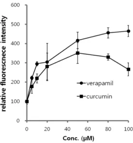
Figure 3. The effects of curcumin on the calcein retention in MCF-7/ADR cells after uptake as calcein-AM. The bar represents means ± SE from three independent experiments.
because it appears to be a sensitive indicator of p-gp activity (Marbeuf-Gueye et al., 2000). In this assay, the cells were incubated with test compounds for 10 min and then calcein-AM was added to be taken up into cells. The calcein-calcein-AM was degraded by intracellular esterase to release calcein which is a p-gp substrate. The activity of the test compounds on p-gp was determined as fluorescence of calcein in the cell. Figure 3 demonstrated the increase in calcein retention after treatment with verapamil (positive control) and curcumin in MCF-7/ ADR cells. Although verapamil showed higher activity, cur-cumin also exhibited a substantial increase in the retention of calcein in MCF-7/ADR cells which overexpress p-gp. Since calcein is known to be a good substrate for p-gp, we concluded that curcumin modulated intracellular calcein levels by inhib-iting p-gp. However, it was not likely that curcumin acted by down-regulating MDR1 gene because the treatment time of curcumin in the experiment was not long (< 1 h).
Effect of curcumin on the reversal of MDR
Curcumin has shown cytotoxicity in cancer cells (Anucha-preeda et al., 2002). Before proceeding to further study, cyto-toxicity of curcumin itself was investigated in MCF-7/ADR. Curcumin showed remarkable cytotoxicity against MCF-7/ ADR in high dose range from 5 µM to 100 µM (Figure 4). To evaluate MDR reversal effect of curcumin, low concentration of curcumin (5µM) was chosen to exclude the cytotoxicity effect of curcumin itself. As shown in Figure 5, the com-bination showed significantly higher cytotoxicity compared to the paclitaxel or curcumin alone which were delivered in DMSO. Because the cytotoxicity of low dose (5µM) cur-cumin was negligible as shown in Figure 4 and 5, the enhance-ment of anticancer activity of paclitaxel by curcumin was likely due to the inhibitory effect of curcumin on p-gp-medi-ated efflux in MDR cells. However, the reversal of MDR by
curcumin was not sufficient considering 1µM of paclitaxel showed much less than 50% cell survival rate in drug-sensitive MCF-7 cells (data not shown). In the further study, paclitaxel was incorporated into lipid nanoparticles to overcome p-gp-mediated efflux efficiently.
Reversal of MDR by incorporation of paclitaxel and com-bined use with curcumin
P-gp inhibition alone by curcumin was not sufficient to reverse MDR in MCF-7/ADR. Paclitaxel was incorporated into lipid nanoparticles to evade the efflux in the present study. The particle size and zeta potential of the resulting lipid nano-particles incorporating paclitaxel (PTX/LN) were 283±36 nm and -39 mV, respectively. Paclitaxel incorporated into lipid nanoparticles (PTX/LN) showed dose dependent anticancer activity in MCF-7 and MCF-7/ADR (Figure 6). PTX/LN showed approximately 0.1µM of IC50 in MCF-7, while larger than 2µM of IC50 in MCF-7/ADR. Based on the results of cytotoxicity, incorporation of paclitaxel into lipid nanoparticles could not fully avoid p-gp-mediated efflux. To investigate the involvement of p-gp mediated efflux, the cells were treated with PTX/LN and 5µM of curcumin. The anticancer activity of paclitaxel was not modulated by low dose of curcumin in MCF-7, which suggested p-gp-mediated efflux would not par-ticipate in MCF-7 and was in accord with the result of western blot assay. On the contrary, curcumin enhanced anticancer activity of MCF-7/ADR significantly, suggesting the involve-ment of p-gp-mediated efflux and susceptibility of incorpo-rated PTX to the efflux pump.
Figure 4. Cytotoxicity of curcumin against MCF-7/ADR cells after 24 and 48 h-incubation in MTT assay. The bar represents mean ± SE from three independent experiments.
Figure 5. The effect of curcumin on the cytotoxicity of paclitaxel (PTX) delivered in DMSO in MCF-7/ADR cells after incubation for 48 hr. The means ± SE from three independent experiments per-formed are shown. *; Significantly different from control group (*; p<0.05).
Conclusion
Curcumin inhibited p-gp mediated efflux in MCF-7/ADR which is MDR cell line overexpressing p-gp and the inhibitory mechanism was not likely through down-regulation of p-gp expression. Low dose of curcumin (<5µM) demonstrated p-gp inhibition with negligible cytotoxicity in MCF-7/ADR and enhanced anticancer activity of paclitaxel which is p-gp sub-strate in MCF-7/ADR. Although the incorporation of pacli-taxel into lipid nanoparticles could reverse MDR to some extent, combined use with p-gp inhibitor was required for additional full recovery of anticancer activity because the incorporated drug was susceptible to the efflux system in MCF-7/ADR.
Acknowledgement
This research was supported by the “Grant from the Korean Ministry of Education, Science and Technology” (The Regional Core Research Program/ Center for Healthcare Tech-nology Development) and also in part by Woosuk University.
References
Anuchapreeda, S., Leechanachai, P., Smith, M.M., Ambudkar, S.V., Limtrakul, P. 2002. Modulation of p-glycoprotein expres-sion and function by curcumin in multidrug-resistant human KB cells. Biochem. Pharmacol. 64, 573-582; Choi, B.H., Kim, C.G., Lim, Y. Shin, S.Y. and Lee, Y.H., 2008. Curcumin down-regulates the multidrug-resistance mdr1b gene by inhibiting the PI3K/Akt/NFêB pathway. Cancer Letters 259, 111-118.
Chavanpatil, M.D., Patil Y., Panyam, J. 2006. Susceptibility of nanoparticles-encapsulated paclitaxel to p-glycoprotein-medi-ated drug efflux. Int. J. Pharm. 320, 150-156.
Chi, K.N., Chia, S.K., Dixon, R., Newman, M.J., Wacher, V.J., Sikic, B., Gelmon, K.A., 2005. A phase I pharmacokinetic study of the P-glycoprotein inhibitor, ONT-093, in combi-nation with paclitaxel in patients with advanced cancer. Invest. New Drugs 23, 311-315.
Choi, B.H., Kim, C.G., Lim, Y., Shin, S.Y., Lee, Y.H., 2008. Cur-cumin down-regulates the multidrug-resistance mdr1b gene by inhibiting the PI3K/Akt/NF kappa B pathway. Cancer Lett. 259(1), 111-118.
Ganta, S., Amiji, M., 2009. Coadministration of paclitaxel and cur-cumin in nanoemulsion formulations to overcome multidrug resistance in tumor cells. Mol. Pharm. 6(3), 928-939. Germano, S., O’Driscoll, L., 2009. Breast cancer: understanding
sensitivity and resistance to chemotherapy and targeted ther-apies to aid in personalized medicine. Curr. Cancer Drug Tar-gets 9(3), 393-418.
Gottesman, M.M., 2002. Mechanisms of cancer drug resistance. Annu. Rev. Med. 53, 615-627.
Labbozzetta, M., Notarbartolo, M., Poma, P., Maurici, A., Inguglia, L., Marchetti, P., Rizzi, M., Baruchello, R., Simoni, D., D'Alessandro, N. 2009. Curcumin as a possible lead com-pound against hormone-independent, multidrug-resistant breast cancer. Ann. N. Y. Acad. Sci. 1155, 278-283.
Lee, E.S., Na, K. and Bae, Y.H. 2005. Doxorubicin loaded pH-sen-sitive polymeric micelles for reversal of resistant MCF-7 tumor. J. Control. Release 103, 405-418.
Longley, D.B., Johnston, P.G., Wilson, M., Mc Ewan, M., Allen, W.L., Mc Dermot, U., Galligan, L., Johnston,P.G. 2005. Molec-ular mechanisms of drug resistance. J. Phathol. 205, 275-292. Marbeuf-Gueye, C., Salerno, M., Quidu, P., Garnier-Suillerot, A., Figure 6. Anticancer activity of paclitaxel incorporated into lipid nanoparticles (PTX/LN) in MCF-7 (A) and MCF-7/ADR (B) cells after incubation for 48 hr. The means ± SE from three independent experiments performed are shown.
2000. Inhibition of the P-glycoprotein- and multidrug resis-tance protein-mediated efflux of anthracyclines and calcein-acetoxymethyl ester by PAK-104P. Eur. J. Pharmacol. 391(3), 207-216.
Molnár, J., Engi, H., Hohmann, J., Molnár, P., Deli, J., Weso-lowska, O., Michalak, K., Wang, Q. 2010. Reveresal of mul-tidrug resistance by natural substances from plants. Curr. Top. Med. Chem. 10(17), 1757-1768.
Qureshi, S., Shah, A.H., Ageel, A.M. 1992. Toxicity studies on Alpinia galangal and Curcuma longa. Planta Med. 58, 124-7. Shapira, A., Livney, Y.D., Broxterman, H.J., Assaraf, Y.G. 2011. Nanomedicine for targeted cancer therapy: towards the over-coming of drug resistance. Drug Resist Updat 14(3), 150-63. Szakács, G., Paterson, J.K., Ludwig, J.A., Booth-Genthe, C. and
Gottesman, M.M. 2006. Targeting multidrug resistance in can-cer. Nature Rev. 5, 219-234.
Toppmeyer, D., Seidman, A.D., Pollak, M., Russell, C., Tkaczuk, K., Verma, S., Overmoyer, B., Garg, V., Ette, E., Hardu, M.W., Demetri, G.D. 2002. Safety and efficacy of the multidrug resis-tance inhibitor in combination with paclitaxel in advanced breast cancer refractory to paclitaxel, Clin. Cancer Res. 8, 670-678.
Tsukamoto, F., Shiba, E., Taguchi, T., Sugimoto, T., Watanabe, T., Kim, S.J., Tanji, Y., Kimoto, K., Izukura, M., Takai, S.I. 1997. Immunochemical detection of P-glycoprotein in breast cancer abd its significance as a prognostic factor. Breast Cancer. 25, 259-263.