Introduction
Cancer is considered to be the leading cause of death worldwide as well the single most im-portant barrier to improve life expectancy in the 21st century [1]. In South Korea, newly diag-nosed patients with cancer more than 220,000 and a quarter of deaths in 2019 were caused by cancer [2]. Colorectal cancer is the third most common malignancy in Korean, which highly increase with age for both men and women [3]. In women, cervical cancer is the fourth most common disease which denote a rising trend with age [3, 4]. Chemotherapy has been the mainstay of cancer treatment in the past 60
years, which is commonly used to treat ad-vanced, metastatic or recurrent disease; however, current regimens suffer from intense side effects, limited response, and drug resistance [5-7]. Therefore, to explore the safer and persistently effective drugs with fewer side effects are neces-sary [8]. Emerging evidence indicates that lots of natural products from plants, microbes and marine organisms have beneficial effects on the prevention and treatment of cancer [9-12]. Marine organisms are important resource of marine natural products, which is a key attribute related to human health in the prevention of canc er and metabolic syndrome related obesity, diabe tes, chronic inflammation and cardiovascular dis
* Corresponding author Phone/Fax: 82-41-660-1550
E-mail: sunnycha@hanseo.ac.kr; sunnyday8109@gmail.com
This is an open-access journal distributed under the terms of the Creative Commons Attribution Non-Commercial License
(http://creativecommons.org/licenses/by-nc/4.0/)
https://doi.org/10.15433/ksmb.2020.12.2.123 ISSN 2383-5400 (Online)
Polysiponia morrowii Extract Inhibits Cancer Growth on CT-26 and Hela cells Chunying Zhang1, Seon-Heui Cha1,2*
1Department of Integrated of Bioindustry,
2Department of Marine Bio and Medical Sciences, Hanseo University, Chungcheongman-do 31962, Republic of Korea
(Received 24 November 2020, Revised 17 December 2020, Accepted 24 December 2020)
Abstract Cancer is an unfavorable human disease, and the treatment commonly have side effects and can be ineffective. Since exploration and development of cancer treatment drugs is particularly demanding, this study aimed to investigate the anticancer activities of Polysiponia morrowii extract s (PME) on CT-26 and HeLa cells. The results showed that PME inhibited cell proliferation in a dose-dependent manner, with IC50 values of 41.04% in CT-26 and 48.51% in HeLa cell cultures. Moreover, cytological observation using Hoechst 33342 staining assay showed typical apoptotic morphology in both cancer cells, and production of sub-G1 DNA was induced by PME treatment in a dose-dependent manner, with 34.41% in CT-26 and 46.01% in HeLa cell cultures. These findings suggest that PME may have potential preventive effects or medicinal value in the treatment of colorectal and cervical cancers.
ease [13-16]. Many studies have shown that the anticancer activity of algae is closely related to the content of antioxidant compounds such as polyphenols and flavonoids [17]. In Chlorophyc eae, the derived extract of Halimeda sp. contains a lot of polyphenols, such as catechins, epicatech ins, gallic acid and epigallocatechin gallate [18]. In Rhodophyceae, the alcoholic extract of Acant
hophora spicifera showed tumoricidal activity in
mouse Ehrlich ascites carcinoma cells [19]. In addition, edible seaweed extracts such as Palmar
ia palmate has been shown to inhibit the prolifer
ation of cancer cells [20]. Furthermore, two bro mophenols isolated from Rhodomela confervoid
es exhibited anticancer activities in vitro prolifer
ation of selected cancer cell lines [21].
Polysiphonia morrowii Harvey (P. morrowii)
is a species of red algae native to Northeast Asia [22]. It has become an invasive species in Europ e, Australia, New Zealand, and South America [23, 24]. P. morrowii derived compounds have been reported some of its functions including ant ioxidant, anti-inflammatory, anticoagulant, antivi ral and anti-microbial activities [25-29]. Howeve r, the functionality of P. morrowii still unclear, more importantly it is not known its anti-cancer effects yet. Therefore, in this study, we applied
P. morrowii extract (PME) to mouse colon carcin
oma cell line (CT-26) cells and human cervical cancer (Hela) cells to investigate its anticancer effects.
Materials and Methods
Chemicals and Reagents
RPMI-1640 medium, fetal bovine serum (FBS), penicillin–streptomycin, phosphate
buffer saline (PBS) and trypsin–EDTA were purchased from Gibco/BRL (Burlington, Ont, Canada).
3-(4,5-Dimethylthiazol-2-yl)-2,5-diphenyltetrazo lium bromide (MTT), Ribonuclease A,
propidium iodide (PI), dimethyl sulfoxide (DMSO), and Hoechst 33342 were purchased from Sigma (St. Louis, MO, USA).
Extract method of PME
P. morrowii was collected from Jeonbuk
buangun at January 2019, rinsed with fresh water to remove the salt, epiphytes, and sand, and stored at -75℃. The frozen samples were lyophilized and finely ground. To prepare the extract, 1 g (dry weight) of the alga was solubilized in 100 mL of 80% methanol for 24 h under continuous shaking at 20℃, and then the extracts were filtered and concentrated under a vacuum in a rotary evaporator (EYELA, Tokyo, Japan) at 40℃.
Cell culture
Hela cells were obtained from the American Type Culture Collection (Manassas, VA, USA) and CT-26 cells were purchased from the KCLB (Korean Cell Line Bank, Seoul, Korea). These cells were grown on DMEM supplemented with 10% (v/v) FBS, 10 μg/mL of adenosine, thymidine, cytidine and guanosine, penicillin (100 μg/mL) and streptomycin (100 µg/mL) at 37°C in a 5% CO2 incubator.
Cell growth inhibitory assay
MTT assay was performed to identify whether the PME inhibit the growth of
cancer cells. Detached cells (CT-26 and Hela cells) were seeded into flat-bottomed 96 well plates at a density of 2 × 10⁵ cells/mL with a final volume of 100 µl/well for 24 h at 37°C. Then, the cells were treated with PME (25, 50, 100 µg/mL) and incubated for the additional 24 h at 37°C. MTT stock solution (final 10%) was added to each well and incubated for 0.5 h at 37°C. The MTT formazan crystals were dissolved by adding dimethylsulfoxide and the optical density was measured at 570 nm by a multi-well scanning spectrophotometer (Multiskan Spectrum, Thermo Electron Co.).
Nuclear staining with Hoechst 33342
The nuclear morphologic changes of CT-26 and Hela cells was performed using the cell-permeable DNA dye Hoechst 33342 (Sigma). The cells were seeded in 24 well culture plates at a concentration of 1.0 × 10⁵ cells/mL. After 24 h, the cells were treated with different concentrations of PME (25, 50, 100 µg/mL) and followed by incubation for 24 h at 37°C. The cells images were observed using a fluorescence microscope (Zeiss, city, Germany).
Flow cytometry analysis
Cell cycle analysis was conducted to determine the population of apoptotic sub-G1
hypodiploid cells. The cancer cells were seeded at a concentration of 2 × 10⁵ cells/mL in a 60-mm dish for 24 h at 37°C, and then treated with various concentrations of PME (25, 50 and 100 μg/mL) for 12 h at 37°C. The cells staining using PI solution
(50 μg/mL) (Sigma, USA) and RNase A (0.2 μg/mL) (Promega, WisconsinUSA). Flow cytometry was conducted using a Beckman
Coulter CytoFLEX Flow Cytometer
(Beckman Coulter).
Statistical analysis
All of measurements were tested in triplicate and values are represented as the mean ± S.E. Significant differences were compared using one-way analysis, and a subsequent multiple comparison test (Tukey) was performed using Graphpad prism version 8.0 (Graphpad software, CA). Differences were considered significant at p < 0.05. Results
PME induced the growth inhibitory effect against CT-26 and Hela cells
In order to determine whether the PME have anti-cancer effect, the different concentrations of PME (25, 50 and 100 μg/mL) were treated to CT-26 and Hela cells for 24 h. PME treatment inhibited the growth of both cancer cells compar ed to that of the control in a dose-dependent man ner. The 50% inhibitory concentration (IC50) val
ue were 41.04 μg/mL and 48.51 μg/mL on CT-2 6 and Hela cells, respectively. Our results showe d that PME compounds showed good inhibitory effect against CT-26 and Hela cells with IC50 val
ue of 41.04 and 48.51 µg/mL, respectively (Figu
Figure 1. Inhibitory effects of PME on growth of CT-26 and Hela cells. The cells (2 × 10⁵ cells/mL) cultured with PME (25, 50, 100 µg/mL) for 24 h and MTT assay was performed. The results are representative of three separate experiments (n = 3). **** P < 0.0001.
PME induced the apoptotic body formation and sub-G1 DNA content in CT-26 and Hela cells
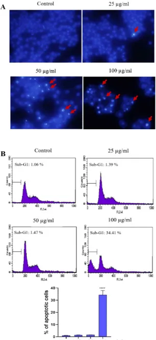
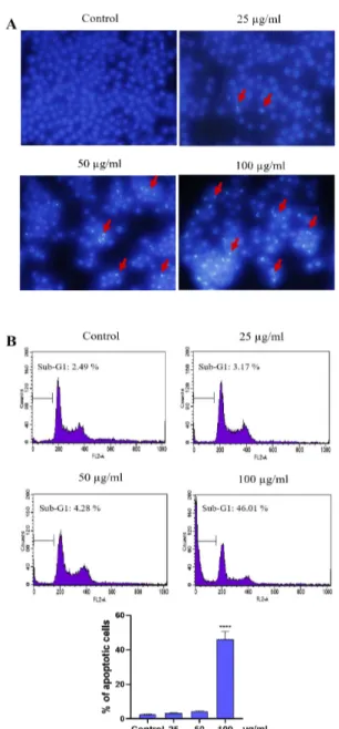
Next, we examined whether PME treatment in duces cell death in CT-26 and Hela cells using Hoechst 33343 staining. The morphological obse rvation in the cell nuclei of CT-26 and Hela cells showed morphological alterations including segr egated and apoptotic bodies formation in a dose-dependent manner (Figure 2A, 3A). And, we exa mined sub-G1 DNA content by treatment of PM E using flow cytometry. PME-treated both CT-2 6 and HeLa cells increased sub-G1 populations (apoptotic cells) compared with that of the contr ol, and the proportions of sub-G1 phase were recorded to 34.41% and 46.01%, respectively in 100 µg/mL PME treatment cells (Figures 2B, 3
B).
Figure 2. PME-induced apoptosis in CT-26 cells. The
cells were incubated with PME (25, 50, 100 µg/mL) for 24 h. (A) Microscopic images of cell apoptosis were measured by Hoechst 33342. The apoptotic cell s are characterized by brighter staining of chromatin condensation and fragments (arrows indicate apoptoti c cells). (B) The proportion of sub-G1 DNA producti on in CT-26 cells were analyzed by flow cytometry and the bar graph presenting the percentage of apopto tic cells. Data were presented as the mean ± SD. **** P < 0.0001.
Figure 3. PME-induced apoptosis in Hela cells. The
cells were incubated with PME (25, 50, 100 µg/mL) for 24 h. (A) Microscopic images of cell apoptosis were measured by Hoechst 33342. The apoptotic cell s are characterized by brighter staining of chromatin condensation and fragments (arrows indicate apoptoti c cells). (B) The proportion of sub-G1 DNA producti on in Hela cells were analyzed by flow cytometry and the bar graph presenting the percentage of apopto tic cells. Data were presented as the mean ± SD. **** P < 0.0001.
Discussion
So far, marine natural products have been rec-ognized as the most valuable candidate of phar-maceutical materials [30, 31]. Seaweeds pre-sented significant antiproliferative activity on various tumor cell lines [32-34]. However, no studies have been concerned to evaluate the anti-cancer activities of fractions or compounds iso-lated from P. morrowii. Previous study found that P. morrowii derived the 80% methanolic ex-tract (3-bromo-4,5-dihydroxybenzaldehyde, BDB) exhibited strong anti-IPNV and an-ti-IHNV activities [35]. The compound of BDB isolated from marine red alga, Rhodomela
con-fervoides, were found to have selective
anti-tumor activities against KB, Bel 7402 and A549 tumor cells [21]. Another study isolated a novel bromophenol [Bis (3-bromo-4,5-dihy-droxybenzyl) ether, BBDE] from P. morrowii which inhibited inflammation in RAW 264.7 macrophage cells [36].
In the present study, PME derived from a red algae of P. morrowii, was found to have in-hibitory effects on the growth of CT-26 and Hela tumor cells, and their IC50 values of 41.04%
(CT-26 cells) and 48.51% (Hela cells) also showed the anticancer properties. Antioxidant compounds such as polyphenols and flavonoids in seaweed are implicated with their anticancer activities [18]. Flavonoids can prevent cancer by affecting the signal transduction pathway of cell proliferation, antioxidant activity, regulating the enzyme activity related to estrogen biosynthesis and the metabolic pathway of carcinogens [37]. Polyphenolic compounds suppressed cancer cells by altering the metabolism of potential
carci-nogens through xenobiotic metabolic enzymes [17]. Another evidences indicated that poly-phenolic compounds change the metabolism of many potential carcinogens by activating a varie-ty of xenobiotic enzymes, interfering the telo-phase stage of mitosis, reducing the mitotic in-dex as well as colony forming unit of tumor cells [38, 39]. However, further studies are necessary to clarify the anticancer component(s) of PME in P. Morrowii and its mechanisms of inhibiting the growth of CT-26 and Hela cancer cells. Cell proliferation and apoptosis are two factor s that deciding the growth of cancer cells [40]. We speculated that the decrease of cell viability after treatment may be due to the PME-induced cell death or attribute to the inhibition of bioche mical or biological functions of cells exposed to PME. In this study, cells treated with PME prese nted typical features of early apoptosis which co uld observed as crescents around the nucleus, or the entire chromatin was appear as one or a grou p of or featureless bright blue spherical beads [41]. Furthermore, the proportion of sub-G1 phas
e (apoptotic cells) were significantly increased by treatment with PME at higher concentrations, our results demonstrated that PME treatment inhi bits the growth of cancer cells through increasin g the formation of apoptotic body and sub-G1
DNA population, eventually lead to subsequent apoptosis in CT-26 and Hela cells (Figure 2, 3). Our study results are similar to previous anticanc er studies [42] that have demonstrated the effect of colon cancer cells growth suppression by inhi biting nuclear transcription factors. Therefore, th is seaweed has potential as an anticancer effect, and is expected to be an important marine resour ce for anticancer function research in the future.
Conclusions
PME could remarkably inhibit the proliferatio n of CT-26 and Hela cancer cell lines in a dose-d ependent manner, which also induce the apoptosi s and increase their sub-G1 populations. These
results suggest that PME has anticancer activity and presents a promising prospect for research and development. However, the development of PME into a clinically effective anti-cancer drug requires further studies.
References
1. Bray, F., et al., Global cancer statistics 2018: GLOBOCAN estimates of incidence and mort ality worldwide for 36 cancers in 185 countrie s. CA Cancer J Clin, 2018. 68(6): p. 394-424. 2. Jung, K.W., et al., Cancer Statistics in Korea:
Incidence, Mortality, Survival, and Prevalenc e in 2015. Cancer Res Treat, 2018. 50(2): p. 303-316.
3. Jung, K.W., et al., Prediction of Cancer Incide nce and Mortality in Korea, 2019. Cancer Res Treat, 2019. 51(2): p. 431-437.
4. Arbyn, M., et al., Estimates of incidence and mortality of cervical cancer in 2018: a world wide analysis. The Lancet Global Health, 202 0. 8(2): p. e191-e203.
5. Monk, B.J., et al., Phase III trial of four cispla tin-containing doublet combinations in stage IVB, recurrent, or persistent cervical carcino ma: a Gynecologic Oncology Group study. J Clin Oncol, 2009. 27(28): p. 4649-55. 6. Johnson, C.A., et al., Cervical Cancer: An Ov
erview of Pathophysiology and Management. Semin Oncol Nurs, 2019. 35(2): p. 166-174. 7. Fridlender, M., Y. Kapulnik, and H. Koltai,
Plant derived substances with anti-cancer acti vity: from folklore to practice. Front Plant Sc i, 2015. 6: p. 799.
Modern Medicine from Natural Products. Mol ecules, 2016. 21(5).
9. Dyshlovoy, S.A. and F. Honecker, Marine Co mpounds and Cancer: 2017 Updates. Mar Dru gs, 2018. 16(2).
10. Khalifa, S.A.M., et al., Marine Natural Produc ts: A Source of Novel Anticancer Drugs. Mar Drugs, 2019. 17(9).
11. Courdavault, V., et al., Towards the Microbial Production of Plant-Derived Anticancer Drug s. Trends Cancer, 2020. 6(6): p. 444-448. 12. Lukasiewicz, K. and M. Fol, Microorganisms
in the Treatment of Cancer: Advantages and Limitations. J Immunol Res, 2018. 2018: p. 2397808.
13. Lowenthal, R.M. and J.H. Fitton, Are seaweed -derived fucoidans possible future anti-cancer agents? Journal of Applied Phycology, 2014. 27(5): p. 2075-2077.
14. Wali, A.F., et al., Natural products against can cer: Review on phytochemicals from marine sources in preventing cancer. Saudi Pharm J, 2019. 27(6): p. 767-777.
15. Wang, Y., et al., Simulated digestion and ferm entation in vitro with human gut microbiota of polysaccharides from Coralline pilulifera. Lwt, 2019. 100: p. 167-174.
16. Sithranga Boopathy, N. and K. Kathiresan, An ticancer drugs from marine flora: an overvie w. J Oncol, 2010. 2010: p. 214186. 17. Alghazeer, R., et al., Anticancer and Antioxidan
t Activities of Some Algae from Western Liby an Coast. Natural Science, 2018. 10(07): p. 232-246.
18. Yoshie, Y., et al., Compositional Difference of Phenolic Compounds between Two Seawe eds, Halimeda. Journal of Tokyo University of Fisheries, 2002. 88: p. 21-24.
19. Vasanthi, H.R., et al., Tumoricidal effect of the red algae Acanthophora spicifera on Ehrli ch’s ascites carcinoma in mice. Seaweed Rese arch and Utilization, 2004. 25: p. 217-224. 20. Yuan, Y. V., et al., Extracts from dulse (Palmari a palmata) are effective antioxidants and inhib itors of cell proliferation in vitro. Food Chem Toxicol, 2005. 43(7): p. 1073-1081.
21. Lijun, H., et al., Isolation and pharmacological activities of bromophenols from Rhodomela
confervoides. Chinese Journal of Oceanology and Limnology, 2005. 23(2): p. 226-229. 22. Geoffroy, A., et al., Patterns of genetic diversi
ty of the cryptogenic red alga Polysiphonia morrowii (Ceramiales, Rhodophyta) suggest multiple origins of the Atlantic populations. Ecol Evol, 2016. 6(16): p. 5635-47.
23. Piñeiro-Corbeira, C., H. Verbruggen, and P. Díaz-Tapia, Molecular survey of the red algal family Rhodomelaceae (Ceramiales, Rhodoph yta) in Australia reveals new introduced speci es. Journal of Applied Phycology, 2019. 32( 4): p. 2535-2547.
24. D’Archino, R., K.F. Neill, and W.A. Nelson, Recognition and distribution of Polysiphonia morrowii (Rhodomelaceae, Rhodophyta) in N ew Zealand. Botanica Marina, 2013. 56(1). 25. Kim, S.Y., et al., In vitro antiviral activity of
red alga, Polysiphonia morrowii extract and its bromophenols against fish pathogenic infe ctious hematopoietic necrosis virus and infect ious pancreatic necrosis virus. J Microbiol, 20 11. 49(1): p. 102-6.
26. Radonic, A., et al., Anionic Polysaccharides From Phototrophic Microorganisms Exhibit A ntiviral Activities to Vaccinia Virus. Journal of Antivirals & Antiretrovirals, 2010. 02(04). 27. Tannin-Spitz, T., et al., Antioxidant activity
of the polysaccharide of the red microalga Por phyridium sp. Journal of Applied Phycology, 2005. 17(3): p. 215-222.
28. Matsui, M.S., et al., Sulfated Polysaccharides from Red Microalgae Have Antiinflammatory Properties In Vitro and In Vivo. Applied Bioc hemistry and Biotechnology, 2003. 104(1): p. 13-22.
29. Jung, W.K., et al., A novel anticoagulant prote in from Scapharca broughtonii. J Biochem M ol Biol, 2002. 35(2): p. 199-205.
30. Gerwick, W.H. and B.S. Moore, Lessons from the past and charting the future of marine natu ral products drug discovery and chemical biol ogy. Chem Biol, 2012. 19(1): p. 85-98. 31. El Gamal, A.A., Biological importance of mar
ine algae. Saudi Pharm J, 2010. 18(1): p. 1-2 5.
32. Yuan, Y.V. and N.A. Walsh, Antioxidant and antiproliferative activities of extracts from a
variety of edible seaweeds. Food Chem Toxic ol, 2006. 44(7): p. 1144-50.
33. Paul, S. and R. Kundu, Antiproliferative activi ty of methanolic extracts from two green alga e, Enteromorpha intestinalis and Rizoclonium riparium on HeLa cells. Daru, 2013. 21(1): p. 72.
34. Murugan, K. and V.V. Iyer, Antioxidant and Antiproliferative Activities of Extracts of Sele cted Red and Brown Seaweeds from the Man dapam Coast of Tamil Nadu. Journal of Food Biochemistry, 2014. 38(1): p. 92-101. 35. Kang, S., et al., In vitro antiviral activities
of Korean marine algae extracts against fish pathogenic infectious hematopoietic necrosis virus and infectious pancreatic necrosis virus. Food Science and Biotechnology, 2008. 17: p. 1074-1078.
36. Choi, Y. K., et al., Bis (3-bromo-4,5-dihydroxy benzyl) ether, a novel bromophenol from the marine red alga Polysiphonia morrowii that suppresses LPS-induced inflammatory respon se by inhibiting ROS-mediated ERK signaling pathway in RAW 264.7 macrophages. Biome d Pharmacother, 2018. 103: p. 1170-1177. 37. Zhao, M., et.al., Immunomodulatory and antica
ncer activities of flavonoids extracted from lit chi (Litchi chinensis Sonn.) pericarp. Internati onal Immunopharmacology, 2007. 7(2): p. 16 2-166.
38. Gawron, A. and I. Kruk, Cytotoxic effect of xanthotoxol (8-hydroxypsoralen) on TCTC ce lls in vitro. Polish journal of pharmacology and pharmacy, 1992. 44 1: p. 51-7.
39. Zhao, M., et al., Immunomodulatory and antic ancer activities of flavonoids extracted from litchi (Litchi chinensis Sonn.) pericarp. Intern ational Immunopharmacology, 2007. 7(2): p. 162-166.
40. Tao, M.Z., et al., Effects of TGF-beta1 on the Proliferation and Apoptosis of Human Cervic al Cancer Hela Cells In Vitro. Cell Biochem Biophys, 2015. 73(3): p. 737-41.
41. Syed Abdul Rahman, S.N., N. Abdul Wahab, and S.N. Abd Malek, In Vitro Morphological Assessment of Apoptosis Induced by Antiprol iferative Constituents from the Rhizomes of Curcuma zedoaria. Evid Based Complement
Alternat Med, 2013. 2013: p. 257108. 42. Gwak, J., et al., Polysiphonia japonica extract
suppresses the Wnt/beta-catenin pathway in c olon cancer cells by activation of NF-kappaB. Int J Mol Med, 2006. 17(6): p. 1005-10.