ISSN 1225-6552, eISSN 2287-7630 https://doi.org/10.7853/kjvs.2021.44.1.45 < Case Report >
Veterinary Service
Available online at http://kjves.org*Corresponding author: Heui-Jin Kim, E-mail. [email protected] ORCID https://orcid.org/0000-0003-2577-1488
젖소 만성 창상성심낭염의 증례보고 및 고찰
김종호1,3ㆍ이경현1ㆍ노수권1ㆍ조헌호2ㆍ소병재1ㆍ김희진1
*
농림축산검역본부 질병진단과1, 부산광역시보건환경연구원2, 전북대학교 수의과대학3
A case study of chronic traumatic pericarditis
(Hardware disease) in a Holstein cattle
Jongho Kim1,3, Kyunghyun Lee1, Su Gwon Roh1, Heon-Ho Jo2, ByungJae So1, Heui-Jin Kim1*
1
Animal Disease Diagnostic Division, Animal and Plant Quarantine Agency, Gimcheon 39660, Korea
2
Busan Metropolitan City Institute of Health and Environment, Busan 46510, Korea
3
College of Veterinary Medicine, Jeonbuk National University, Iksan 54596, Korea
(Received 30 January 2021; revised 23 March 2021; accepted 23 March 2021)
Abstract
A 23-month-old Holstein cattle showed excess salivation and reluctance of walking and suddenly died after forced sudden movements. Grossly, numerous fibrous adhesions were present within cranial ab-dominal cavity including the reticulum and diaphragm and thoracic cavity involving lungs, pericardial sac, and heart. A perforation made by a 10 cm-long sharp-ended wire was detected in the reticulum. Histopathologically, fibrous suppurative epicarditis and myocardial necrosis were observed. Fibrosis with neovascularization were found in lungs, spleen, and liver. And granulomatous reticulitis was observed. For differential diagnosis, no pathogenic bacteria were detected through microbiological tests and PCR results were also negative for bovine susceptible pathogenic antigens. Based on the gross and histo-pathological examination, we diagnosed this case as chronic traumatic pericarditis. Cattle are inquisitive and prone to swallow various kinds of metallic foreign bodies since they do not use their lips. There-fore, avoiding ingestion of metallic objects in animal feed and animal areas by careful environmental management of farms is required and farmers should give the adequate minerals and vitamins into the feeds not to lick or shallow foreign bodies in case of mineral deficiency. For veterinary practitioners, physical examination, blood tests, and diagnostic imaging (X-ray and Ultrasonography) are required for an exact diagnosis. Furthermore, placing the magnets in rumen would be effective for prophylactic administrations.
Key words : Traumatic pericarditis, Metallic objects, Hardware disease, Holstein, Pathology
서 론
철물병(hardware disease)은 동물이 우연히 사료와 함께 철사, 못, 깡통과 같은 금속성 물질을 섭취한 뒤 발생하는 질환을 모두 포함하는 질병이며 창상성2위 복막염(Traumatic reticuloperitonitis, TRP)과 창상성심낭 염(Traumatic pericarditis, TP)이 발생할 수 있다(Anteneh
와 Ramswamy, 2015; Máinez 등, 2016; Braun 등, 2018). 대표적인 증상으로 상복부 통증, 경정맥노장, 식욕부 진, 과한 침흘림, 유량감소 및 기립불능 등이 있다 (Mohamed, 2010; Ziegler 등, 2013; Anteneh와 Ramswamy, 2015). TP의 경우 심낭염으로 인한 심외막과 심낭의 유착, 심낭 내 액체저류로 심장기능이 떨어져 울혈성 심부전(congestive heart failure)이 발생할 수 있고 금속 성 물질이 심장을 뚫은 경우 갑작스럽게 폐사할 수 있 다(Anteneh와 Ramswamy, 2015). 기존 연구결과에 따
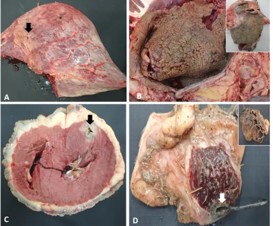
Fig. 1. Gross lesions at necropsy. (A) Yellow material (Black arrow) deposited on the lung surface. (B) Thick yellowish white material de-position on the pericardium and thick greenish yellow material de-position on the epicardial surface of heart (Inset). (C) Section of the heart showing 2 cm yellowish dis-coloration (Black arrow) inside the myocardium. (D) An adhesion be-tween the spleen and the reticulum (Inset, opposite side) penetrated by a sharp ended wire (White arrow).
12월에서 4월에 상대적으로 빈발하는 것으로 보고되 었다(Braun 등, 2018). 국내의 TRP 혹은 TP 발생률 조 사결과는 없었으나 도축과정에서 창상성제2위질병(trau-matic reticular diseases, TRD)의 분포 조사한 결과를 보면 5.5% (171/3,121) 정도로 확인되어 TRP의 발생률 과 큰 차이를 보이지 않는 것을 확인할 수 있었다 (Byeon 등, 2011). 반추동물은 벌집형태인 2위의 해부학적 특징 때문 에 2위 내에 금속성 물질이 잔존할 확률이 높으며 2 위가 횡격막 뒤에 위치하여 복압이 크게 상승하는 경 우 고정된 금속성 물질이 2위를 뚫고 상복강 장기 및 흉강 장기에 손상을 유발할 수 있다(Mohamed, 2010; Byeon 등, 2011). 소의 경우 호기심이 많고 볏짚 섭취 시 입술을 사용하지 못하여 양과 염소에 비해 금속성 물질을 상대적으로 쉽게 섭취하는 경향이 있다(Anteneh 와 Ramswamy, 2015). 본 증례에서는 젖소에서 발생한 철물병의 증상 및 병리학적 특징을 소개하고 이에 대해 고찰하여 철물 병에 대한 이해 및 대책을 세우는데 기여하고자 한다. 병력 및 육안병리학적 검사 2019년 12월 부산 강서구의 젖소 농가에서 23개월 령 육우 한 마리가 기립불능과 침흘림 증상을 보였다. 축주가 환축을 일으키기 위해서 밧줄로 견인을 시도 하였고 그 과정에서 급사하였다. 부검을 진행하였을 때 육안적으로 폐의 표면에 황색 막편(Fig. 1A, 화살 표)이 부착되어 있었고 폐엽 일부는 흰색의 증식된 물 질에 의해 심낭과 유착되어 관찰되었다(Fig. 1A, 1B). 심낭은 1 cm의 정도 두께의 흰색 조직이 다량 증식되 어 있었다(Fig. 1B). 심장 단면상 심외막에는 1 cm 정 도의 표면은 초록색을 띈 노란색 물질이 부착되어 있 었고, 심근에 직경 2 cm의 반점(Fig. 1C, 화살표)이 관 찰되었으며 중앙부는 비어있었다(Fig. 1C). 복강 장기 에서는 2위와 횡격막 주변에 흰색의 증식된 조직이 관찰되었고 철사에 의한 2위 천공을 확인할 수 있었 다. 천공된 주위 조직 주변으로 농성물질이 관찰되었 으며 2위에는 유착된 비장일부가 관찰되었다(Fig. 1D). 2위를 절개하였을 때 길이 10 cm 정도의 철사가 2개 관찰되었다. 그 외 장기에서 특이병변은 관찰할 수 없 었다.
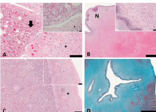
Fig. 2. Histopathological findings of the lesions. (A) Suppurative pneu-monia (black arrow, inset) and fib-rosis with neovascularization (as-terisk, inset) on lung surface. He-matoxylin and eosin stain (H&E stain). Scale bar=200 µm; inset scale bar=20 µm. (B) Myocardial necrosis (N) and magnification of myocardial necrosis (inset). H&E stain. Scale bar=2 mm; inset scale bar=100 µm. (C) Fibrosis with ne-ovascularization (asterisk, inset) on liver. H&E stain. Scale bar=200 µm; inset scale bar=100 µm. (D) Fibrosis (blue area) and myocar-dial necrosis around the injured myocardium. Masson’s trichrome stain. Scale bar=5 mm.
조직병리학적검사 및 병원체 감사
병리조직학적 검사를 위하여 조직을 10% 중성 완 충 포르말린에 고정하였으며 일반적인 조직처리 과정 을 거쳐 파라핀 포매한 후, 2 µm 두께로 조직절편을 제작하였다. Leica SelecTech reagents (Hematoxylin 560 and Alcoholic Eosin-Y 515; Leica Biosystems, Wetzlar, Germany)를 사용하여 헤마톡실린-에오신 염 색(hematoxylin-eosin staining)을 실시하였다. 또한, 조 직의 섬유화된 정도를 파악하기 위하여 흉강장기 및 상복부 실질장기를 대상으로 Roche Trichrome III Blue Staining kit (Roche, Basel, Switzerland)를 사용하 여 Masson’s trichrome 염색을 실시하였다. 세균 및 바 이러스 원인체 감별진단을 위해 육안병변이 있던 장 기를 대상으로 5% sheep blood agar (Asan Pharm. Co., Ltd., Seoul, South Korea)와 MacConkey agar (Becton Dickinson, Sparks, MD, USA)를 사용해 세균 분리를 시도하였고 주요 호흡기(Bovine herpesvirus 1, Bovine respiratory syncytial virus) 및 소화기(Bovine coronavi-rus, Bovine rotavicoronavi-rus, Bovine Viral Diarrhea virus) 바이 러스를 대상으로 LiliF IBR PCR kit, LiliF BD-Multi RT-PCR Kit (iNtRON Biotechnology, Seongnam, South Korea)를 사용하고 기존 문헌의 방법을 이용하여 유전 자 검사를 실시하였다(Alegre 등, 2001; Socha 등, 2009). 조직병리학적 검사결과 폐 흉막은 섬유 조직 증식
이 관찰되었으며, 폐포강 내에도 호중구(neutrophils)의 침윤(Fig. 2A, 화살표)이 관찰되었다(Fig. 2A). 심외막 의 육안소견에서 관찰된 노란색 막편은 지방 조직과 증식된 섬유 조직에 괴사된 호중구가 침윤되어 있었 고 심근내에 관찰된 노란색의 반점은 괴사된 심근 조 직에 호중구 등의 괴사된 염증세포가 침윤된 것으로 확인되었다(Fig. 2B). 천공된 2위는 대식세포(macro-phages)가 침윤되어 있었고 일부 비장 조직이 섬유 조 직을 통해 유착되어 있었으며 철사에 의한 2위 조직 괴사 및 손상부위를 조직학적으로 확인할 수 있었다. 간과 비장의 조직병리학적 검사결과 두 장기 모두 장 막에 섬유 조직 증식과 함께 혈관신생이 관찰되었다 (Fig. 2C). Masson’s trichrome을 이용한 특수염색 결과 괴사된 심근의 섬유 조직 증식 및 심외막의 섬유 조직 증식이 확인되었으며 흉막, 간과 비장 장막에서도 섬유 조직 증식을 관찰할 수 있었다(Fig. 2D). 감별진단을 위한 실험실적 검사 결과 병원성 세균 이 미분리 되었고 병원성 소 바이러스도 모두 검출되 지 않았다.
고 찰
본 증례는 조직병리학적으로 심근괴사 및 심한 섬2위의 육아종성2위염, 간과 비장의 장막부위 혈관신 생을 동반한 섬유화로 진단하였으며 부검소견 및 원 인체 검사 결과를 종합하여 창상성심낭염으로 최종진 단하였다. 여러 장기 장막 부위의 섬유화와 혈관신생 소견으 로 볼 때, 본 증례에서는 철사의 자극에 의해 만성적 으로 병변이 진행된 것임을 알 수 있었다. 육안적으로 병변이 관찰되지 않았던 간과 비장도 장막부위 섬유 화와 혈관신생을 확인할 수 있어 철사에 의한 오랜 자 극이 이 장기들에도 있었음을 추정할 수 있었다. 정확한 진단이 철물병의 빠른 치료와 무의미한 지지 치료(supportive care)를 방지하는 데 중요하다(Mohamed, 2010). 진단방법은 임상소견, 통증 검사(pain tests), 혈 청학적검사, 영상학적검사(방사선촬영, 초음파검사) 등 이 존재한다. 축주나 임상수의사들이 쉽게 사용할 수 있는 임상소견관찰과 pain test (pole test, pinching of the withers, pain percussion)의 경우, 기존 연구결과를 보았을 때 철물병의 진단률이 높지 않았다(Braun 등, 2007; Braun 등, 2018). 503건의 TRP 증례에서도 행동 에 있어 눈에 띄는 이상이 관찰된 증례는 13마리(2.6%) 에 불과했다(Braun 등, 2018). 28마리의 TP가 있던 증 례 중 대다수의 증례에서 자세의 이상이나 앞다리의 외전은 관찰되지 않았고 뚜렷하게 전신상태에 이상이 있던 개체는 4마리(14.3%)였으며 경정맥노장이 관찰 된 개체는 15마리(53.6%)였다(Braun 등, 2007). Pain test 와 같은 통증을 유도한 검사결과 TRP 증례에서 진단 율은 각각 43%, 39%, 24%이었고 40건의 버팔로 TP 증례의 통증검사 진단율은 10% 이내였다(Mohamed, 2010; Braun 등, 2018). 혈액을 이용한 혈액, 혈청학적 검사결과는 철물병 의 진단에 있어 도움이 된다 알려져 왔고 피브리노겐 (fibrinogen), 총혈장단백질(total plasma protein)이 증가 된다고 보고되었다(Anteneh와 Ramswamy, 2015; Braun 등, 2018). 하지만, 503두의 TRP 증례 분석에서 피브 리노겐과 총혈장단백질 증가는 각각 69%, 64% 확인 되었고 28건의 TP 증례 분석에서는 피브리노겐 증가 만 67.9% 확인되어 이런 혈청검사만으로 철물병을 진 단하기는 어려울 것으로 판단된다(Braun 등, 2007; Braun 등, 2018).
2위 주변 외측방사선촬영(lateral plain radiography) 와 초음파 진단은 각각 금속성물질의 확인과 섬유소 성 물질 및 농 축적 같은 병변을 확인하는데 유용할 수 있다(Khalphallah 등, 2016). 방사선 촬영의 경우 금 X-ray 투과성 물질의 경우 발견하기 어려운 단점이 있 고 염증성 변화에 의한 병변을 놓칠 수 있다는 단점이 있다(Khalphallah 등, 2016; Eo 등, 2017). 초음파 진단 의 경우 복수의 확인, 2위 형태나 윤곽 관찰을 통한 병변의 진행정도 파악 등이 용이한 장점이 있으나 2 위를 뚫은 이물을 찾기 어려운 단점이 존재한다 (Khalphallah 등, 2016). 각각의 진단법의 장점과 단점 이 존재하므로 각각의 방법을 종합적으로 활용한 진 단이 필요할 것으로 생각된다. TRD와 TRP의 경우라면 1위절개술(rumenotomy)과 항생제 처치 그리고 재발 방지를 위해 자석(magnet)을 투여하는 방법이 유용할 수 있다(Anteneh와 Ramswamy, 2015; Devi Prasad 등, 2017). 하지만 TP의 경우 예후 가 불량하고 치료에 대한 생존율도 좋지 않아 도태시 키는 것이 낫다고 알려져 있다(Braun 등, 2007). 철물병의 예방을 위해서는 소가 금속성 물질을 섭 취 못하도록 금속성 물질의 제거를 포함하는 주의 깊 은 축사관리가 중요하다. 국내 도축우를 대상으로 TRD 분포를 조사한 결과를 보면 다른 나라에선 보고 되지 않은 이물인 용접봉이 13.3%를 차지하여 축사를 신축하거나 개조할 경우 건축 부자재 특히 뾰족한 못 등에 대한 관리 등이 필요함을 시사했다(Byeon 등, 2011). 또한 미네랄과 비타민이 결핍된 경우 이물 섭 취가 증가할 수 있어 적절한 미네랄 블록과 비타민 공 급이 중요하다(Anteneh와 Ramswamy, 2015). 끝으로 철물병이 발생하기 쉬운 환경이거나 발생이 의심스럽 다면 예방적으로 자석을 위에 투여하는 것이 질병의 악화를 막는 방법이 될 수 있다(Anteneh와 Ramswamy, 2015).
결 론
기립불능과 침흘림 증상을 보인 젖소 한 마리가 밧 줄로 견인하는 과정에서 폐사하였다. 부검 및 조직병 리학적 검사 결과와 감염성 원인체 감별진단을 종합 하여 철물병에 의한 폐사로 진단하였다. 철물병의 진 단율과 예후에 관한 기존의 문헌을 참고하였을 때 정 확하고 빠른 대응을 위해서는 임상소견(병력)을 고려 한 신체검사, 혈청학적검사, 영상학적검사(방사선촬 영, 초음파검사)가 필요하며 질병 예방을 위해 농장 관리자들의 주의깊은 축사관리가 필수적이라 할 수 있다.감사의 글
본 증례보고는 농림축산검역본부 농림축산검역검사 기술개발 시험연구비(과제코드 B-1543069-2019-21-02) 의 지원을 받아 수행되었습니다.
CONFLICT OF INTEREST
No potential conflict of interest relevant to this article was reported.
ORCID
Jongho Kim, https://orcid.org/0000-0002-3719-860X Kyunghyun Lee, https://orcid.org/0000-0002-3113-2781 Su Gwon Roh, https://orcid.org/0000-0002-3597-6615 Heon-Ho Jo, https://orcid.org/0000-0002-5191-6663 ByungJae So, https://orcid.org/0000-0002-6125-6873 Heui-Jin Kim, https://orcid.org/0000-0003-2577-1488
REFERENCES
Alegre M, Nanni M, Fondevila N. 2001. Development of a Multiplex Polymerase Chain Reaction for the Differen-tiation of Bovine Herpesvirus‐1 and‐5. J Vet Med 48: 613-621.
Anteneh M. and Ramswamy V. 2015. Hardware Disease in Bovine (Review Article). Acad J Anim Diseases 4(3):
146-159.
Braun U, Lejeune B, Schweizer G, Puorger M, Ehrensperger F. 2007. Clinical findings in 28 cattle with traumatic pericarditis. Vet Rec 161: 558-563.
Braun U, Warislohner S, Torgerson P, Nuss K, Gerspach C. 2018. Clinical and laboratory findings in 503 cattle with traumatic reticuloperitonitis. BMC Vet Res 14: 66. Byeon HS, Park SG, Lee SM, Quak HK, Kwon KM, Ahn BW.
2011. A survey of traumatic reticular diseases in Korea and the effects on beef quality grade. Korean J Vet Res 51(2): 93-99.
Devi Prasad V, Ravi Kumar P, Harikrishna NVV, Bhagyaraju D. 2017. Traumatic reticulitis, reticulo-peritonitis and peri-carditis (Foreign body syndrome) in bovines. J Livestock Sci 8: 98-102.
Eo KY, Lee HH, Lee SK, Jung YM, Yeo YG, Ryu JS, Kang SG, Kwak D, Kwon OD. 2017. Traumatic pericarditis caused by a bamboo twig in captive waterbuck (Kobus ellipsi-prymnus). J Vet Med Sci 79(9): 1556-1558.
Khalphallah A, Abu-Seida AM, Abdelhakiem M, Elmeligy E, Mahmoud U. 2016. Laboratory, radiographic and ultra-sonographic findings of acute traumatic reticuloper-itonitis in buffaloes (Bubalus bubalis). Asian J Anim Vet Adv 11: 675-683.
Máinez M, Rosell J, Such R, Cardona T, Juan-Sallés C. 2016. Traumatic (foreign body) pericarditis in a Toco Toucan (Ramphastos Toco). J Zoo Wildl Med 47: 1097-1100. Mohamed T. 2010. Clinicopathological and ultrasonographic find-ings in 40 water buffaloes (Bubalus bubalis) with trau-matic pericarditis. Vet Rec 167(21): 819-824.
Socha W, Larska M, Rola J. 2009. Molecular characterisation of the first polish isolates of bovine respiratory syncytial virus. Bull Vet Inst Pulawy 53: 569-574.
Ziegler J, Parish S, Snekvik K, Barrington G. 2013. Traumatic gastroperitonitis (Hardware disease) in an alpaca (Vicugna pacos). J Zoo Wildl Med 44: 163-166.