저작자표시-동일조건변경허락 2.0 대한민국 이용자는 아래의 조건을 따르는 경우에 한하여 자유롭게 l 이 저작물을 복제, 배포, 전송, 전시, 공연 및 방송할 수 있습니다. l 이차적 저작물을 작성할 수 있습니다. l 이 저작물을 영리 목적으로 이용할 수 있습니다. 다음과 같은 조건을 따라야 합니다: l 귀하는, 이 저작물의 재이용이나 배포의 경우, 이 저작물에 적용된 이용허락조건 을 명확하게 나타내어야 합니다. l 저작권자로부터 별도의 허가를 받으면 이러한 조건들은 적용되지 않습니다. 저작권법에 따른 이용자의 권리는 위의 내용에 의하여 영향을 받지 않습니다. 이것은 이용허락규약(Legal Code)을 이해하기 쉽게 요약한 것입니다. Disclaimer 저작자표시. 귀하는 원저작자를 표시하여야 합니다. 동일조건변경허락. 귀하가 이 저작물을 개작, 변형 또는 가공했을 경우 에는, 이 저작물과 동일한 이용허락조건하에서만 배포할 수 있습니다.
Master's Thesis
in the Department of biomedical sciences
Senescent fibroblasts regulate
skin pigmentation
Ajou University Graduate School
Cancer Biology
Senescent fibroblasts regulate
skin pigmentation
Tae Jun Park, Advisor
I submit this thesis as the
Master's thesis in the Department of Biomedical Sciences
February, 2019
Ajou University Graduate School
Cancer Biology
Major
i
- Abstract –
Senescent fibroblasts regulate skin pigmentation
Cutaneous ageing is an important extrinsic process that modifies the pigmentary system. Because cellular senescence is a fundamental ageing mechanism, we examined the role of senescent cells in ageing pigmentation.
Methods: Biopsies obtained from senile lentigo and perilesional normal skin were
assayed for a marker of cellular senescence, p16INK4A. To determine the secretory phenotypes of senescent fibroblasts, we performed microarray, RNA sequencing and methylation array analyses in senile lentigo and senescent fibroblasts.
Results: In vivo, senescent fibroblasts accumulated at the sites of age-related
pigmentation. Phenotype switching of the cells resulted in the repression of stromal-derived factor 1 (SDF1) by promoter methylation. SDF1 induced melanocyte
differentiation via stromal-epithelial interactions, ultimately driving skin pigmentation. Furthermore, the elimination of senescent fibroblasts from pigmented skin using radiofrequency was accompanied by skin lightening, rendering it a potential target for treatment.
Conclusion: Aged pigmented skin contains an increasing proportion of senescent
fibroblasts. Cells with phenotype switching exhibited a loss of SDF1, which stimulates the melanogenic process and thereby contributes to ageing pigmentation. These data may promote the development of new therapeutic paradigms, such as a stroma-targeting therapy for pigmentary disorders.
ii
Table of contents
ABSTRACT‧‧‧‧‧‧‧‧‧‧‧‧‧‧‧‧‧‧‧‧‧‧‧‧‧‧‧‧‧‧‧‧‧‧‧‧‧‧‧‧‧‧‧‧‧‧‧‧‧‧‧‧‧‧‧‧‧‧‧‧‧ i TABLE OF CONTENTS ‧‧‧‧‧‧‧‧‧‧‧‧‧‧‧‧‧‧‧‧‧‧‧‧‧‧‧‧‧‧‧‧‧‧‧‧‧‧‧‧‧‧‧‧‧‧‧‧‧ ii LIST OF FIGURES ‧‧‧‧‧‧‧‧‧‧‧‧‧‧‧‧‧‧‧‧‧‧‧‧‧‧‧‧‧‧‧‧‧‧‧‧‧‧‧‧‧‧‧‧‧‧‧‧‧‧‧‧‧ iii LIST OF TABLES ‧‧‧‧‧‧‧‧‧‧‧‧‧‧‧‧‧‧‧‧‧‧‧‧‧‧‧‧‧‧‧‧‧‧‧‧‧‧‧‧‧‧‧‧‧‧‧‧‧‧‧‧‧‧ iii I. INTRODUCTION ‧‧‧‧‧‧‧‧‧‧‧‧‧‧‧‧‧‧‧‧‧‧‧‧‧‧‧‧‧‧‧‧‧‧‧‧‧‧‧‧‧‧‧‧‧‧‧‧‧‧‧‧‧ 1 II. MATERIALS AND METHODS ‧‧‧‧‧‧‧‧‧‧‧‧‧‧‧‧‧‧‧‧‧‧‧‧‧‧‧‧‧‧‧‧‧‧‧‧‧‧‧‧ 2
1. Cell culture ‧‧‧‧‧‧‧‧‧‧‧‧‧‧‧‧‧‧‧‧‧‧‧‧‧‧‧‧‧‧‧‧‧‧‧‧‧‧‧‧‧‧‧‧‧‧‧‧‧‧‧‧‧‧‧‧‧ 2 2. Enzyme-linked immunosorbent assay (ELISA) ‧‧‧‧‧‧‧‧‧‧‧‧‧‧‧‧‧‧‧‧‧‧‧‧‧‧‧ 2 3. Institutional review board statement ‧‧‧‧‧‧‧‧‧‧‧‧‧‧‧‧‧‧‧‧‧‧‧‧‧‧‧‧‧‧‧‧‧‧‧‧‧ 3 4. RNA sequencing ‧‧‧‧‧‧‧‧‧‧‧‧‧‧‧‧‧‧‧‧‧‧‧‧‧‧‧‧‧‧‧‧‧‧‧‧‧‧‧‧‧‧‧‧‧‧‧‧‧‧‧‧ 3 5. Senescence Associated β-Galactosidase (SA-β-Gal) Staining ‧‧‧‧‧‧‧‧‧‧‧‧‧‧‧‧‧‧ 3 6. Sequencing analysis of bisulfite-treated DNA ‧‧‧‧‧‧‧‧‧‧‧‧‧‧‧‧‧‧‧‧‧‧‧‧‧‧‧‧‧‧ 3 7. In vitro model of senescent fibroblasts ‧‧‧‧‧‧‧‧‧‧‧‧‧‧‧‧‧‧‧‧‧‧‧‧‧‧‧‧‧‧‧‧‧‧‧ 4 8. Promoter analysis ‧‧‧‧‧‧‧‧‧‧‧‧‧‧‧‧‧‧‧‧‧‧‧‧‧‧‧‧‧‧‧‧‧‧‧‧‧‧‧‧‧‧‧‧‧‧‧‧‧‧‧‧ 4 9. Ex vivo skin organ culture and pigmentation assays in cultured skin ‧‧‧‧‧‧‧‧‧‧‧‧ 5 10. Melanin contents and tyrosinase activity assay ‧‧‧‧‧‧‧‧‧‧‧‧‧‧‧‧‧‧‧‧‧‧‧‧‧‧‧‧ 5 11. Lentivirus production ‧‧‧‧‧‧‧‧‧‧‧‧‧‧‧‧‧‧‧‧‧‧‧‧‧‧‧‧‧‧‧‧‧‧‧‧‧‧‧‧‧‧‧‧‧‧‧‧ 6 12. Real-time PCR analysis ‧‧‧‧‧‧‧‧‧‧‧‧‧‧‧‧‧‧‧‧‧‧‧‧‧‧‧‧‧‧‧‧‧‧‧‧‧‧‧‧‧‧‧‧‧‧ 6 13. Western blot analysis ‧‧‧‧‧‧‧‧‧‧‧‧‧‧‧‧‧‧‧‧‧‧‧‧‧‧‧‧‧‧‧‧‧‧‧‧‧‧‧‧‧‧‧‧‧‧‧‧ 7 14. Immunocytochemistry and immunohistochemical analysis ‧‧‧‧‧‧‧‧‧‧‧‧‧‧‧‧‧‧ 7 15. Microscope image acquisition ‧‧‧‧‧‧‧‧‧‧‧‧‧‧‧‧‧‧‧‧‧‧‧‧‧‧‧‧‧‧‧‧‧‧‧‧‧‧‧‧‧ 8 16. Statistical analysis ‧‧‧‧‧‧‧‧‧‧‧‧‧‧‧‧‧‧‧‧‧‧‧‧‧‧‧‧‧‧‧‧‧‧‧‧‧‧‧‧‧‧‧‧‧‧‧‧‧‧ 8
III. RESULT ‧‧‧‧‧‧‧‧‧‧‧‧‧‧‧‧‧‧‧‧‧‧‧‧‧‧‧‧‧‧‧‧‧‧‧‧‧‧‧‧‧‧‧‧‧‧‧‧‧‧‧‧‧‧‧‧‧‧‧ 9
1. Senescent fibroblasts in senile lentigo ‧‧‧‧‧‧‧‧‧‧‧‧‧‧‧‧‧‧‧‧‧‧‧‧‧‧‧‧‧‧‧‧‧‧‧‧ 9 2. Senescent fibroblasts promote skin pigmentation‧‧‧‧‧‧‧‧‧‧‧‧‧‧‧‧‧‧‧‧‧‧‧‧‧‧‧ 14 3. SDF1 level was downregulated in senescent fibroblasts and senile lentigo‧‧‧‧‧‧‧ 19 4. Senescent fibroblasts involve SDF1 deficiency under promoter methylation ‧‧‧‧‧ 25 5. SDF1 and CXCR4 expression in skin cells ‧‧‧‧‧‧‧‧‧‧‧‧‧‧‧‧‧‧‧‧‧‧‧‧‧‧‧‧‧‧‧ 31 6. Effects of SDF1 in skin pigmentation‧‧‧‧‧‧‧‧‧‧‧‧‧‧‧‧‧‧‧‧‧‧‧‧‧‧‧‧‧‧‧‧‧‧‧‧ 34 7. CXCR4 inhibition by AMD3100 increased melanin synthesis ‧‧‧‧‧‧‧‧‧‧‧‧‧‧‧‧ 29 8. Effects of SDF1 in skin pigmentation‧‧‧‧‧‧‧‧‧‧‧‧‧‧‧‧‧‧‧‧‧‧‧‧‧‧‧‧‧‧‧‧‧‧‧‧ 39
IV. DISCUSSION ‧‧‧‧‧‧‧‧‧‧‧‧‧‧‧‧‧‧‧‧‧‧‧‧‧‧‧‧‧‧‧‧‧‧‧‧‧‧‧‧‧‧‧‧‧‧‧‧‧‧‧‧‧‧ 42 V. REFERENCES ‧‧‧‧‧‧‧‧‧‧‧‧‧‧‧‧‧‧‧‧‧‧‧‧‧‧‧‧‧‧‧‧‧‧‧‧‧‧‧‧‧‧‧‧‧‧‧‧‧‧‧‧‧‧ 46
iii
국문요약 ‧‧‧‧‧‧‧‧‧‧‧‧‧‧‧‧‧‧‧‧‧‧‧‧‧‧‧‧‧‧‧‧‧‧‧‧‧‧‧‧‧‧‧‧‧‧‧‧‧‧‧‧‧‧‧‧‧‧‧‧‧ 50
LIST OF FIGURES
▶ Figure 1. Cellular senescence in ageing pigmented skin ‧‧‧‧‧‧‧‧‧‧‧‧‧‧‧‧‧‧‧‧‧‧‧ 11 ▶ Figure 2. Cellular senescence in ageing pigmented skin. ‧‧‧‧‧‧‧‧‧‧‧‧‧‧‧‧‧‧‧‧‧‧ 13 ▶ Figure 3. Senescent fibroblasts model ‧‧‧‧‧‧‧‧‧‧‧‧‧‧‧‧‧‧‧‧‧‧‧‧‧‧‧‧‧‧‧‧‧‧‧‧‧ 15 ▶ Figure 4. Senescent fibroblasts promote skin pigmentation ‧‧‧‧‧‧‧‧‧‧‧‧‧‧‧‧‧‧‧‧ 17 ▶ Figure 5. Effect of senescent fibroblasts in melanocyte proliferation ‧‧‧‧‧‧‧‧‧‧‧‧‧ 18 ▶ Figure 6. Real-time PCR analysis of target genes ‧‧‧‧‧‧‧‧‧‧‧‧‧‧‧‧‧‧‧‧‧‧‧‧‧‧‧‧21 ▶ Figure 7. SDF1 expression level in senescent fibroblasts and senile lentigo ‧‧‧‧‧‧‧‧23 ▶ Figure 8. SDF1 promoter activity ‧‧‧‧‧‧‧‧‧‧‧‧‧‧‧‧‧‧‧‧‧‧‧‧‧‧‧‧‧‧‧‧‧‧‧‧‧‧‧‧ 26 ▶ Figure 9. SDF1 deficiency under DNA methylation ‧‧‧‧‧‧‧‧‧‧‧‧‧‧‧‧‧‧‧‧‧‧‧‧‧‧ 27 ▶ Figure 10. SDF1 expression regulation by p53 ‧‧‧‧‧‧‧‧‧‧‧‧‧‧‧‧‧‧‧‧‧‧‧‧‧‧‧‧‧‧ 29 ▶ Figure 11. NF-B signaling in senescent fibroblasts ‧‧‧‧‧‧‧‧‧‧‧‧‧‧‧‧‧‧‧‧‧‧‧‧‧ 30 ▶ Figure 12. Gene expression in normal skin cells ‧‧‧‧‧‧‧‧‧‧‧‧‧‧‧‧‧‧‧‧‧‧‧‧‧‧‧‧‧ 32 ▶ Figure 13. Fibroblast derived SDF1 inhibit melanogenesis ‧‧‧‧‧‧‧‧‧‧‧‧‧‧‧‧‧‧‧‧‧35 ▶ Figure 14. SDF1 decreased forskolin-induced cAMP levels in melanocytes ‧‧‧‧‧‧‧ 37 ▶ Figure 15. Influence of AMD3100 on the paracrine of melanocytes ‧‧‧‧‧‧‧‧‧‧‧‧‧ 38 ▶ Figure 16. The inhibitory effect of SDF1 on cutaneous pigmentation ‧‧‧‧‧‧‧‧‧‧‧‧ 40 ▶ Figure 17. SDF1 decreased forskolin-induced cAMP levels in melanocytes ‧‧‧‧‧‧‧ 41
List of Tables
▶Table 1. RNA sequencing analysis of senescent fibroblasts and controls ‧‧‧‧‧‧‧‧‧‧‧‧ 21 ▶Table 2. RNA array analysis of senile lentigo ‧‧‧‧‧‧‧‧‧‧‧‧‧‧‧‧‧‧‧‧‧‧‧‧‧‧‧‧‧‧‧‧‧‧‧ 24
1
I. INTRODUCTION
Pigmentation is an outcome of the interplay between melanocytes and neighboring cells such as keratinocytes and fibroblasts (Bastonini et al., 2016). UV radiation is a well-known stimulus that regulates this intricate process. Several paracrine factors secreted from these cells types upon UV exposure regulate melanogenesis, causing skin tanning and hyperpigmentation (Kim et al., 2016). Cutaneous ageing is another important extrinsic process that modifies the pigmentary system. Senile lentigo, also known as age spots, is one of the major changes associated with laxity and wrinkling during the ageing of skin. It is characterized by the presence of hyperpigmented spots in the elderly.
Cellular senescence is a fundamental ageing mechanism. Senescent cells and those with the related senescence-associated secretory phenotype (SASP) are known to be the main drivers of the age-related phenotype (Coppé et al., 2010; Tchkonia et al., 2013; Kim et al., 2017). During intrinsic and extrinsic skin ageing, the skin can contain senescent cells in epidermal and dermal compartments (Mine et al., 2008; Coppé et al., 2010; Velarde et al., 2016; Ghosh et al., 2016). Cellular senescence has been studied in dermal fibroblasts, which secrete factors that contribute to skin wrinkling (Waldera Lupa et al., 2015). For example, the chronic secretion of matrix metalloproteinases by senescent cells is an important contributor to the degradation of collagen and other extracellular matrix components in dermal tissue (Fisher et al., 2002).
A decrease in the expression of transforming growth factor type II receptor appeared to be a critical event in age-related skin thinning (Quan et al., 2001). However, despite the important role exerted by neighbouring cells on the regulation of melanocyte biology, few studies have examined how senescent cells are involved in skin pigmentation (Salducci et al., 2014; Kovacs et al., 2010),and it remains unclear whether senescent cells affect nearby epidermal melanocytes and influence ageing pigmentation.
Here, we show that in vivo, senescent fibroblasts accumulate at the sites of age-related pigmentation and that senescent fibroblasts alter melanocyte differentiation via stromal-epithelial interactions during ageing. We present an integrated study aimed at increasing our understanding of the key roles of senescent fibroblasts and their secretory phenotype in driving ageing pigmentation. These data might also promote the development of new therapeutic paradigms, such as a stroma-targeting therapy for pigmentary disorders.
2
II. MATERIALS AND METHODS
1. Cell culture
Normal human melanocytes, keratinocytes and fibroblasts were isolated from foreskin. In these experiments, melanocytes at passages 2-7 were maintained in F12 medium containing 10% heat-inactivated foetal bovine serum (FBS, Invitrogen, Carlsbad, CA), 24 μg/mL 3-isobutyl-1-methylxanthine, 80 nM 12-O-tetradecanoylphorbor 13-acetate, 1.2 ng/mL basic fibroblast growth factor and 0.1 μg/mL cholera toxin (All from Sigma-Aldrich, St. Louis, MO). Keratinocytes at passages 2-3 were grown in Epilife medium supplemented with human keratinocyte growth supplement (HKGS; Gibco-BRL, Bethesda, MD). Fibroblasts at passages 3-7 were maintained in Dulbecco's modified Eagle's medium (DMEM) (Gibco-BRL) supplemented with 10% FBS. SDF1-overexpressing or knockdown fibroblasts (3.5×10⁴) were seeded in the inserts of Transwell chambers (Corning, Tewksbury, MA), and melanocytes (1×10⁵) were seeded at the bottom of 6-well plates. After 24 h, the insert chambers were moved into the melanocyte-seeded 6-well plates, and the cultures were maintained in melanocyte culture medium for 5 days. The inserts with fibroblasts were changed to fresh ones at 3 days.
2. Enzyme-linked immunosorbent assay (ELISA)
Cells (1×10⁵) were seeded in 6-well plates and incubated for 48 h, after which the media were harvested. SDF1 secretion into the cell culture media was measured using SDF1 ELISA kits (R&D Systems, Minneapolis, MN) according to the manufacturer's instructions. To measure cAMP levels (R&D Systems), 1×10⁵ melanocytes were treated with recombinant human SDF1α (rhSDF1, 100 ng/mL, R&D System) for 24 h or infected with an SDF1-containing lentivirus for 2 days and then analysed to determine cAMP levels in the cell lysates. For the forskolin treatments, melanocytes were treated with rhSDF1 (0 to 200 ng/mL) 10 min prior to treatment with forskolin (700 ng/mL) and then cultured for 30 min. They were then analysed to determine the cAMP level. These experiments were
3
performed according to the manufacturer's instructions.
3. Institutional review board statement
This study was approved by the institutional review board of Ajou University Hospital (IRB numbers: AJIRB-DEV-DE3-15-491 and AJIRB-BMR-SMP-14-387). All participants were informed of the study goals and procedures, and signed written informed consent forms were obtained prior to their participation.
4. RNA sequencing
Total RNA was extracted from young, replicative senescent, sham irradiated and UVA-induced senescent fibroblasts using Macherey-Nagel RNA kits (Macherey-Nagel GmbH & Co. KG, Düren, Germany). Briefly, the sample quality was checked using a Bioanalyzer RNA chip (Agilent Technologies) and RNA sequencing was carried out with a Nextseq 500 device (Illumina, San Diego, CA). RNA sequencing data was deposited in SRA (SRP131607 and SRP131659).
5. Senescence Associated β-Galactosidase (SA-β-Gal) Staining
The cells or frozen tissue slides were fixed with 10% formalin for 1 min and then incubated with SA-β-Gal solution (X-gal, 1 mg/mL; citric acid/sodium phosphate, pH 5.8, 40 mM; potassium ferrocyanide, 5 mM; potassium ferricyanide, 5 mM; NaCl, 150 mM; MgCl2, 2 mM) for 12 h at 37 °C. After PBS washing, SA-β-Gal-positive cells were analyzed under light microscopy.
6. Sequencing analysis of bisulfite-treated DNA
The human SDF1 promoter region in genomic DNA isolated from young, replicative-senescent, sham-irradiated, UVA-induced senescent fibroblasts, perilesional normal skin and SL skin were sequenced after bisulfite treatment. The genomic DNA (1 μg) was treated with bisulfite, and PCR was then carried out. The PCR conditions were as follows: one
4
cycle at 94 °C for 3 min; 35 cycles at 94 °C for 30 s, 52 °C for 40 s, and 72 °C for 30 s; and one cycle at 72 °C for 3 min. Primers were designed for the SDF1 CpG island promoter (-741 to -477). The forward 5′-GTTTGTGATTAGTTTATTTTATTA-3′ and reverse 5′-CTAAATAAAAACCAATAAAAAAC-3′ sequences were used. The PCR product was ligated into blunt TOPO vectors (Mgmed, Seoul, Korea), and 10 (cells) or 5 (tissues) colonies were extracted and analysed by sequencing for CpG island methylation.
7. In vitro model of senescent fibroblasts
A primary culture of human dermal fibroblasts (HDFs) was prepared and maintained in our laboratory in DMEM (high-glucose) supplemented with 10% FBS. The number of population doublings (PDs) of HDFs was calculated based on the equation PDs log(A/BC)/log2, where A, B, and C indicate the number of collected cells, the number of plated cells and the attachment efficiency, respectively. The doubling time of HDF was measured with PDs, and the young cells used in this study represent cells with doubling times of approximately 24 h. The doubling time of replicative senescent fibroblasts was 14 days. For the UVA-induced senescent fibroblasts, HDF samples were washed once with PBS and placed in fresh PBS. The cells were treated with 8-methoxy-psoralen (8-MOP; Sigma-Aldrich) at 25 ng/mL for 16 h and irradiated with UVA (wavelength 320-400 nm, maximum peak 350 nm) using a LZC-1 photoreactor system (Luzchem Research Inc. Ontario, Canada). Several doses of UVA were tested according to methods described in previous reports (Salducci M et al., 2014; Kovacs D et al., 2010), and a dose of 5 J/cm2 was chosen for the experiment because it maintained the fibroblasts in a fully senescent state in which more than 75% show SA-β-Gal staining without cell death. Sham-irradiated HDF was rinsed and placed into an irradiator box without UV irradiation. After irradiation, the cells were maintained in DMEM for 7 days.
8. Promoter analysis
A reporter construct containing LEF/TCF luciferase was generated in our laboratory. A total of 6 copies of a response element were inserted into the lentivirus pGF1 vector
5
(pGF1-LEF/TCF, System Biosciences, Mountain View, CA) to generate lentivirus particles in HEK 293TN cells. Melanocytes were infected with LEF/TCF or with a control luciferase lentivirus. The cells were treated with 100 ng/mL of human recombinant SDF1α for 12 h, and luciferase activity was then analysed. The human SDF1 promoter (2 Kb) was cloned in our laboratory and inserted into a pGF1 vector (pGF1-SDF1, System Bioscience) to generate lentivirus particles in HEK 293TN cells. Young, replicative-senescent, sham-irradiated and UVA-sham-irradiated fibroblasts were infected with the SDF1 promoter or a control luciferase lentivirus for 3 days in the presence of puromycin (3.5 μM), and luciferase activity was then analysed using a Synergy 2 luminometer (BioTek, Winooski, VT) according to the luciferase reporter assay system instructions (Promega, Madison, WI). All luciferase assays were carried out in triplicate.
9. Ex vivo skin organ culture and pigmentation assays in cultured skin
Normal human skin samples obtained during minor skin surgery were placed on a sterilized stainless-steel grid in a culture dish containing DMEM supplemented with 5% FBS. The senescent fibroblasts were seeded in the bottom of the culture dish. After 3 days of culture in an incubator at 37 °C with 5% CO2, the specimens were fixed in 10%
formalin and embedded in paraffin before they were cut into sections. Melanin pigments were detected with Fontana-Masson staining. An image analysis was performed using Image Pro Plus Version 4.5 (Media Cybernetics Co., Rockville, MD), and the pigmented area per epidermal area (PA/EA) was measured.
10. Melanin contents and tyrosinase activity assay
The cells were lysed with a 0.1 M phosphate buffer (pH 6.8) containing 1% Triton X-100 with a protease inhibitor cocktail (Roche, Basel, Switzerland). The supernatants were measured to determine the protein concentration using the Lowry assay system. Pellets were solubilized in 100 μL of 1 N NaOH for 3 h at 60 °C and the absorbance was measured at 490 nm to determine the melanin content relative to a standard curve using
6
synthetic melanin (Sigma-Aldrich). For the assay of the tyrosinase activity, each sample was incubated with 2 mM L-DOPA (Sigma-Aldrich) in a 0.1 M phosphate buffer (pH 6.8) for 90 min at 37 °C. After incubation, the tyrosinase activity was measured at 490 nm.
11. Lentivirus production
The human SDF1 cDNA was cloned from normal fibroblasts in our laboratory. cDNA was inserted into the pCDH-CMV-MCS-EF1-Puro lentivirus vector (System Biosciences). For the knockdown of SDF1 expression, shRNA was prepared in a pLKO lentiviral vector (Sigma-Aldrich). Fibroblasts were plated and grown in 6 cm culture dishes. After culturing overnight, they were infected with the lentivirus and the cells were then selected with 3.5 μM puromycin for one week. The shRNA sequences were as follows: sh-SDF1 #1: 5′- CG CCAACGTCAAGCATCTCAAA-3′; sh-SDF1 #2: 5′- ACATCTCAAAATTCTCAACACA -3′.
To generate lentiviral particles, HEK-293TN cells were transfected with plasmid DNA (pGag-pol, pVSV-G, and pCDH-SDF1 or shSDF1) using Lipofectamine (Invitrogen). Viral supernatant was collected after 48 h and transduced into normal human melanocytes and fibroblasts.
12. Real-time PCR analysis
First-strand cDNA was synthesized by a reverse-transcription reaction using oligo-dT primers from 1 μg of total cellular RNA (Thermo Fisher Scientific, Waltham, MA). Real-time PCR was carried out with the Power SYBR Green PCR Master Mix (Bio-Rad, Hercules, CA) using the following conditions: initial activation at 95 °C for 5 min, followed by 40 cycles of 95 °C for 15 s and 60 °C for 1 min. The primers used for real-time PCR as follows: MITF: 5'-AGAACAGCAACGCGCAAAAGAAC-3', 5'-TGATGAT CCGATTCACCAAATCTG-3', Tyrosinase: CACCACTTGGGCCTCAA TTTC-3', 5'-AAAGCCAAACTTGCAGTTTCCAC-3', SDF1: 5'-TGCCAGAGCCAAC GTCAAG-3', 5'-CAGCCGGGCTACAATCTGAA-3', CXCR4: 5'-GCCTTATCCTGCC TGGTATTGTC-3', 5'-GCGAAGAAAGCCAGGATGAGGAT-TGGTATTGTC-3', human 18S: 5'-CGGCTACCACATCCAA
7
GGAA-3', 5'-GCTGGAATTACCGCGGCT-3', β-actin: 5'-CCCTGG CACCCAGCAC-3', GCCGATCCACACGGAGTAC-3', Apol6: CAGATTTGCTG CCACAGAG-3', 5'-GTGACATAGTCTGCCTTCTC-3', Col5a3: 5'-TTCAGCTCTTCT CGAGCGGGATTT-3', 5'-TCAAAGCCTCAGCACCAAATGCAC-3', LIF1: 5'-TATCACCATCTGTGCCTTTGC TGC-3', 5'-TCTGCCAGATTGTTCCTATGCCCA-3', p16INK4A: 5'-CCCAACGCACCG AATAGTTA-3', 5'-ACCAGCGTGTCCAGGAAG-3'.
13. Western blot analysis
Cells were lysed in RIPA buffer (1% NP-40, 150 mM NaCl, 10 mM Tris-HCl at pH 8.0, 1 mM EDTA) with a complete protease inhibitor (Sigma-Aldrich). The proteins were separated by SDS-polyacrylamide gel and transferred to PVDF membranes (Millipore, Billerica, MA). The antibody against MITF was purchased from Abcam (ab12039, Cambridge, UK), those for pCREB (9191s) and CREB (9197s) were purchased from Cell Signalling Technology (Danvers, MA), and the antibodies for tyrosinase (sc-7833) and p53 (sc-126) were purchased from Santa Cruz Biotechnology (Dallas, TX).
14. Immunocytochemistry and immunohistochemical analysis
For immunocytochemistry, cells grown in Lab-Tek chambers (Nalge Nunc International, Rochester, NY) were fixed in 4% paraformaldehyde for 15 min at room temperature and permeated with 0.2% Triton X-100. Non-specific antibody binding was blocked by 1% BSA for 1 h, and the cells were then incubated with adequate primary antibodies overnight at 4 °C. All fluorescence photographs were taken using a fluorescence microscope (Carl Zeiss, Oberkochen, Germany). Immunohistochemical staining was performed with primary antibodies on 4 μm-thick representative tissue sections obtained from formalin-fixed paraffin-embedded tissue samples in a Benchmark XT automated immunohistochemistry stainer (Ventana Medical Systems Inc., Tucson, AZ). The primary antibodies used were as follows: p16INK4A, predilution (725-4713, Ventana Medical Systems, Inc.); anti-human Ki67 antigen, clone MIB-1, 1:100 (Dako Denmark A/S, Glostrup, Denmark); CXCR4,
8
1:100 (MAB172, R&D System); SDF1, 1:100 (MAB350, R&D System); p53, 1:1000 (sc-126, Santa Cruz); cleaved caspase 3, 1:1000 (9661s, Cell Signaling); LIF1, 1:1000 (AF-250, R&D System); vimentin, 1:100 (ab92547, Abcam); FSP-1, 1:100 (ab27957, Abcam); and procollagen type 1, 1:50 (M-38, DSHB, Iowa city, IA). Detection was performed using a Ventana Optiview DAB Kit (Ventana Medical Systems). Immunohistochemical staining was scored by an experienced pathologist (JHK). For the SDF1 or procollagen expression analysis, SDF1- or procollagen-positive fibroblasts in two of the most highly labelled areas (hot fields) under 400x magnification were counted and graded as none (no SDF1-or procollagen-positive cells), weak (1-10 positive cells), moderate (11-30 positive cells), and strong (more than 30 positive cells).
15. Microscope image acquisition
Image acquisition during the histology and immunohistochemical staining processes was performed using a ScanScope® CS system (Aperio Technologies, Inc., Vista, CA) at room temperature. The cell images were acquired using an Olympus microscope mounted onto an Olympus DP70 digital camera with DP-Manager software (Olympus Microscope Corp., Tokyo, Japan) at room temperature. Immunofluorescence images were collected on a Zeiss LSM 510 microscope and analyzed with Zeiss Axio Imager software (Carl Zeiss) at room temperature.
16. Statistical analysis
Data are presented as the mean ± SD of independent determinations and were analysed using Wilcoxon or paired Student's t-tests with a p value < 0.05 considered significant. The statistical analysis of SDF1 staining in SL was performed using the Chi-square test (χ2 test). IBM SPSS ver. 23 (IBM Corp., Armonk, NY) was used for all statistical analyses.
9
III. Results
1. Senescent fibroblasts in senile lentigo
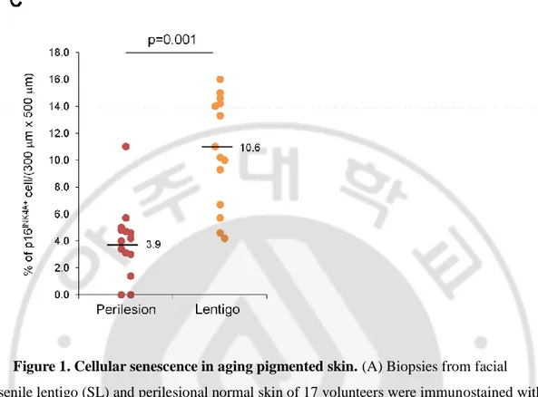
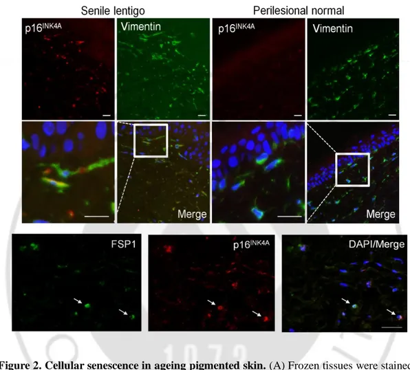
To characterize cellular senescence in ageing pigmented skin, biopsies obtained from the senile lentigo (SL) and perilesional normal skin of 17 volunteers were assayed for a marker of cellular senescence, p16INK4A (Dimri GP et al., 1995). A significantly higher number of p16INK4A-positive cells were observed in SL dermal skin than in perilesional normal skin (Figure 1A-C). The senescent cells were located near the dermo-epidermal junction, including the superficial dermis, but were rarely detected in the deep dermis (93.8 vs. 6.2%) (Figure 1B). Perilesional normal skin contained fewer p16 INK4A-positive cells than were observed in lesional skin but showed a similar distribution, with 92.3% of the positive cells in the superficial dermis. An in vivo marker of senescence, senescence-associated galactosidase (SA- β-Gal), was also observed in the corresponding p16INK4A-positive cells (Figure 2A). Because cellular senescence is characterized by stable cell cycle arrest, Ki67 immunostaining was performed and demonstrated an absence of proliferation in the p16INK4A-positive cells (Figure 2B). Ki67-positive cells were observed in the epidermis in both SL and perilesional normal skin samples. Double-immunostaining for p16INK4A/vimentin or p16INK4A/fibroblast-specific protein 1 (FSP1) revealed that the senescent cells were fibroblasts (Figure 2C and Figure 2D). These results indicate that ageing pigmented skin is characterized by the accumulation of senescent fibroblasts.
10
11
Figure 1. Cellular senescence in aging pigmented skin. (A) Biopsies from facial
senile lentigo (SL) and perilesional normal skin of 17 volunteers were immunostained with p16INK4A. Proportion of p16 INK4A -positive cells according to the depth of the skin. The bar graph indicates the percentage of p16 INK4A cells in the dermis. Superficial dermis is defined as within 500 mm of the epidermal-dermal junction. (B) The number of p16 INK4A -positive cells is presented as the dotted graph. Lines in the graph indicate the mean values. (p value, Wilcoxon test). (C) Senescent cells in senile lentigo (SL). Fourteen cases of SL were stained with p16 INK4A and positive spindle-shaped cells were counted and presented as a dotted graph (% of p16 INK4A -positive spindle-shaped cells/total spindle-shaped cells). Numbers indicate the mean of percentages in each case.
13
Figure 2. Cellular senescence in ageing pigmented skin. (A) Frozen tissues were stained
with SA-β-Gal/nuclear fast red (NFR) or p16INK4A (n=4). (B) Ki67 and p16INK4A co-immunostaining. Arrow heads and arrows indicate Ki67+/p16INK4A- keratinocytes and Ki67-/p16INK4A+ fibroblasts, respectively. (C) Immunostaining of vimentin and p16INK4A (n=6). (Scale bars, 50 mm). Senescent fibroblasts in senile lentigo and perilesional normal. Immunostaining of fibroblast-specific protein 1 (FSP1, green) and p16INK4A (red). The scale bar indicates 50 m.
14
2. Senescent fibroblasts promote skin pigmentation
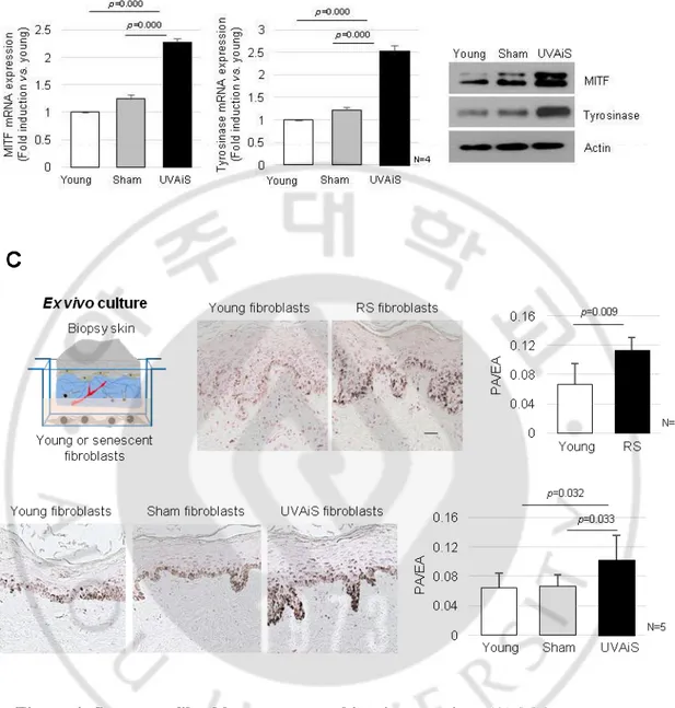
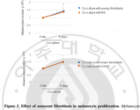
To investigate the regulatory role played by senescent fibroblasts in skin pigmentation, in vitro models of senescent fibroblasts, i.e., replicative-senescent (RS) and UVA-induced senescent (UVAiS) fibroblasts, were established (Figure 3) (Salducci M et al., 2014; Kovacs D et al., 2010; Kim HS et al., 2003). The senescent fibroblasts were then co-cultured with normal human melanocytes using Transwell plates. Melanin levels and tyrosinase activity were significantly higher in the senescent fibroblasts than in the controls (young or sham-irradiated fibroblasts) (Figure 4A). In addition, the mRNA and protein expression levels of melanogenesis-associated proteins, microphthalmia-associated transcription factor (MITF), and tyrosinase were significantly upregulated (Figure 4B). Melanocyte proliferation was unaffected by the senescent fibroblasts (Figure 5). The effects of the senescent fibroblasts on pigmentation were further evaluated using ex vivo human skin samples. Fontana-Masson staining that epidermal pigmentation was significantly increased by the presence of senescent fibroblasts (Figure 4C). An increase in the pigmented area/epidermal area (PA/EA) ratio was observed in an image analysis. Taken together, these data indicate that senescent fibroblasts promote skin pigmentation.
15
Figure 3. Senescent fibroblasts model. (A) Replicative senescent (RS) and
UVA-induced senescent (UVAiS) fibroblasts. Primary human fibroblasts were sub-cultured for 6 months or irradiated with 5 J/cm2 UVA or sham for 7 days, respectively. Two hundred cells were counted in each experiment and the numbers of SA-β-Gal positive fibroblasts are presented as percentages. Real-time PCR analysis of p16INK4A mRNA.
17
Figure 4. Senescent fibroblasts promote skin pigmentation. (A) Melanocytes were
co-cultured with senescent fibroblasts (RS or UVAiS) or control cells (Young or Sham). The melanin content and tyrosinase activity were measured. (B) Expression levels of mRNA and proteins of MITF and tyrosinase. (C) Pigmentation of ex vivo human skin visualized by Fontana-Masson staining (Left). The pigmented area/epidermal area (PA/EA) ratio was measured by an image analysis (Right). Data are mean ± SD (p value, t-test). (Scale bars, 50 mm).
18
Figure 5. Effect of senescent fibroblasts in melanocyte proliferation. Melanocytes
(1x10⁵) were co-cultured with senescent fibroblasts or controls for five days. The numbers of melanocytes were analyzed (left panel). BrdU incorporation assay. Melanocytes were co-cultured with senescent or young fibroblasts for three days and BrdU incorporation was analyzed by ELISA (middle panel). FACS analysis. Melanocytes were co-cultured with senescent or young fibroblasts for three days. The sub-G1 population was analyzed by FACS (right panel). (p value, t-test)
19
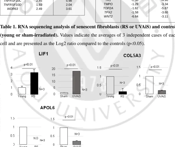
3. SDF1 level was downregulated in senescent fibroblasts and senile lentigo
To identify the secretory phenotype of the senescent fibroblasts that promoted pigmentation, a gene expression profiling analysis was performed using RNA sequencing. Among the 43 genes that were differentially expressed between the senescent fibroblasts and controls, 4 encoded secreted proteins: senescent fibroblasts expressed higher levels of LIF1 and lower levels of COL5A3, APOL6 and SDF1 than were observed in normal fibroblasts. (Table 1). The corresponding mRNA expression levels were detected by real-time PCR. (Figure 6). The full RNA sequencing data are available at the Sequencing Read Archive (SRA; SRP131607 and SRP131659). Of these, we selected stromal derived factor-1 (SDFfactor-1) as an intriguing candidate gene. SDFfactor-1, also known as C-X-C motif chemokine ligand 12 (CXCL12), is a widely expressed constitutive chemokine that regulates tissue homeostasis and inflammatory responses and is the only known ligand for C-X-C chemokine receptor type 4 (CXCR4) (Brenner M et al., 2009; Teixidó J et al., 2017; Zgraggen S et al., 2014) While no reports have directly linked SDF1 to pigmentation, SDF1 and its receptor CXCR4 are known to inhibit the cAMP signalling pathway, and cAMP signalling is also known to be involved in the production of melanin (Ruiz EJ et al., 2014). Thus, we investigated whether SDF1 may be one of the proteins responsible for age-related pigmentation.
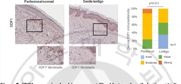
The expression of the SDF1 mRNA was suppressed in senescent fibroblasts but present at high levels in normal fibroblasts. ELISA data showed that normal fibroblasts but not senescent fibroblasts secreted a considerable level of SDF1 (Figure 7A). To further clarify SDF1 downregulation in vivo, biopsies obtained from SL and perilesional normal skin were assayed for SDF1 expression. Immunohistochemical staining detected very low levels of the SDF1 protein in a few fibroblasts obtained from the SL, whereas fibroblasts obtained from perilesional normal skin contained high levels of SDF1 expression (Figure 7B-C). Consistent with these results, the expression of the SDF1 mRNA was significantly lower in SL than in perilesional normal skin (Log2 ratio, -0.527) (Table 2). These data indicate that in ageing pigmented skin, senescent fibroblasts are deficient in SDF1. We also characterized the expression of 3 other secreted proteins identified by RNA sequencing
20
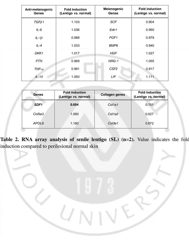
(LIF1, COL5A3, and APOL6) and genes known to control melanogenesis, such as melanocyte stimulating hormone (α-MSH), stem cell factor (SCF), and TGFβ, but none of these genes were differentially expressed. It is likely that relative weak changes in the LIF1, APOL6 and COL5A3 transcript expressions in SL skin samples compared to senescent fibroblasts are most likely due to their expressions in keratinocytes (Figure 6). However, collagen-related genes (COLA1, COLA2 and COL3A1) were down-regulated, as expected. The full microarray data are available at GEO (GSE109778).
21
Genes Log2 Ratio (Young vs. RS) Log2 Ratio (Sham vs. UVAiS) APLP1 1.65 3.10 BTG2 2.01 3.05 C12orf5 1.47 1.97 C1QTNF1 1.80 2.17 CYFIP2 2.83 4.22 DCBLD2 1.58 1.86 FUCA1 3.11 3.11 GDF15 2.39 5.47 GM2A 2.16 2.20 HIST1H2AC 2.67 3.24 HIST1H2BK 2.11 2.63 HIST2H2BE 2.51 2.74 LIF1 2.94 3.26 MDM2 1.41 2.31 MTRNR2L8 1.54 1.67 PARD6G 2.85 3.07 PSG2 2.43 3.21 SLC14A1 4.16 3.59 TNFRSF10C 2.45 5.98 TNFRSF10D 1.93 2.04 WDR63 2.49 3.61
Table 1. RNA sequencing analysis of senescent fibroblasts (RS or UVAiS) and controls (young or sham-irradiated). Values indicate the averages of 3 independent cases of each
cell and are presented as the Log2 ratio compared to the controls (p<0.05).
Figure 6. Real-time PCR analysis of target genes target genes. N.D : not detected. (p value, t-test)
Genes Log2 Ratio (Young vs. RS) Log2 Ratio (Sham vs. UVAiS) ANLN -1.21 -5.54 APOL6 -1.17 -3.28 ASPM -1.79 -5.50 BIRC5 -1.99 -5.40 C1R -2.28 -3.19 CDC20 -2.52 -5.72 CDKN2C -1.46 -3.52 CENPF -1.59 -5.30 COL5A3 -1.90 -3.39 SDF1 -3.55 -2.47 HR -3.43 -2.98 KIF18B -1.62 -4.76 KIF2C -2.01 -5.27 KIFC1 -2.12 -5.05 NUSAP1 -1.44 -5.82 OLFML2B -1.57 -3.37 PHGDH -1.80 -2.39 TENM3 -3.84 -2.17 TK1 -2.36 -4.90 TMPO -1.29 -3.34 TOP2A -1.62 -5.67 TPX2 -1.58 -3.60 WNT2 -6.64 -3.11
23
Figure 7. SDF1 expression level in senescent fibroblasts and senile lentigo. (A)
Real-time PCR and ELISA analyses of SDF1. N.D.: not detected. (B) SDF1 Immunohistochemical staining of SL and perilesional normal skin. SDF1 (red) were visualized (scale bars, 50 mm). (C) Immunohistochemical analysis of SDF1 expression level in perilesional and lentigo lesion skin. The scale bar indicates 50 m. (D) SDF1 immunostaining of SL (Scale bars: upper, 100 mm; lower, 50 mm). The expressions are graded as none, weak, moderate, or strong (p value, c2 test).
24 Anti-melanogenic Genes Fold induction (Lentigo vs. normal) Melanogenic Genes Fold induction (Lentigo vs. normal) TGF1 1.103 SCF 0.904 IL-6 1.036 Edn1 0.990 IL-1 0.988 FGF1 0.979 IL-4 1.033 BMP6 0.940 DKK1 1.017 HGF 1.027 PTN 0.969 NRG-1 1.055 TNF- 0.981 CSF2 0.917 IL-10 1.050 LIF 1.111
Table 2. RNA array analysis of senile lentigo (SL) (n=2). Value indicates the fold
induction compared to perilesional normal skin
Genes Fold induction
(Lentigo vs. normal) Collagen genes
Fold induction (Lentigo vs. normal)
SDF1 0.694 Col1a1 0.701
Col5a3 1.090 Col1a2 0.627
25
4. Senescent fibroblasts involve SDF1 deficiency under promoter methylation.
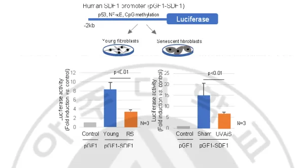
SDF1 promoter activity was lower in senescent fibroblasts than in control fibroblasts, indicating that SDF1 expression is transcriptionally regulated (Figure 8). It was previously reported that ageing causes distinct epigenetic changes in human skin and that age-related changes in DNA methylation contribute to the phenotypic changes associated with skin ageing (Grönniger E et al., 2010; Koch CM et al., 2011; Qian H and Xu X, 2014; Moulin L et al., 2017). DNA methylation is also recognized as an important mechanism involved in the regulation of SDF1 expression (Wendt MK et al., 2006).
A subsequent methylation-specific PCR analysis performed after bisulfite treatment showed that the CpG island methylation of the SDF1 promoter was markedly reduced in senescent fibroblasts (Figure 9A). The reduced SDF1 expression observed in the senescent fibroblasts was restored in the presence of a demethylating agent, 5-aza 2-deoxycytidine (5-Aza) (Figure 9B). DNA methylation of the SDF1 promoter was also observed in ageing pigmented skin. A methylation-specific PCR analysis showed that the level of methylation of the SDF1 promoter was significantly increased in SL (Figure 9C). SDF1 expression is regulated by several types of transcription factors, such as p53, NF-κB, SP1 and AP1, and p53 and NF-κB, which are commonly upregulated in senescent cells. p53 was found to suppress SDF1 production in cultured stromal fibroblasts, while NF-κB is an inducer of SDF expression (Moskovits N et al., 2006; Maroni P et al., 2007 Therefore, we also evaluated the ability of p53 and NF-κB to regulate SDF1 transcription in senescent fibroblasts (Figure 10 and 11). However, these data were excluded because the dermal fibroblasts in SL skin samples rarely expressed p53 and NF-κB was activated in senescent fibroblasts. Taken together, these findings indicate that in ageing-related pigmentation, senescent fibroblasts exhibit SDF1 deficiency as a result of changes in DNA promoter methylation.
26
Figure 8. SDF1 promoter activity. (A) The cells were treated with 100 ng/ml of human
recombinant SDF1 for 12 hr, and the luciferase activity was then analyzed. The human SDF1 promoter (2 kb) was cloned in our laboratory and inserted into a pGF1 vector (pGF1-SDF1, System Bioscience), generating lentivirus particles in HEK 293TN cells.
27
28
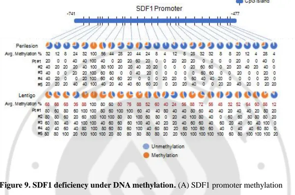
Figure 9. SDF1 deficiency under DNA methylation. (A) SDF1 promoter methylation
analysis in senescent fibroblasts (RS and UVAiS). Ten clones from each cell were analyzed 26 CpG human SDF1 promoter (-741 to -477) islands. Blue circles indicate an
unmethylated status and red circles denote methylation. The methyation % of each CpG island was presented and red numbers indicate more than 1.5 times increase in CpG island methylation. (B) Real-time PCR analysis of SDF1 mRNA in the presence of
5-aza-deoxycytidine (5-Aza). (C) DNA methylation status of the SDF1 promoter in SL patients (n=5). Pt # indicates the patient number.
29
Figure 10. SDF1 expression regulation by p53. The p53 expression levels were analyzed by western blotting in young, replicative and UVA-induced senescent fibroblasts (upper panel). Young fibroblasts were infected with Ad-LacZ (control) or a p53-expressing adenovirus (Ad-p53) for two days. The SDF1 mRNA expression levels were analyzed by real-time PCR. The lower panel shows the p53 immunostaining of SL skin samples (scale bar, 50 m).
30
Figure 11. NF-B signaling in senescent fibroblasts. Immunocytochemical staining of
p65 (RelA) localization (upper panel, scale bar, 10 m) and results of a western blotting analysis of IBa degradation (lower panel). (p value, t-test)
31
5. SDF1 and CXCR4 expression in skin cells.
The biological role of SDF1 in controlling skin pigmentation was then investigated. The endogenous expression of SDF1 and its receptor CXCR4 was examined in skin cells in vitro and in vivo (Figure 12A-C). The mRNA and protein expressions of SDF1 was strongest in the fibroblasts among the tested cells (Figure 12A-C). Melanocytes scarcely expressed SDF1, instead expressing CXCR4, indicating possible paracrine crosstalk between fibroblasts and melanocytes through SDF1. The SDF1/vimentin double-positive fibroblasts and melanocytes expressing SDF1 and CXCR4 were consistently observed in in
vivo normal human skin (Figure 12D). Keratinocytes scarcely expressed SDF1 or CXCR4 in vitro or in vivo.
33
Figure 12. Gene expression in normal skin cells. SDF1 and CXCR4 expressions in
cultured melanocytes (MC), keratinocytes (KC) and fibroblasts (FB). (A) Real-time PCR (B) ELISA, and (C) immunocytochemical (Scale bar, 10 mm) analyses. N.D., not detected. (D) Immunohistochemical staining of normal human skin. SDF1 (red)/vimentin (green) double-immunostained fibroblasts and CXCR4 (green)/MITF (red) stained melanocytes were visualized (scale bars, 50 mm).
34
6. Effects of SDF1 in skin pigmentation.
To investigate the paracrine effect of fibroblast-derived SDF1 on pigmentation, melanocytes were co-cultured with fibroblasts infected with the sh-SDF1 or SDF1 lentivirus using Transwell plates (Figure 13A). Melanin levels and tyrosinase activity were higher in the SDF1-knockdown fibroblasts than in the controls. The mRNA and protein expression levels of MITF and tyrosinase were significantly upregulated. (Figure 13B). Consistent with these results, SDF1-overexpressing fibroblasts exhibited decreased melanogenesis (Figure 13C). Inducing SDF1 overexpression or knockdown in fibroblasts did not affect melanocyte proliferation (Figure 14). To further investigate the effects of exogenous SDF1 on melanocytes, melanocytes were treated with recombinant human SDF1α (rhSDF1, 100 ng/mL). Exogenous SDF1 reduced pigmentation (Figure 13D), and this effect was reversed by treatment with a selective CXCR4 antagonist, AMD 3100 (Figure 15A). We further confirmed that SDF1 acts as a paracrine factor in melanocytes. Conditioned media (CM) was obtained from young or RS fibroblasts incubated with melanocytes in the presence or absence of AMD3100. The mRNA expression levels of MITF and tyrosinase were higher in melanocytes treated with AMD3100 and CM obtained from young fibroblasts. However, the senescent fibroblast-derived CM exerted no effect (Figure 15B).
36
Figure 13. Fibroblast derived SDF1 inhibit melanogenesis. (A) Fibroblasts were
infected with SDF1 and shSDF1 lenti-virus for 3days, and the SDF1 expression was analyzed by real time PCR. (B) Pigmentation of melanocytes co-cultured with fibroblasts infected with shSDF1 lentivirus or control. (C) Pigmentation of melanocytes co-cultured with SDF1-overexpressing fibroblasts. (D) Effect of recombinant human SDF1a (rhSDF1, 100 ng/ml) treatment on melanocytes.
37
Figure 14. SDF1 decreased forskolin-induced cAMP levels in melanocytes.
Melanocytes were treated with rhSDF1 (0 to 200 ng/ml) for 10 min before a forskolin treatment (700 ng/ml) for 30 min. The cAMP levels were then analyzed. (p value, t-test)
38
Figure 15. Influence of AMD3100 on the paracrine of melanocytes. Human primary
melanocytes were treated with CXCR4 antagonist AMD3100 (1 M) and co-cultured with human primary fibroblasts for three days. (A) The MITF and tyrosinase mRNA expression levels were analyzed by real-time PCR. (p value, t-test) (B) Melanocytes were cultured with conditioned media (CM) from young or RS fibroblasts with/without 1 mM of AMD3100 for 2 days and then analyzed for MITF and tyrosinase mRNA expression.
39
8. Effects of SDF1 in skin pigmentation.
Finally, the inhibitory effect of SDF1 on cutaneous pigmentation was explored in ex vivo human skin samples. In samples grown in the presence of rhSDF1, the amount of epidermal pigmentation was significantly reduced, as shown by Fontana-Masson staining (Figure 16A). Furthermore, in senescent fibroblasts, SDF1 overexpression reversed their stimulatory effects on melanogenesis (Figure 16B). To determine the molecular mechanism by which SDF1 inhibited melanogenesis, we next examined the SDF1/cAMP signalling pathway (Teixidó J et al., 2017). Treatment with rhSDF1 was associated with decreased levels of CREB phosphorylation (Figure 16C). Consistent with these results, treatment with rhSDF1 or SDF1 overexpression decreased cAMP formation (Figure 16C). Furthermore, rhSDF1 decreased forskolin-induced cAMP levels (Figure 17). These data suggest that SDF1 exerts an anti-melanogenic function by down-regulating cAMP/pCREB/MITF/tyrosinase signalling in melanocytes.
41
Figure 16. The inhibitory effect of SDF1 on cutaneous pigmentation. (A)
Pigmentation of ex vivo human skin treated with rhSDF1 (Scale bar, 100 μm). (B) SDF1 overexpression in senescent fibroblasts reversed their stimulatory effects on melanogenesis.
(C) Expressions of phospho-CREB and cAMP formation in melanocytes treated with
rhSDF1 or SDF1 lentivirus. Data are the mean ± SD (p value, t-test). N.S indicate not significance.
Figure 17. SDF1 decreased forskolin-induced cAMP levels in melanocytes.
Melanocytes were treated with rhSDF1 (0 to 200 ng/ml) for 10 min before a forskolin treatment (700 ng/ml) for 30 min. The cAMP levels were then analyzed. (p value, t-test).
42
IV. Discussion
Melanogenesis, the formation of melanin pigment, is regulated by cytokines and growth factors in a highly orchestrated manner (Bastonini et al., 2016). In this study, we defined what we believe to be a novel mechanism in which aged fibroblasts contribute to local regulation of melanogenesis. We show that the aged pigmented skin contains an increasing proportion of senescent fibroblasts.Phenotype switching of the cells resulted in the loss of SDF1 and SDF1 deficiency appears to be a potent stimulus of melanogenic process that contribute to the uneven pigmentation. These changes might be epigenetic and the hypermethylation of the SDF1 promoter was remarkably distinct in hyperpigmented skin compared to perilesional skin. Although what drives the initiation of this epigenetic change is not well understood, it has been known that the natural and photoageing-related epigenetic changes contribute to the phenotypic changes associated with skin ageing (Grönniger et al., 2010; Koch et al., 2011; Qian et al., 2014; Moulin et al., 2017).
Human skin, unlike other organs undergoes photoageing superimposed natural ageing process which attribute to ageing pigmentation (Moulin et al., 2017). Both processes are cumulative, and therefore, the most noticeable age-related changes may occur on superficial layer of the skin. The present study showed that cellular senescence occurs especially in the fibroblasts located in upper dermis of pigmented skin. The senescent fibroblasts were expected to influence melanocytes with cross-talk readily occurring through damaged basement membrane (Goyarts et al., 2007). We showed that the senescent fibroblasts played a stimulatory role in pigmentation and upregulated the expression of melanogenesis regulators, MITF and tyrosinase, in melanocytes, which are in agreement with the previous results (Salducci et al., 2014; Kovacs et al., 2010). Moreover, the impact of senescent fibroblasts on skin pigmentation was directly demonstrated when eliminating senescent cells with an intervention which reduced pigmentation. Together these findings suggest the crosstalk between melanocytes and senescent fibroblasts during ageing process plays an important role in the stimulation of melanogenesis and subsequent ageing-related pigmentation.
43
Shin et al., 2015) and had p16INK4A-positive keratinocytes in 10 of 25 samples (Shin et al., 2015). In the present study, p16INK4A-positive keratinocytes were observed in 5 of 17 samples. The number of p16INK4A-positive keratinocytes did not appear to be correlated with the number of p16INK4A-positive fibroblasts. It was suggested that SL is most likely initiated by keratinocyte proliferation, followed by the quiescence of enlarged senescent keratinocytes with a higher melanin burden (Shin et al., 2015; Chen et al., 2010). According to the literature, increased expressions of pigmentary proteins were consistently shown regardless of the degree of lentigo progression (Chen et al., 2010). We therefore speculate that SL is a disorder affecting both melanocytes and keratinocytes in which the melanocyte activation may be controlled by intricate interactions with senescent fibroblasts during the ageing process. Keratinocyte senescence may contribute to the increased epidermal thickness in SL cases. Nevertheless, it should be noted that a role of senescent keratinocytes in controlling pigmentation could not be completely ruled out and therefore requires additional investigations.
Growing data in the literature are focusing on the important role of melanogenic factors secreted from the senescent phenotype of fibroblasts in favoring the hyperpigmentation linked to ageing (Salducci et al., 2014; Kovacs et al., 2010; Chen et al., 2010, 36). In particular, the ability of these cells to release high amount of pro-pigmenting factors, such as HGF, KGF, SCF have highlighted, which could act in promoting the pigmentation. Moreover, crucial role of these factors in the development of age spots was demonstrated with immunohistochemical studies showing positive reactivates of KGF and/or HGF (Kovacs et al., 2010; Chen et al., 2010; Hasegawa et al., 2015). However, in our RNA sequencing data, none of these factors appear significantly modulated in the senescent cell models. The expressions of the melanogenic factors of HGF and KGF varied and were not consistent between replicative and UVA-induced senescent fibroblasts. Instead, only the LIF1 transcript was significantly upregulated in all samples. Moreover, many kinds of genes were differentially expressed in age spots, but melanocyte pigment genes were not among them (array data deposit in GSE109778), that is consistent with previous observation (Choi et al., 2017). Those results led us to hypothesize that the hyperpigmentation in age spots results from decrease of the expression of
anti-44
melanogenic factor, SDF1.
In the skin, dysregulation of SDF1 signal was previously linked to autoimmune and chronic inflammatory dermatological disorders, such as psoriasis, atopic dermatitis and lupus (Zgraggen et al., 2014). Studies of vitiligo animal models showed melanocyte-derived SDF1 plays an important role in the activation of melanocyte-specific autoimmunity resulting in continuous loss of the melanocytes (Speeckaert et al., 2017). The present study suggests that beyond the case of vitiligo, where an immune response is thought to be responsible for the destruction of normal melanocytes, SDF1 plays an inhibitory role in controlling cutaneous pigmentation in vivo. Among many cytokines affecting melanocytes, several (TGFβ1, IFN-γ, DKK1, and clusterin) are known to have hypopigmentation effects and are able to modulate the expression levels of tyrosinase and related enzymes independently (Lee et al., 2017; Natarajan et al., 2014). We found that normal fibroblasts secrete considerable amounts of SDF1 and SDF1 inhibits pigmentation of melanocytes expressing CXCR4. In ageing skin, the SDF1 level was markedly reduced and the SDF1 deficient ageing skin showed increased pigmentation. The role of fibroblast-derived SDF1 on cutaneous pigmentation in vivo requires further investigation. Nevertheless, the inhibitory action of SDF1 to control pigmentation suggests that SDF1 is a possible therapeutic target for the development of skin-lightening agents to treat hyperpigmentation. As the SDF1 expression was found to be epigenetically controlled and epigenetic modifications such as DNA methylation could be reversed, treatment involving senile lentigo using epigenetic “drugs” could be explored.
It would be noted that the cell culture model of senescent fibroblasts used in our study may not necessarily represent cells which impact melanogenesis in the facial area, as the fibroblasts are from the foreskins of neonates or young adults of skin phototype III or IV. It is also be noted that for induction replicative senescence, dermal fibroblasts were cultured at a high serum concentration to induce cell proliferation. This may not normally be seen in
in vivo, where they are in general quiescent cells (Mignon et al., 2017). Indeed, the ageing
process of dermal fibroblasts in their physiological tissue environment has only been partially elucidated in in vitro aged dermal fibroblasts (Tigges et al., 2014). Therefore, the intricate role of known melanogenic factors in promoting pigmentation independently of
45
the expression of SDF1 during the development of SL would be considered, and further investigations are necessary.
46
V. REFERENCES
1. Bastonini E, Kovacs D, Picardo M. Skin pigmentation and pigmentary disorders: Focus on epidermal/dermal cross-talk. Ann Dermatol. 2016;28:279-89
2. Kim M, Han JH, Kim JH, Park TJ, Kang HY. Secreted frizzled-related protein 2 (sFRP2) functions as a melanogenic stimulator; The role of sFRP2 in UV-induced hyperpigmentary disorders. J Invest Dermatol. 2016;136:236-44
3. Coppé JP, Desprez PY, Krtolica A, Campisi J. The senescence-associated secretory phenotype: The dark side of tumor suppression. Annu Rev Pathol. 2010;5:99-118
4. Tchkonia T, Zhu Y, van Deursen J, Campisi J, Kirkland JL. Cellular senescence and the senescent secretory phenotype: Therapeutic opportunities. J Clin Invest. 2013;123:966-72 5. Kim YH, Choi YW, Lee JH, Soh EY, Kim JH, Park TJ. Senescent tumor cells lead the
collective invasion in thyroid cancer. Nat Commun. 2017;8:15208
6. Mine S, Fortunel NO, Pageon H, Asselineau D. Aging alters functionally human dermal papillary fibroblasts but not reticular fibroblasts: A new view of skin morphogenesis and aging. PLoS One. 2008;3:e4066
7. Coppé JP, Patil CK, Rodier F, Krtolica A, Beauséjour CM, Parrinello S. et al. A human-like senescence-associated secretory phenotype is conserved in mouse cells dependent on physiological oxygen. PLoS One. 2010;5:e9188
8. Velarde MC, Demaria M. Targeting senescent cells: Possible implications for delaying skin aging. Gerontology. 2016;62:513-8
9. Ghosh K, Capell BC. The senescence-associated secretory phenotype: Critical effector in skin cancer and aging. J Invest Dermatol. 2016;136:2133-9
10. Waldera Lupa DM, Kalfalah F, Safferling K, Boukamp P, Poschmann G, Volpi E. et al. Characterization of skin aging-associated secreted proteins (SAASP) produced by dermal fibroblasts isolated from intrinsically aged human skin. J Invest Dermatol. 2015;135:1954-68
11. Fisher GJ, Kang S, Varani J, Bata-Csorgo Z, Wan Y, Datta S. et al. Mechanisms of photoaging and chronological skin aging. Arch Dermatol. 2002;138:1462-70
47
transforming growth factor beta by downregulating its type II receptor and inducing Smad7. J Biol Chem. 2001;276:26349-56
13. Salducci M, André N, Guéré C, Martin M, Fitoussi R, Vié K, Cario-André M. et al. Factors secreted by irradiated aged fibroblasts induce solar lentigo in pigmented reconstructed epidermis. Pigment Cell Melanoma Res. 2014;27:502-4
14. Kovacs D, Cardinali G, Aspite N, Cota C, Luzi F, Bellei B. et al. Role of fibroblast-derived growth factors in regulating hyperpigmentation of solar lentigo. Br J Dermatol. 2010;163:1020-7
15. Dimri GP, Lee X, Basile G, Acosta M, Scott G, Roskelley C. et al. A biomarker that identifies senescent human cells in culture and in aging skin in vivo. Proc Natl Acad Sci. 1995;92:9363-7
16. Kim HS, Song MC, Kwak IH, Park TJ, Lim IK. Constitutive induction of p-Erk1/2 accompanied by reduced activities of protein phosphatases 1 and 2A and MKP3 due to reactive oxygen species during cellular senescence. J Biol Chem. 2003;278:37497-510 17. Brenner M, Coelho SG, Beer JZ, Miller SA, Wolber R, Smuda C. et al. Long lasting
molecular changes in human skin after repetitive in situ UV irradiation. J Invest Dermatol. 2009;129:1002-11
18. Teixidó J, Martínez-Moreno M, Díaz-Martínez M, Sevilla-Movilla S. The good and bad faces of the CXCR4 chemokine receptor. Int J Biochem Cell Biol. 2017;95:121-31 19. Zgraggen S, Huggenberger R, Kerl K, Michael D. An important role of the
SDF-1/CXCR4 axis in chronic skin inflammation. PLoS One. 2014;9:e93665
20. Ruiz EJ, Oeztuerk-Winder F, Ventura JJ. A paracrine network regulates the cross-talk between human lung stem cells and the stroma. Nat Commun. 2014;5:3175
21. Grönniger E, Weber B, Heil O, Peters N, Stäb F, Wenck H. et al. Aging and chronic sun exposure cause distinct epigenetic change in human skin. PLoS Genet. 2010;6:e1000971 22. Koch CM, Suschek CV, Lin Q, Bork S, Goergens M, Joussen S. et al. Specific
age-associated DNA methylation changes in human dermal fibroblasts. PLoS One. 2011;6:e16679
23. Qian H, Xu X. Reduction in DNA methyltransferases and alteration of DNA methylation pattern associate with mouse skin ageing. Exp Dermatol. 2014;23:357-9
48
24. Moulin L, Cenizo V, Antu AN, André V, Pain S, Sommer P. et al. Methylation of LOXL1 promoter by DNMT3A in aged human skin fibroblasts. Rejuvenation Res. 2017;20:103-10
25. Wendt MK, Johanesen PA, Kang-Decker N, Binion DG, Shah V, Dwinell MB. Silencing of epithelial CXCL12 expression by DNA hypermethylation promotes colonic carcinoma metastasis. Oncogene. 2006;25:4986-97
26. Moskovits N, Kalinkovich A, Bar J, Lapidot T, Oren M. p53 attenuates cancer cell migration and invasion through repression of SDF-1/CXCL12 expression in stromal fibroblasts. Cancer Res. 2006;66:10671-6
27. Maroni P, Bendinelli P, Matteucci E, Desiderio MA. HGF induces CXCR4 and CXCL12-mediated tumor invasion through Ets1 and NF-kappaB. Carcinogenesis. 2007;28:267-79
28. el-Domyati M, el-Ammawi TS, Medhat W, Moawad O, Brennan D, Mahoney MG. et al. Radio frequency facial rejuvenation: Evidence-based effect. J Am Acad Dermatol. 2011;64:524-35
29. Seo KY, Yoon MS, Kim DH, Lee HJ. Skin rejuvenation by microneedle fractional radiofrequency treatment in Asian skin: Clinical and histological analysis. Lasers Surg Med. 2012;44:631-6
30. Na J, Zheng Z, Dannaker C, Lee SE, Kang JS, Cho SB. Electromagnetic initiation and propagation of bipolar radiofrequency tissue reactions via invasive non-insulated microneedle electrodes. Sci Rep. 2015;5:16735
31. Spandau DF, Lewis DA, Somani AK, Travers JB. Fractionated laser resurfacing corrects the inappropriate UVB response in geriatric skin. J Invest Dermatol. 2012;132:1591-6
32. Goyarts E, Muizzuddin N, Maes D, Giacomoni PU. Morphological changes associated with aging: Age spots and the microinflammatory model of skin aging. Ann N Y Acad Sci. 2007;1119:32-9
33. Yonei N, Kaminaka C, Kimura A, Furukawa F, Yamamoto Y. Two patterns of solar lentigines: A histopathological analysis of 40 Japanese women. J Dermatol. 2012;39:829-32
49
34. Shin J, Park JY, Kim SJ, Kang HY. Characteristics of keratinocytes in facial solar lentigo with flattened rete ridges: Comparison with melasma. Clin Exp Dermatol. 2015;40:489-94
35. Chen N, Hu Y, Li WH, Eisinger M, Seiberg M, Lin CB. The role of keratinocyte growth factor in melanogenesis: A possible mechanism for the initiation of solar lentigines. Exp Dermatol. 2010;19:865-72
36. Goorochurn R, Viennet C, Tissot M, Locatelli F, Granger C, Varin-Blank N. et al. Differential morphological and functional features of fibroblasts explanted from solar lentigo. Br J Dermatol. 2017;177:109-11
37. Hasegawa K, Fujiwara R, Sato K, Shin J, Kim SJ, Kim M. et al. Possible involvement of keratinocyte growth factor in the persistence of hyperpigmentation in both human facial solar lentigines and melasma. Ann Dermatol. 2015;27:626-9
38. Choi W, Yin L, Smuda C, Batzer J, Hearing VJ, Kolbe L. Molecular and histological characterization of age spots. Exp Dermatol. 2017;26:242-8
39. Speeckaert R, Ongenae K, van Geel N. Alterations of CXCL12 in serum of patients with vitiligo. J Invest Dermatol. 2017;137:1586-8
40. Lee J, Kim M, Park TJ, Kang HY. Fibroblast-derived clusterin negatively regulates pigmentation. J Invest Dermatol. 2017;137:1812-5
41. Natarajan VT, Ganju P, Singh A, Vijayan V, Kirty K, Yadav S. et al. IFN-γ signaling maintains skin pigmentation homeostasis through regulation of melanosome maturation. Proc Natl Acad Sci USA. 2014;111:2301-10
42. Mignon C, Uzunbajakava NE, Raafs B, Botchkareva NV, Tobin DJ. Photobiomodulation of human dermal fibroblasts in vitro: Decisive role of cell culture conditions and treatment protocols on experimental outcome. Sci Rep. 2017;7:2797 43. Tigges J, Krutmann J, Fritsche E, Haendeler J, Schaal H, Fischer JW. et al. The
50 -국문 요약 노화 섬유아세포의 색소 조절 기전 연구 피부 노화는 색소분비체계가 변형된 중요한 외부 과정이다. 그러한 이유로 근본적인 노화 기전을 노화 색소 침착에서 노화된 세포의 역할을 조사하였다. 방법 : 노인성 흑색종과 그 주변피부에서 얻은 생검에서 세포노화 표지자인 p16INK4A에 대하여 분 석하였다. 노화된 섬유 아세포에서 분비되는 표현형을 밝히기 위하여 노인성 흑색종과 노화된 섬 유 아세포에서 마이크로 어레이, RNA 시퀀싱, 그리고 메틸화 어레이 분석을 진행하였다. 결과 : In vivo 상에서 노화와 연관된 부위에 노화된 섬유아세포가 축적되어 있었다. 세포의 표현 형 변환으로 프로모터 메틸화에 의한 SDF1의 감소가 나타났다. 이에 따라 간질- 상피 상호작용을 통하여 멜라닌 세포의 분화가 유도되었고, 궁극적으로 피부 색소 침착을 유도했다. 결론 : 노화된 피부는 노화 된 섬유 아세포의 비율이 증가하여있다. 표현형 변환을 가진 세포는 SDF1의 발현이 감소되어 있었으며, 이는 멜라닌 생성과정에 작용하여 색소 침착을 감소시킨다. 이 자료는 색소 침착에 대하여 간질 표적치료와 같은 새로운 치료적 패러다임을 제시한다.