INTRODUCTION
Lignin is the second most abundant and aromatic rigid bio-polymer in the world. Lignin provides mechanical strength and structural support as a matrix component for the structure of lignocellulose chemically bonded with cellulose and hemi-cellulose. In the paper pulp industries, about 70 million tons year-1lignin as a co-product are generated (Satheesh et al. 2009). Lignin is composed of three different phenyl propane monomer units: p-coumaryl, coniferyl and sinapyl alcohol (Thevenot et al. 2010; Pandey et al. 2011). Structurally, lignin consists of aromatic rings with side chains and -OH and -OCH3groups which are linked by various strong cova-lent bonds such as alkyl-aryl ether and C-C bonds (Hirose et al. 2002). Lignin is an amorphous polymer that has graft-ing and crosslinkgraft-ing abilities. Thus, Lignin has been widely used in polymer composites and adhesive applications and
has served as a precursor in the synthesis of carbon and car-bon fibers (Hayashi et al. 2000; Jin et al. 2012; Hainal et al. 2012). It is supposed that about 30% of the organic carbon in the earth’s biosphere is generated from lignin and lignin structure ensures a high carbon level in the ratio of elements; the high utility of this high carbon content has motivated the application of lignin as a precursor in the production of car-bon fibers and carcar-bon nanotubes (Boerjan et al. 2003).
An electron beam is composed of a narrow stream of elec-trons in the form of waves; electron beams are widely used in the medical and polymer industries, pollutant detoxifica-tion, food manufacturing, and the agricultural fields (Ragan et al. 2011; Imbraguglio et al. 2012; Senna et al. 2012). In a recent study, electron beam irradiation was used as a phy-sical pretreatment process for bioethanol production. Further-more, ionizing radiation can easily penetrate the chemical structure of lignin to produce free radicals that can modify the lignin structure and break down dispensable bonds (Gonugunta et al. 2012).
In the present study, different absorbed doses of electron
Journal of Radiation Industry 7 (2~3) : 167~170 (2013)
─ ─ 167 ──
Electron Beam Effects on Lignin Stabilization during Carbonization
Byoung-Min Lee, Phil-Hyun Kang and Joon-Pyo Jeun*Research Division for Industry & Environment, Korea Atomic Energy Research Institute, Jeongeup 580-185, Korea
Abstract -- Lignin can be a valuable natural chemical resource. Structurally, lignin is a three-dimensional polymer made up of condensed C-C bonds and some ether linkages, most of which are not readily degraded. In this study, lignin carbonization under various electron beam pretreat-ment conditions was characterized through a thermogravimetric analysis (TGA), X-ray diffraction (XRD) and Raman spectroscopy. Lignin stabilization was controlled by various doses of electron beam irradiation corresponding to 50, 100, 200, 500 and 1,000 kGy; the carbonization process was performed under a nitrogen gas atmosphere at 1000��C for 1 h. The TGA results showed that a 1,000 kGy lignin dose increased the residue weight from 39.96% to 45.23%, compared to non-irradiated lignin. This observation is in agreement with the XRD and Raman spectroscopy results, in which the two theta degrees and the degree of crystallization were improved by increasing the electron beam irradiation.
Key words : Electron beam, Lignin, Carbonization, Irradiation
* Corresponding author: Joon-Pyo Jeun, Tel. +82-62-570-3063, Fax. +82-62-570-3098, E-mail. [email protected]
beam irradiation were used to study the influence of process-ing parameters on lignin carbonization. A thermogravimet-ric analysis (TGA) was used to study the effect on the degree of carbonization in the lignin, depending on the absorbed dose of the electron beam. The crystalline structure of the carbonized lignin was analyzed by X-ray diffraction (XRD) and Raman spectroscopy.
EXPERIMENTALS
MaterialsIn this study, alkali lignin is used as a starting material for the carbonization. Alkali lignin was purchased from Sigma Aldrich (Mw: 28,000).
Lignin Carbonization
Electron beam irradiation (EBI) was used to stabilize the lignin at EB-tech (Daejeon, Korea). EBI was carried out at the absorbed doses of 50, 100, 200, 500 and 1,000 kGy using a dose rate of 50 kGy scan-1at beam current of 30 mA and
beam energy of 1.14 MeV. The electron beam irradiated lignins were carbonized in a tubular furnace for 1 hour after the temperature reached to 1000�C at a heating rate of 10�C min-1from ambient temperature under a nitrogen atmosph-ere. And then, the tubular furnace was cooled at room tem-perature.
Characterization
The effect of an electron beam in thermally stabilizing lignin was investigated through a Thermogravimetric analy-sis (TGA) using a TA Instruments SDT-Q600 at a 10�C min-1 heating rate, over a temperature range of 40 to 1000�C by purging nitrogen gas. X-ray diffractometer (XRD, D/MAX-2500, Rigaku) was used to produce x-ray diffractograms of the carbon atoms and to determine the crystalline structure of carbonized lignin. The XRD analysis was carried out over a temperature range of 5�to 80�(2 theta) at a step speed of 3�min-1and a step size of 0.01�. The Raman spectra of the carbonized lignin were recorded with Raman spectrometer (Nanofinder 30, Tokyo Instruments) using 488 nm lasers. The peaks were fitted with Gaussian curves using a linear background obtained from the Raman spectra in the 500 to
2,000 cm-1region.
RESULTS AND DISCUSSION
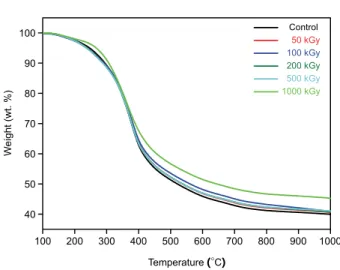
Thermogravimetric analysisFig. 1 shows the thermal properties of stabilized lignin under a nitrogen atmosphere. These results indicate that a large weight loss occurred at approximately 350�C which was mainly due to lignin degradation. Meanwhile, the residue weight increased from 39.96% to 45.23% owing to an increase in the irradiated dose of the electron beam from 0 to 1000 kGy. Before carbonization, the lignin stabilized at doses of 1000 kGy and higher doses improved the thermal stability (Cho et al. 2007).
X-ray diffraction analysis
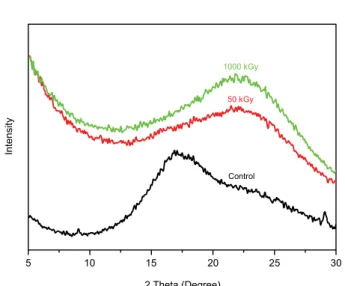
X-ray diffraction analysis can be used to characterize hard carbons and crystal structure. In Fig. 2, The appearance of broad (002) diffraction peak at 22�was evidence for short range ordering of adjacent aromatic rings, which make dis-tribute a preferential interatomic distance between carbon atoms. It can be observed that increasing with absorbed doses of electron beam irradiation leads to gradually increase the intensities of (002) diffraction peak. This indicates that the electron beam irradiation has been attributed to the existence of both of highly ordered graphite and less ordered graphite (Jones et al. 1991).
Byoung-Min Lee, Phil-Hyun Kang and Joon-Pyo Jeun 168
Fig. 1. TGA thermograms measured under nitrogen gas for lignin
Raman spectra
Raman spectrometry is used to characterize a carbon mate-rial because of the drastically spectral changes not only due to the kind of available allotropic carbon but the well-defined structural changes of the each allotrope. Raman spectra of the carbonized lignin are shown in Fig. 3. In general, two sharp peaks of G-band around 1,580 cm-1and D-band around
1,350 cm-1were appeared in carbon materials. The first-order
region of the Raman spectrum at approximately 1,580 cm-1
(G-band) can be attributed to the E2gmode of the graphite lattice vibration. The D-band appeared at approximately 1,350 cm-1, corresponding to the A
igvibration mode, which can be attributed to lattice defects, edges of graphite layers, and structurally disordered carbons (Kim et al. 2000). The resulting Raman spectra all exhibit bands at approximately 1,350 and 1,580 cm-1; upon increasing the absorbed dose of
the electron beam, the 1,350 cm-1band remained almost
con-stant, whereas the 1,580 cm-1band shifted toward a lower
frequency. In the second-order region of the spectra, from 2,200 to 3,400 cm-1, carbonized lignin exhibited bands at
2,700 cm-1and 2,900 cm-1, resulting from the change in the
absorbed dose are shown in Fig. 3(B). These bands were an over tone of the D′-band and a combination of the D-band and D′-band. Generally, highly crystalline samples cause the band at 2,700 cm-1to split into two bands. The spectra were used to calculate the intensity ratio of the D to G-band (ID/IG), which decreased with an increase of the stabilization absorb-ed dose of the electron beam (Fig. 4). Thus, radiation treat-Electron Beam Effects on Lignin Stabilization during Carbonization 169
Fig. 2. X-ray diffractograms for carbonized lignin at 1000�C at heat-ing rates of 10�min-1following stabilization by an electron
beam.
Fig. 4. The ratio of the intensity of the D-band versus the intensity
of the G-band from the raman spectra of carbonized lignin under the same conditions as in Fig. 2.
Fig. 3. Raman spectra of carbonized lignin in (A) the first-order
re-gion and (B) the second-order rere-gion, under the same condi-tions as in Fig. 2.
ment resulted in a decrease in the disorder amount and num-ber of defects (Rodriguez-Mirasol et al. 1996; Ishimaru et
al. 2007; Panapoy et al. 2008; Song et al. 2009).
CONCLUSION
We reported the effects of electron beam irradiation on the stabilization component of lignin carbonization. TGA results showed that the residue weight of the stabilized lignin increased by approximately 5%, depending on the absorbed dose of the electron beam, as high radiation energy produced thermally stable lignin. During carbonization, the lignin struc-ture transformed from the phenolic compound to graphite in stages depending on the carbonization temperature, caused by the previous electron beam irradiation step. The XRD and Raman analyses indicated that the degree of graphite increas-ed with the amount of electron beam treatment. Remarkable increases in the intensity of the (002) diffraction peaks were observed, and the ratio of the intensity of the D-band to the intensity of the G-band of ratio also increased. The degree of carbonization can be easily controlled by electron beam irradiation.
ACKNOWLEDGEMENTS
This study was supported by the Nuclear R&D program of the Korea Science and Engineering Foundation, which is funded by the Ministry of Science, ICT and Future Planning of the Republic of Korea.
REFERENCES
Boerjan W, Ralph J and Baucher M. 2003. Lignin Biosynthesis. Annu. Rev. Plant Biol. 54:519-546.
Cho CW, Cho D, Ko YG, Kwon OH and Kang IK. 2007. Stabi-lization, carbonization, and characterization of PAN precur-sor webs processed by electrospinning technique. Carbon Letters 8:313-320.
Gonugunta P, Vivekanandhan S, Mohanty AK and Misra M. 2012. A Study on Synthesis and Characterization of Bio-based Carbon Nanoparticles from Lignin. World J. of Nano Sci. & Eng. 2:148-153.
Hainal AR, Capraru AM, Volf I and Popa VI. 2012. Lignin as a carbon source for the cultivation of some Rhodotorula
species. Cellulose Chem. Technol. 46:87-96.
Hayashi J, Kazehaya A, Muroyama K and Watkinson AP. 2000. Preparation of activated carbon from lignin by chemical activation. Carbon 38:1873-1878.
Hirose T, Fujino T, Fan T, Endo H, Okabe T and Yoshimaru M. 2002. Effect of carbonization temperature on the structural changes of woodceramics impregnated with liquefied wood. Carbon 40:761-765.
Imbraguglio D, Giovannozzi AM, Nastro A and Rossi AM. 2012. Submicron machining and biomolecule immobilization on porous silicon by electron beam. Nanoscale Research Letters 7:1-8.
Ishimaru K, Hatu T, Bronsveld P, Meier D and Imamura Y. 2007. Spectroscopic analysis of carbonization behavior of wood, cellulose and lignin. J. Mater. Sci. 42:122-129.
Jin XJ, Yu ZM and Wu Y. 2012. Preparation of activated carbon from lignin obtained by straw pulping by KOH and K2CO3 chemical activation. Cellulose Chem. Technol. 46:79-85. Jones LE and Thrower PA. 1991. Influence of boron on carbon
fiber microstructure, physical properties, and oxidation be-havior. Carbon 29:251-269.
Kim YM, An KL, Kim C, Choi YO, Park SH, Yang KS and Lee WE. 2000. Raman spectroscopical evaluations of carboniza-tion and graphitizacarboniza-tion of coal tar pitch. Carbon Science 1: 22-26.
Panapoy M, Dankeaw A and Ksapabutr B. 2008. Electrical con-ductivity of PAN-based carbon nanofibers prepared by elec-trospinning method. Thamasat Int. J. Sc. Tech. 13:11-17. Pandey MP and Kim CS. 2011. Lignin Depolymerization and
Conversion: A Review of Thermochemical Methods. Chem. Eng. Technol. 34:29-41.
Ragan S and Megonnell N. 2011. Activated carbon from renew-able resources - lignin. Cellulose Chem. Technol. 45:527-531.
Rodriguez-Mirasol J, Cordero T and Rodriguez JJ. 1996. High-temperature carbons from kraft lignin. Carbon 34:43-52. Satheesh Kumar MN, Mohanty AK, Erickson L and Misra M.
2009. Lignin and its applications with polymers. J Biobased Mater. Bioeng. 3:1-24.
Senna MMH, Moneam YKA, Hakiem AAA and Said HM. 2012. Characterization of plasticized maize starch/chitosan blends irradiated with an electron beam. J. Polym. Res. 19:1-11. Song C, Wang T, Qiu Y, Qiu J and Cheng H. 2009. Effect of
carbonization atmosphere on the structure changes of PAN carbon membranes. J. Porous Mater. 16:197-203.
Thevenot M, Dignac MF, Rumpel C. 2010. Fate of lignins in soils: A review. Soil Biol. & Biochem. 42:1200-1211.
Manuscript Received: October 25, 2013 Revised: November 1, 2013 Revision Accepted: November 21, 2013 Byoung-Min Lee, Phil-Hyun Kang and Joon-Pyo Jeun