INTRODUCTION
The management of colorectal cancer, especially that of rectal cancer, has changed in many aspects during the last few decades (1-3). Previous studies based on patients’ clini-cal outcome and hospital data showed that a surgeon’s oper-ation volume and the level of the hospital influenced the treatment outcome, and the surgeon’s operation volume has been suggested to be the most important contributing fac-tor to the outcome (4-8). The volume-outcome association is more pronounced in rectal cancer than in colon cancer due to the high complexity of rectal surgery (6). Although the difference in surgical procedures has been known to be the most important influencing factor (6), more detailed differ-ences have not been well evaluated. In the present study, therefore, we attempted to unravel the differences of rectal cancer management and then to compare the differences, based on the surgeons and the hospitals.
MATERIALS AND METHODS
Questionnaires describing preoperative evaluation, opera-tive procedure, and postoperaopera-tive surveillance in 39 major
categories were sent out to the members of the ‘Korean Soci-ety of Coloproctology’, including the surgeons at 30 univer-sity hospitals, in August 2004. The surgeons who operated rectal cancer in their hospitals were requested to respond to the questionnaires. Sixty responses were received during the three months’ period. After the data were coded blindly into the database, the results were compared according to the sur-geon’s annual operation volume, the level of the hospital, and the surgeon’s age. Statistical analyses were performed by chai-square test using SPSS 11.0�(SPSS Inc., Chicago, IL, U.S.A.).
RESULTS
Respondents
Thirty three respondents (55.0%) operated more than 50 rectal cancer cases (high-volume surgeons), and 37 respon-dents (61.6%) worked at university hospitals or in other ter-tiary care facilities (high-level hospitals) (Fig. 1). Twenty res-pondents (33.3%) were 40-yr old or younger (young surge-ons), and the respondents’ age was not significantly different according to the surgeon’s operation volume or the level of the hospital.
Sun-Il Lee, Yoon-Ah Park*, Seung-Kook Sohn*
Department of Surgery, Korea University College of Medicine, Seoul; Department of Surgery*, Yonsei University College of Medicine, Seoul, Korea
Address for correspondence Seung-Kook Sohn, M.D.
Department of Surgery, Yongdong Severance Hospital, Yonsei University College of Medicine, 146-92 Dogok-dong, Gangnam-gu, Seoul 135-720, Korea
Tel : +82.2-2019-3370, Fax : +82.2-3462-5994 E-mail : sksohn@yumc.yonsei.ac.kr
*This study is supported by a Korea University Grant.
S86
A Survey on the Impact of Operation Volume on Rectal Cancer
Management
The rectal cancer management can be influenced by the surgeon’s practice and the hospital. This study was to evaluate the differences according to the surgeon’s operative volume and the level of the hospital. Questionnaires were sent out to the members of the ‘Korean Society of Coloproctology’, and the responses were eval-uated according to the surgeon’s operation volume, the surgeon’s age, and the level of the hospital. Sixty responses were received during the three months’ peri-od (from August to October 2004). Thirty three respondents (55%) operated more than 50 cases of rectal cancer per year (high-volume surgeons), and 37 respon-dents (61%) worked at university hospitals or tertiary care facilities (high-level hos-pitals). The preoperative evaluation with endorectal ultrasonography (ERUS) was significantly different according to the surgeon’s operation volume and the level of the hospital, whereas magnetic resonance imaging and positron emission tomogra-phy (PET) was significantly different only for the surgeon’s operation volume. The preoperative radiation therapy was significantly different according to the surgeon’s operation volume, the surgeon’s age, and the level of the hospital. However, there was no significant difference found on the operative procedures or postoperative surveillance. The preoperative loco-regional evaluation and the preoperative radi-ation therapy could be considered as the factors that influence the volume-outcome relationship in rectal cancer treatment.
Key Words : Rectal Neoplasm; Surgery; Preoperative Care; Postoperative Care; Korea
Received : 11 January 2007
Preoperative evaluation
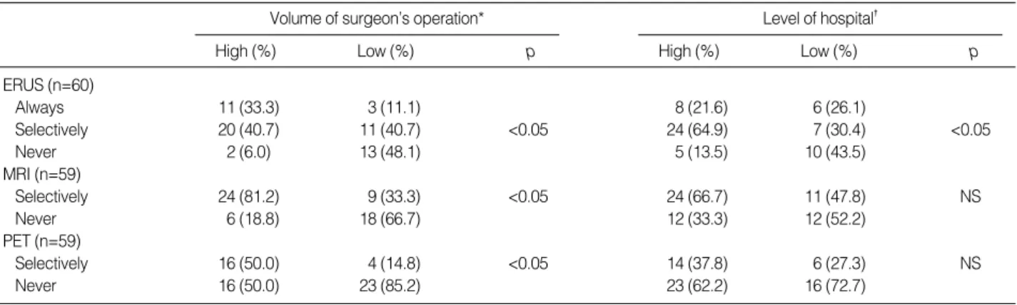
Preoperative rigid sigmoidoscopy was used routinely by 40.0% of the respondents and selectively by 36.6% of the respondents, whereas 23.3% did not use the examination. Colonoscopy, however, was performed routinely by 88.3% of the respondents and selectively by 10.0% of the respon-dents. The surgeons participated in colonoscopic examina-tion alone or together with endoscopist at 50% of the respon-dents’ hospitals. Only 21.7% of the respondents routinely used barium enema, whereas 16.6% did not use this exami-nation for preoperative evaluation. One respondent had a limitation for usage of magnetic resonance imaging (MRI) and positron emission tomography (PET) during their eval-uation, while others performed the examinations without limitation in their hospital or neighboring institution. Liver imaging by computerized tomography (CT) was done by 96.7% of the respondents, and abdominal ultrasound was additionally used by 30% of the respondents. Significant differences were found in endorectal ultrasound (ERUS),
pelvic MRI, and 18F-fluorodeoxyglucose-PET: ERUS and MRI were significantly different according to the surgeon’s operation volume, and ERUS was also significantly different according to the level of the hospital (Table 1). The selective use of PET was significantly different according to the sur-geon’s operation volume (Table 1).
Preoperative radiation therapy
Preoperative radiation therapy was given fractionatedly in 4 to 6 weeks with the radiation dose ranging from 4,500 to 5,500 cGy. Forty six respondents (76.7%) performed preop-erative radiation therapy, which was significant according to the surgeon’s operation volume (90.9% of high-volume sur-geons vs. 22.2% of low-volume sursur-geons), the level of the hospital (89.1% of high-level hospitals vs. 56.5% of low-level hospitals), and the surgeon’s age (95% of young surgeons vs. 67.5% of aged surgeons). As for the purpose of preoperative radiation therapy, 80.4% of the 46 surgeons performed it for the purpose of sphincter preservation.
Operation
Total mesorectal excision (TME) was considered as the principle in rectal cancer surgery by 96.6% of the respon-dents, and 68.9% of them performed it only for mid and low rectal cancer. Inferior mesenteric artery was ligated at its root routinely by 26.6% and selectively by 68.3% of the respon-dents. Rectal irrigation was performed before dividing below a tumor by 81.6% of the respondents. During an operation, 2 cm of distal mucosal resection margin was considered as safe by 62.0% of the respondents; and 1 cm or less by 29.3% of the respondents. Paraaortic lymph node dissection was performed routinely (5%), selectively (75%), or never (20%); and lateral pelvic lymph node dissection was performed selec-tively (93.3%) or never (6.6%). Coloanal anastomosis was performed by 88.3% of the respondents, and the most
pre-Number of surgeons 20 15 10 5 0 <11 11-30 31-50 51-100 >100
Annual operative volume (cases/yr) High-level hospital
Fig. 1.Distribution of surgeons according to operation volume and level of hospital. High-level hospital: university hospitals or tertiary care facilities.
Low-level hospital
Volume of surgeon’s operation*
High (%) Low (%) p Level of hospital� High (%) Low (%) p ERUS (n=60) Always 11 (33.3) 3 (11.1) 8 (21.6) 6 (26.1) Selectively 20 (40.7) 11 (40.7) <0.05 24 (64.9) 7 (30.4) <0.05 Never 2 (6.0) 13 (48.1) 5 (13.5) 10 (43.5) MRI (n=59) Selectively 24 (81.2) 9 (33.3) <0.05 24 (66.7) 11 (47.8) NS Never 6 (18.8) 18 (66.7) 12 (33.3) 12 (52.2) PET (n=59) Selectively 16 (50.0) 4 (14.8) <0.05 14 (37.8) 6 (27.3) NS Never 16 (50.0) 23 (85.2) 23 (62.2) 16 (72.7)
Table 1.Preoperative evaluation of rectal cancer according to the surgeon volume and the level of hospital: endorectal ultrasound (ERUS), pelvic magnetic resonance imaging (MRI), and positron emission tomography (PET)
*High volume, who performs more than 50 operations of rectal cancer cases annually; �High level, university hospitals and tertiary care facilities. NS, not significant.
ferred type was straight form (77.4%), J-pouch (15.1%) and coloplasty (7.5%). Diverting stoma was performed routinely (20.8%) or selectively (54.7%) during coloanal anastomosis, however, 24.5% did not fashion diverting stoma during co-loanal anastomosis.
Operative procedures were not significantly different accor-ding to the surgeon’s operation volume, the age, or the level of the hospital.
Postoperative management
Adjuvant chemotherapy was given by surgeons in 73.3% of the respondents’ hospitals and by medical oncologists in 15.0% of the respondents’ hospitals. In 11.7% of the respon-dents’ hospitals, chemotherapy was given by surgeons or medical oncologists according to the patients. Postoperative radiation therapy was given according to the stage: 21.6% of the respondents routinely for T3N0 and 50.0% for T3N1 or T3N2.
Carcinoembryonic antigen (CEA), abdominopelvic CT, chest radiography, and colonoscopy were the main surveil-lance tools after operation. There were no significant differ-ences in the use of these tools according to the surgeon’s opera-tion volume, the level of the hospital, or the surgeon’s age (Table 2). Colonoscopy was performed annually (66%) or biannually (28%) by 95% of the respondents for the fllow-ing five years after operation. On the other hand, rigid sig-moidoscope was used only by 43.3% of the respondents. While the regular follow-up during the 5 yr postoperative period was not significant, the regularity of follow up after the initial 5 yr was significantly different showing 90% of high-volume surgeons and 62% of low-volume surgeons regularly performed it every one or two years (p<0.05).
DISCUSSION
This study examined the differences in the rectal cancer management by the surgeons in Korea. Rectal cancer has been
increasing in Korea, including 5,865 new cases reported in 2002. We strongly believe that more than 4000 operations of rectal cancer cases are performed annually by the surgeons who are included in this study. Therefore, considering resec-tability (9), it is quite possible that this study covers more than 70% of the rectal cancer operations in Korea.
The previous studies with oncologic outcome defined the minimal volume for the experienced surgeon as one or two operations per month, and the cutoff value of other studies was about 15 cases or less (4-7). On the other hand, the stud-ies with operative technique and outcome defined the cutoff value as 50 or 75 operations per year (10-12). In this study, the high operation volume was defined as more than 50 ations per year, and the majority of the surgeons (75%) oper-ated more than 30 rectal cancer cases per year. It was possi-ble that may surgeons with a low annual volume were not included in this study.
Recent advances in the management of rectal cancer include improved preoperative pelvic imaging, preoperative chemo-radiation therapy, and improved rectal dissection (1, 2, 9, 13). In the present study, significant differences have been found in the preoperative evaluation with ERUS, MRI, and PET. ERUS has been used for more than 20 yr and is known to be accurate for the early stage. On the other hand, MRI is more informative for the advanced stage and especially for the mesorectal fascia which is important for keeping safe circumferential resection margin during pelvic dissection (14-17). PET is increasingly used for both local evaluation and distant metastasis (18). In the present study, these pre-operative evaluations have been found to be different accord-ing to the surgeon’s operation volume, yet only ERUS has been found to be significantly different according to the level of the hospital.
There were general agreements among the surgeons on the operative procedure (more than 75% of the respondents agreed): TME as the primary technique in rectal cancer sur-gery; selective use of paraaortic and pelvic lymph node dis-section; rectal irrigation; and coloanal anastomosis for sphinc-ter preservation. On the other hand, the operation of upper rectal cancer was controversial among the surgeons, which was also noted in other studies (1, 19-21). Controversies on diverting stoma for low rectal cancer operation (20, 22, 23) were also noted in this study. Regardless of the general agree-ments or the controversies, a significant difference was not found according to the surgeon’s operation volume, the sur-geon’s age, or the level of the hospital.
Unlike the operative procedure, preoperative radiation therapy was significantly different according to the surgeon’s operation volume, the level of the hospital, and even the sur-geon’s age, and it was the only significant factor according to the surgeon’s age. Postoperative surveillance varied among surgeons and was performed differently according to the patient’s condition. Colonoscopy was frequently used rather than sigmoidoscopy because of the comparable expense of CT, computed tomography; CEA, carcinoembryonic antigen.
Surgeons (%) Examinations
(per year) First 2 yr 3 to 5 yr
Abdominal CT Once 42 81 Twice 52 12 Chest X-ray Once 19 47 Twice or more 76 45 CEA Once 1 26 Twice or more 98 70
Table 2.Postoperative surveillance until the 5th postoperative year
these medical examinations in Korea (24). The surveillance system is affected by socioeconomical factors or by insurance systems, which could be different according to the country. These days, intensive surveillance is increasing for the early detection of metastatic or recurrent diseases, and the exami-nations were found more frequent in this study than in other recommendation (25).
This study was conducted by using a questionnaire, and several limitations were present. The questionnaire was com-pleted by the surgeons themselves and many low-volume surgeons did not reply. All the complexities and the individ-ualized treatments could not be assessed only by this ques-tionnaire. The relationship between the surgeon’s actual prac-tice and the clinical outcome could not be proved with this study. Nevertheless, the surgeon’s intention and behavior during the treatment of rectal cancer have accurately been evaluated by this study, and the recent trends of rectal can-cer operation were also revealed.
In conclusion, preoperative loco-regional evaluation was significantly different according to the surgeon’s operation volume, and preoperative radiation therapy was significant-ly different according to the surgeon’s operation volume and the level of the hospital. These results could be considered as the factors to influence volume-outcome relationship in rectal cancer treatment.
REFERENCES
1. MacFarlane JK, Ryall RD, Heald RJ. Mesorectal excision for rectal
cancer. Lancet 1993; 341: 457-60.
2. Hool GR, Church JM, Fazio VW. Decision-making in rectal cancer
surgery: survey of North American colorectal residency programs. Dis Colon Rectum 1998; 41: 147-52.
3. Botterill ID, Blunt DM, Quirke P, Sebag-Montefiore D, Sagar PM, Finan PJ, Chalmers AG. Evaluation of the role of pre-operative
mag-netic resonance imaging in the management of rectal cancer. Col-orectal Dis 2001; 3: 295-303.
4. Hermanek P, Hohenberger W. The importance of volume in
colorec-tal cancer surgery. Eur J Surg Oncol 1996; 22: 213-5.
5. Harmon JW, Tang DG, Gordon TA, Bowman HM, Choti MA, Kauf-man HS, Bender JS, Duncan MD, Magnuson TH, Lillemoe KD, Cameron JL. Hospital volume can serve as a surrogate for surgeon
volume for achieving excellent outcomes in colorectal resection. Ann Surg 1999; 230: 404-11.
6. Schrag D, Panageas KS, Riedel E, Cramer LD, Guillem JG, Bach PB, Begg CB. Hospital and surgeon procedure volume as
predic-tors of outcome following rectal cancer resection. Ann Surg 2002; 236: 583-92.
7. Meyerhardt JA, Tepper JE, Niedzwiecki D, Hollis DR, Schrag D, Ayanian JZ, O'Connell MJ, Weeks JC, Mayer RJ, Willett CG, Mac-Donald JS, Benson AB 3rd, Fuchs CS. Impact of hospital procedure
volume on surgical operation and long-term outcomes in high-risk curatively resected rectal cancer: findings from the Intergroup 0114
Study. J Clin Oncol 2004; 22: 166-74.
8. McGrath DR, Leong DC, Gibberd R, Armstrong B, Spigelman AD.
Surgeon and hospital volume and the management of colorectal can-cer patients in Australia. ANZ J Surg 2005; 75: 901-10.
9. Glimelius B. Chemoradiotherapy for rectal cancer--is there an
opti-mal combination? Ann Oncol 2001; 12: 1039-45.
10. Hannan EL, Wu C, Walford G, King SB 3rd, Holmes DR Jr, Amb-rose JA, Sharma S, Katz S, Clark LT, Jones RH. Volume-outcome
relationships for percutaneous coronary interventions in the stent era. Circulation 2005; 112: 1171-9.
11. Kelly J, Tarnoff M, Shikora S, Thayer B, Jones DB, Forse RA, Hut-ter MM, Fanelli R, Lautz D, Buckley F, Munshi I, Coe N. Best
prac-tice recommendations for surgical care in weight loss surgery. Obes Res 2005; 13: 227-33.
12. Geubbels EL, Wille JC, Nagelkerke NJ, Vandenbroucke-Grauls CM, Grobbee DE, de Boer AS. Hospital-related determinants for
surgi-cal-site infection following hip arthroplasty. Infect Control Hosp Epi-demiol 2005; 26: 435-41.
13. Ortholan C, Francois E, Thomas O, Benchimol D, Baulieux J, Bosset JF, Gerard JP. Role of radiotherapy with surgery for T3 and
resec-table T4 rectal cancer: evidence from randomized trials. Dis Colon Rectum 2006; 49: 302-10.
14. Kim NK, Kim MJ, Yun SH, Sohn SK, Min JS. Comparative study
of transrectal ultrasonography, pelvic computerized tomography, and magnetic resonance imaging in preoperative staging of rectal cancer. Dis Colon Rectum 1999; 42: 770-5.
15. Garcia-Aguilar J, Pollack J, Lee SH, Hernandez de Anda E, Mell-gren A, Wong WD, Finne CO, Rothenberger DA, Madoff RD.
Accu-racy of endorectal ultrasonography in preoperative staging of rec-tal tumors. Dis Colon Rectum 2002; 45: 10-5.
16. Mackay SG, Pager CK, Joseph D, Stewart PJ, Solomon MJ.
Assess-ment of the accuracy of transrectal ultrasonography in anorectal neoplasia. Br J Surg 2003; 90: 346-50.
17. Bipat S, Glas AS, Slors FJ, Zwinderman AH, Bossuyt PM, Stoker J.
Rectal cancer: local staging and assessment of lymph node involve-ment with endoluminal US, CT, and MR imaging--a meta-analysis. Radiology 2004; 232: 773-83.
18. Gearhart SL, Frassica D, Rosen R, Choti M, Schulick R, Wahl R.
Improved staging with pretreatment positron emission tomography/ computed tomography in low rectal cancer. Ann Surg Oncol 2006; 13: 397-404.
19. Lopez-Kostner F, Lavery IC, Hool GR, Rybicki LA, Fazio VW. Total
mesorectal excision is not necessary for cancers of the upper rectum. Surgery 1998; 124: 612-7.
20. Law WL, Chu KW. Anterior resection for rectal cancer with
meso-rectal excision: a prospective evaluation of 622 patients. Ann Surg 2004; 240: 260-8.
21. Phang PT. Total mesorectal excision: technical aspects. Can J Surg
2004; 47: 130-7.
22. Gastinger I, Marusch F, Steinert R, Wolff S, Koeckerling F, Lippert H; Working Group ‘Colon/Rectum Carcinoma’. Protective
defunc-tioning stoma in low anterior resection for rectal carcinoma. Br J Surg 2005; 92: 1137-42.
clin-ical anastomotic leak after a low anterior resection: a prospective, comparative study. Dis Colon Rectum 2005; 48: 2076-9.
24. Park SM, Yun YH, Kwon S. Feasible economic strategies to improve
screening compliance for colorectal cancer in Korea. World J Gas-troenterol 2005; 11: 1587-93.
25. Desch CE, Benson AB 3rd, Somerfield MR, Flynn PJ, Krause C, Loprinzi CL, Minsky BD, Pfister DG, Virgo KS, Petrelli NJ.
Amer-ican Society of Clinical Oncology. Colorectal cancer surveillance: 2005 update of an American Society of Clinical Oncology practice guideline. J Clin Oncol 2005; 23: 8512-9.