저작자표시-비영리-변경금지 2.0 대한민국 이용자는 아래의 조건을 따르는 경우에 한하여 자유롭게 l 이 저작물을 복제, 배포, 전송, 전시, 공연 및 방송할 수 있습니다. 다음과 같은 조건을 따라야 합니다: l 귀하는, 이 저작물의 재이용이나 배포의 경우, 이 저작물에 적용된 이용허락조건 을 명확하게 나타내어야 합니다. l 저작권자로부터 별도의 허가를 받으면 이러한 조건들은 적용되지 않습니다. 저작권법에 따른 이용자의 권리는 위의 내용에 의하여 영향을 받지 않습니다. 이것은 이용허락규약(Legal Code)을 이해하기 쉽게 요약한 것입니다. Disclaimer 저작자표시. 귀하는 원저작자를 표시하여야 합니다. 비영리. 귀하는 이 저작물을 영리 목적으로 이용할 수 없습니다. 변경금지. 귀하는 이 저작물을 개작, 변형 또는 가공할 수 없습니다.
A new anti-proliferative drug induces
mitotic arrest and apoptosis in human
pancreatic cancer cell lines
Cheong Bi Kim
Department of Medical Science
The Graduate School, Yonsei University
[UCI]I804:11046-000000522968
[UCI]I804:11046-000000522968
A new anti-proliferative drug induces
mitotic arrest and apoptosis in human
pancreatic cancer cell lines
Directed by Professor Jae Myun Lee
The Master’s Thesis
submitted to the Department of Medical Science,
the Graduate School of Yonsei University
in partial fulfillment of the requirements for the degree of
Master of Medical Science
Cheong Bi Kim
This certifies that the Master's Thesis of
Cheong Bi Kim is approved.
---
Thesis Supervisor : Jae Myun Lee
---
Thesis Committee Member#1 : Jeon Han Park
---
Thesis Committee Member#2 : Seung Woo Park
The Graduate School
Yonsei University
Acknowledgements
지난 학위 과정 동안 저의 곁에서 함께해주신 분들께 감사의 인사를 전하고 싶습니다. 학부를 졸업하고 대학원에 진학하면서 다짐한 것이 있습니다. 많은 것을 배우자는 태도로 임하는 것이었습니다. 지금 생 각해보면, 저와 함께해주신 분들 덕분에 그 당시에 제가 생각했던 지 식적인 것 뿐만 아니라 정말 다양한 방면으로 많은 것을 배울 수 있었 던 것 같습니다. 먼저, 많이 부족하고 느렸던 제가 성장 할 수 있도록 옆에서 지도해 주신 이재면 교수님께 깊은 감사의 마음을 표합니다. 교수님의 따듯한 관심과 믿음 덕분에 지난 2년을 잘 보내고 학위를 마무리 할 수 있었습 니다. 그리고 바쁘신 중에도 아낌없는 조언을 해주시며 항상 많은 가르 침을 주시는 박전한 교수님께 감사드립니다. 또한, 귀중한 시간을 내어 심사해 주신 박승우 교수님께 진심으로 감사드립니다. 다음으로, 미생물학교실 선생님들께 감사의 말을 전하고 싶습니다. 바쁜 와중에도 시간 내주시면서 소중한 조언과 따듯한 격려 해주신 박필 구 박사님, 심두희 박사님, 차혜란 박사님, 김민정 박사님, 조성규 박사 님, 김혜미 박사님 감사드립니다. 여러가지로 많은 도움을 받았습니다. 같은 방을 쓰며 저를 응원해준 설지희 선생님께도 감사드립니다. 실험실 생활에 있어서 기쁠 때나 슬플 때나 가장 많은 시간을 바로 옆에서 함께 해준 수진이 언니에게도 감사합니다. 힘든 시기 저에게 활력소가 되어 즐거운 실험실 생활을 하게 해준 유진언니, 원태오빠, 광희오빠에게도 고마운 마음을 전합니다. 신촌에 머물러있는 저를 배려해주며 진실된 마음으로 응원해주던 소중한 친구들에게도 진심으로 고맙다는 말을 하고 싶습니다. 잊지 않겠습 니다. 이 글에서 미처 언급하지 못했지만, 저를 아끼고 격려해 주셨던 모든 분들께도 진심으로 감사의 마음을 전합니다. 끝으로, 저를 한결같은 마음으로 묵묵히 지지하고 믿어 주신 아버지 김승남, 어머니 성희송, 그리고 세상에 둘 밖에 없는 소중한 나의 언니 들, 유연이언니, 수림이언니에게 이 논문을 받칩니다. 막내를 넘치는 사 랑으로 키워주셔서 감사합니다. 사랑합니다. 2019년 겨울, 김청비 올림
TABLE OF CONTENTS
ABSTRACT
···
·
1
I. INTRODUCTION
···
·
3
II. MATERIALS AND METHODS
1. Cell culture ···
9
2. Cell proliferation assay ···
10
3. Colony formation assay ···10
4. Cell cycle analysis by flow cytometry ···
11
5. Immunofluorescence assay ···
11
6. In vitro tubulin polymerization assay ···
12
7. Apoptosis analysis by flow cytometry ···
13
8. Western blot analysis ···
13
III. RESULTS
1. #765 shows anti-proliferative effect in human pancreatic cancer cells
···
16
2. #765 induces mitotic arrest by regulating cell cycle-regulatory proteins
in human pancreatic cancer cells ··· 20
3. #765 promotes tubulin depolymerization ···
25
4. #765 induces apoptosis of human pancreatic cancer cells ···
28
5. Activation of JNK is associated with #765-induced apoptosis ···
32
IV. DISCUSSION ···
34
V. CONCLUSION
···
38
REFERENCES ···
39
LIST OF FIGURES
Figure 1. Effect of #765 and gemcitabine on pancreatic cancer
cell proliferation ···
17
Figure 2. Effect of #765 on colony formation of pancreatic cancer
cells ···
19
Figure 3. Analysis of cell cycle in #765-treated pancreatic cancer
cells ··· 22
Figure 4. Analysis of cell cycle-related protein expression in #765
-treated pancreatic cancer cells ···
23
Figure 5. Effect of #765 on microtubule dynamics ··· 26
Figure 6. Effect of #765 on apoptosis in pancreatic cancer cells
and HPDE ···
29
Figure 7. Analysis of apoptosis-related protein expression in #765
-treated pancreatic cancer cells ···
31
Figure 8. Effect of JNK on #765-induced apoptosis in Mia PaCa
-2 cell ···
33
LIST OF TABLE
Table 1. Half-maximal inhibitory concentration (IC
50) of #765
1
ABSTRACT
A new anti-proliferative drug induces mitotic arrest and apoptosis in human pancreatic cancer cell lines
Cheong Bi Kim
Department of Medical Science The Graduate School, Yonsei University
(Directed by Professor Jae Myun Lee)
Pancreatic cancer is one of the most lethal diseases, which has 5-year survival rate of under 8% and the incidence almost equals to the mortality. Although many efforts to develop effective anti-pancreatic cancer drugs have been made, there are still limited therapeutic options for pancreatic cancer patients. Therefore,
2
there is a clinical need to develop effective anti-pancreatic cancer drugs. In this study, the potential therapeutic effect of #765, a novel small molecule, was investigated in human pancreatic cancer cell lines. #765 inhibited proliferation of pancreatic cancer cells with nanomolar IC50 values. #765 exhibited more potent
anti-proliferative effect than gemcitabine, which is used as a first-line drug for treating pancreatic cancer. #765 also suppressed colony formation of pancreatic cancer cells. Furthermore, #765 disrupted microtubule dynamics by promoting tubulin depolymerization and further led to mitotic arrest in pancreatic cancer cells. #765 caused phosphorylation of Histone H3 (Ser10), a mitosis-specific marker, and dephosphorylation of phospho-Cdc2 (Tyr15), a key regulatory step in promoting G2/M transition. Additionally, it was shown that #765 induced apoptosis and caused
cleavage of caspase-3 and poly-(ADP-ribose) polymerase (PARP) in pancreatic cancer cells. #765 induced activation of c-Jun-NH2 terminal kinase (JNK) and
treatment of JNK inhibitor SP600125 significantly reversed #765-induced cleavage of PARP. These results collectively indicate that #765 induces mitotic arrest by disrupting microtubule dynamics and further leads to JNK-mediated apoptosis in human pancreatic cancer cells. Thus, the current study suggests that #765 might be a potent novel anti-pancreatic cancer drug in the future.
Key words: Pancreatic cancer, mitotic arrest, tubulin depolymerization, apoptosis, JNK pathway
3
A new anti-proliferative drug induces mitotic arrest and apoptosis in human pancreatic cancer cell lines
Cheong Bi Kim
Department of Medical Science The Graduate School, Yonsei University
(Directed by Professor Jae Myun Lee)
I. INTRODUCTION
Pancreatic cancer is one of the most lethal diseases in the world, with almost equal incidence and mortality. The overall 5-year survival rate is under 8%, which has remained unchanged for the last two decades in contrast to other types of
4
cancer.1 There are several reasons why almost little progress has been made.
Patients who have this malignant tumor show few symptoms and are typically diagnosed at an advanced stage. In addition, pancreatic cancer has highly metastatic characteristics.2 For these reasons, only 10-20% of pancreatic cancer patients can
receive surgical treatment at the time of diagnosis. Even they can be surgically resectable, there is a high rate of recurrence in the pancreatic cancer patients who had surgical treatment.3 To make matters worse, the chemotherapeutic treatments
currently available for this disease are ineffective. Due to these dismal situations, many efforts to develop new and effective anti-pancreatic cancer drugs have been made. Gemcitabine, erlotinib, FOLFIRINOX (a combination of folinic acid, 5-fluorouracil, irinotecan and oxaliplatin), and albumin-bound paclitaxel plus gemcitabine were introduced for the chemotherapy of pancreatic cancer. However, these drugs have some problems such as chemoresistance, minimal improvement over gemcitabine alone, and considerable toxicities including nausea, fatigue and neuropathy.4-8 In other words, there are still limited therapeutic options for
pancreatic cancer patients. Thus, there is an urgent clinical need to develop effective anti-cancer drugs for treatment of pancreatic cancer.
Pancreatic tumorigenesis has several risk factors: tobacco smoking, obesity, diabetes, chronic pancreatitis, and accumulation of genetic mutations. Pancreatic cancers have four major altered genes: KRAS, CDKN2A, TP53, and SMAD4. The activation of an oncogene, KRAS, and the inactivation of tumor suppressor genes,
5
CDKN2A, TP53, and SMAD4, are the most important events associated with
malignant transformation of pancreatic cancer.9 KRAS is mutated in almost 90% of
pancreatic cancer patients and the mutation is occurred in early stage of the pancreatic cancer malignant transformation. KRAS plays a key role in mitogenic signaling. When growth factor like EGF binds to receptor tyrosine kinase (RTK), KRAS-guanosine diphosphate (GDP) changes to KRAS-guanosine triphosphate (GTP) leading to activation of downstream signaling related to cell proliferation and apoptosis.10 CDKN2A has a role in cell cycle regulation, which encodes p16 and
p14. If there are mutations in CDKN2A, they can not do their role resulting in the inability to arrest the cell cycle even though there is DNA damage or oncogenic activation signal. TP53 plays a key role in cellular stress response by regulating gene expression related to cell cycle arrest and apoptosis resulting in suppression of uncontrolled cell proliferation.11 Accumulation of these genetic alterations in
pancreatic cancer totally leads to dysregulation of cell cycle and apoptosis resulting in uncontrolled proliferation of cancer cells.
Antimitotic agents have been used broadly in the clinic in patients with a variety of malignancies, including pancreatic cancer, breast cancer, and lung cancer.12 Antimitotic agents usually target microtubule dynamics and cell-cycle
regulatory proteins, whose main function is to regulate cell division, and thus result in cell cycle dysregulation (mitotic arrest) followed by cell death.13,14
6
Cell cycle is controlled by cyclin and cyclin-dependent kinase (Cdk). Activation of Cdk1/cyclin B1 complex is required for the progression of cell cycle from G2 to M phase. Phosphorylation of Cdk1 at different sites regulates activity of
the Cdk1/cyclin B1 complex and thus enables cells to enter mitosis.15 If there are
some problems such as disrupted microtubule dynamics or defective mitosis kinase, mitotic arrest can be occurred by the action of spindle assembly checkpoint (SAC) machinery at the onset of anaphase. The SAC acts as a surveillance mechanism that senses whether the chromosomes were attached correctly to the mitotic spindle.16
Microtubules, key components of the cytoskeleton in all eukaryotic cells, play their numerous roles by polymerizing and depolymerizing of tubulins. These highly dynamic properties of microtubules enable them to play pivotal roles in cell shape, motility, transport, and mitosis. Thus, when the microtubule dynamics are disrupted, SAC is activated resulting in mitotic arrest.17
Prolonged mitotic arrest is generally followed by cell death most typically by apoptosis.18 It was reported that apoptotic cascade can be activated in mitosis. When
a cell faces mitotic arrest, the molecular machinery of apoptosis is activated leading to activation of caspase cascade and, eventually, cell death. For example, Bcl-2 family was reported to have a role on controlling the death of mitotically arrested cells and recognized as substrates of mitotic kinase. Mitotic kinase phosphorylates Bcl-2 causing its proteasomal degradation leading to apoptosis. In addition, when cell cycle is arrested in mitotic phase, microtubule-bound pro-apoptotic protein Bim
7
releases from microtubule leading to apoptosis. Phosphorylation of Mcl-1 by Cdk1/cyclin B1 and its APC/CCdc20-mediated destruction initiates apoptosis when a
cell fails to resolve the situation of mitotic arrest.19
Apoptosis, the well-known form of programmed cell death, characterized by morphological changes including cytoplasmic shrinkage, chromatin condensation and DNA fragmentation. It is mainly induced by activation of caspase, which is triggered via the extrinsic or intrinsic apoptotic pathways. The extrinsic apoptotic pathway is initiated by members of the tumor necrosis factor (TNF) receptor superfamily and spreads to other apoptotic signal transduction cascades. The intrinsic apoptotic pathway is characterized by loss of mitochondrial outer membrane permeabilization (MOMP) and is regulated by the Bcl-2 family composed of anti- and pro-apoptotic proteins. At the end of the caspase cascade, caspase-3 is activated and cleaves poly (ADP-ribose) polymerase (PARP) resulting in apoptotic cell death.20
C-Jun-NH2 terminal kinase (JNK), a member of the mitogen-activated kinase
(MAPK) family, is activated by a variety of stimuli and has roles in the cellular stress response including cell survival, cell differentiation, and apoptosis.21 It was
reported that microtubule-interfering agents, including vincristine, paclitaxel, and nocodazole, activate JNK signaling after mitotic arrest leading to apoptosis.22 In
8
activate JNK signaling to downregulate Bcl-2 and thus further promote apoptosis.23,24 Also, there is a report that approximately one-third of the total
MAPKs interact with microtubules directly or indirectly through motor proteins such as dynein.25 These reports indicate that changes in microtubule dynamics and
organization are critical to MAPK signal transduction.
This study focused on the anti-proliferative effect of #765, an indole-derivative novel small molecule, against pancreatic cancer. #765 was first discovered by high-throughput screening of molecules which inhibits lysyl oxidase-2 (LOXL2). Among synthesized LOXL2-interactive small molecules that were identified, #765 was selected as a candidate for an anti-pancreatic cancer drug.
In the present study, the inhibitory effect of #765 on human pancreatic cancer cell proliferation was examined. Furthermore, #765 was found to induce mitotic arrest and apoptosis in Mia PaCa-2 and PANC-1 cells. With these results, I suggest that #765 might be a potential therapeutic agent against human pancreatic cancer.
9
II. MATERIALS AND METHODS
1. Cell culture
Four human pancreatic cancer cell lines, Mia PaCa-2, PANC-1, BxPC-3, and AsPC-1, were purchased from American Type Culture Collection (ATCC; Manassas, VA, USA) and hTERT-immortalized human pancreatic ductal epithelial cells (HPDE) were provided by professor Seungmin Bang (Yonsei University College of Medicine, Seoul, Korea). Mia PaCa-2 cells were cultured in Dulbecco’s modified Eagle’s medium (DMEM; Hyclone, Logan, UT, USA) supplemented with 2.5% horse serum (Gibco, New York, NY, USA), 10% heat-inactivated fetal bovine serum (FBS; Hyclone), and 1% penicillin/ streptomycin (p/s; Hyclone). PANC-1 cells were cultured in DMEM supplemented with 10% FBS and 1% p/s. BxPC-3 and AsPC-1 cells were cultured in RPMI (Hyclone) supplemented with 10% FBS and 1% p/s. HPDE were cultured in keratinocyte serum-free medium supplemented with human recombinant epidermal growth factor (rEGF), bovine pituitary extract (Gibco), and 1% p/s. All cells were grown and maintained at 37℃ in 5% CO2
10
2. Cell proliferation assay
The effect of #765 on cell proliferation was determined by using the Cell counting kit-8 (CCK-8; Dojindo Molecular Technologies Inc., Kumamoto, Japan) followed by instructor’s manual. Cells were seeded at a density of 2.5 x 10³ cells
per well in a 96-well plate and incubated at 37℃ for 24 hours. Then cells were treated with various concentrations of #765 in triplicate. DMSO was used as a solvent for #765 and gemcitabine, and the final concentration of DMSO was less than 1%. After the treatment with #765 for 72 hours, CCK-8 solution (2-[2-methoxy-4-nitrophenyl]-3-[4-nitrophenyl]-5-[2, 4-disulfophenyl]-2H-tetrazolium) with 1/10 volume of the culture medium in a well was added to each well of the plate. The 96-well plate was incubated for 1~2 hours until the color appears. The absorbance was measured at 450 nm using an Epoch Microplate Spectrophotometer (BioTek Instruments Inc., Winooski, VT, USA).
3. Colony formation assay
To examine the effect of #765 on the clonogenicity of cancer cells, colony formation assay was performed.26 Cells were seeded at a density of 500 cells per
well in a 6-well plate and incubated at 37℃ for 24 hours. Then cells were treated with indicated concentrations of #765 for 48 hours. After treatment, cells were released into fresh medium and incubated for about 9 days. When the cells form
11
visible colonies, they were fixed with 100% ethanol and stained using 0.5% crystal violet.
4. Cell cycle analysis by flow cytometry
Cell cycle distribution was analyzed by flow cytometry.27 Cells were seeded at
a density of 2~3 x 10⁵ cells per well in a 6-well plate and incubated at 37℃ for 24
hours. Then cells were treated with #765 at different concentrations for 24 hours. After #765 treatment, cells were harvested, washed with cold FACS buffer (PBS with 1% FBS) twice and fixed in 70% ethanol at -20℃ overnight. Ethanol was then washed twice with cold FACS buffer. Staining by 7-AAD with RNase A for 30 minutes was conducted in dark at room temperature. DNA content was determined by flow cytometry (FACSCelesta, BD Biosciences, San Jose, CA, USA).
5. Immunofluorescence assay
For immunofluorescence, cells were plated at a density of 6.25 x 10⁴ cells per
well in a 4-well chamber slide and incubated at 37℃ for 24 hours. Then cells were treated with indicated compounds. At the specified time point, cells were washed with PBS, fixed in methanol for 20 minutes at 4℃, washed with 0.1% PBS-T (PBS with Triton X-100), and blocked with 20% normal goat serum (0.1% PBS-T with
12
normal goat serum) for 1 hour at room temperature. After blocking, cells were incubated overnight with anti-phospho-Histone H3 antibody (Ser10; #JBC1377950; Upstate, Billerica, MA, USA) or anti-α-tubulin antibody (#sc-32293; Santa Cruz Biotechnology, Dallas, TX, USA) diluted in 20% normal goat serum at 4℃. After incubation, cells were washed with 0.1% PBS-T three times and incubated with Rhodamine Red-X-conjugated rabbit IgG or Alexa Flour 488-conjugated anti-mouse IgG for 1 hour at room temperature. After incubation, cells were washed with 0.1% PBS-T three times. Finally, DAPI staining and mounting was performed using VECTASHIELD Antifade mounting Medium with DAPI (Vector Laboratories Inc., Burlingame, CA, USA). Then slides were imaged using a confocal microscope (Olympus FlouView FV1000; Olympus, Waltham, MA, USA).
6. In vitro tubulin polymerization assay
Microtubule assembly was assessed using the Tubulin Polymerization Assay Kit (BK006P; Cytoskeleton Inc., Denver, CO, USA) followed by manufacturer’s instructions. Purified porcine tubulin proteins (>99% purity) were suspended in Tubulin polymerization buffer containing 80 mM PIPES pH 6.9, 2 mM MgCl2, 0.5
mM EGTA, 1 mM GTP, and 10.2% glycerol in the absence or presence of indicated compounds at 4℃. The mixture was immediately transferred to pre-warmed 96-well plates, and absorbance was measured at 340 nm every 1 minute for 60 minutes
13
using a 37℃ plate reader (Flexstation3; Molecular Devices Inc., Sunnyvale, CA, USA).
7. Apoptosis analysis by flow cytometry
To quantify apoptotic cells, AnnexinⅤ/7-AAD staining was performed.28 Cells
were seeded in 6-well plates with a density of 1~2 x 10⁵ cells per well and incubated
at 37℃ for 24 hours. Then cells were treated with #765 at different concentrations for 48 hours. After #765 treatment, the cells were harvested, washed twice and resuspended with cold 1X AnnexinⅤ binding buffer. Then the cells were stained with APC-conjugated AnnexinⅤ (BD Pharmingen, Sparks, MD, USA) and 7-AAD (BD Pharmingen) for 20 minutes in the dark at room temperature. The cells were analyzed using the FACS Celesta.
8. Western blot analysis
Cells were seeded in culture dishes at appropriate density and incubated at 37℃ for 24 hours. Cells were harvested from dishes by scraping after #765 treatment for 24 hours or 48 hours. The cells were washed twice with ice-cold PBS before being lysed in a RIPA buffer (50 mM Tris-Cl [pH 7.5], 150 mM NaCl, 1% NP-40, 0.5% sodium deoxycholate, 0.1% sodium dodecyl sulfate [SDS]) containing 1X protease
14
inhibitor cocktail and 0.5% sodium orthovanadate as a phosphatase inhibitor and incubated on ice for 30 minutes. The lysates were centrifuged at 12,000 rpm for 20 minutes at 4℃, and the supernatant fraction was used. Protein concentration was determined using a BCA assay kit (Thermo Fisher, Waltham, MA, USA). Whole-cell extracts were boiled in 4X SDS sample buffer (2% SDS, 50 mM Tris-HCl [pH 6.8], 0.4 mM ethylenediaminetetraacetic acid [EDTA, pH 8.5], 10% glycerol, 0.002% pyronin Y) supplemented with 2-mercaptoethanol. Equal amounts of protein were separated by SDS polyacrylamide gel electrophoresis (SDS-PAGE) and transferred to 0.45-μm nitrocellulose membranes (Invitrogen, Carlsbad, CA, USA) by semidry transfer. Transferred protein was confirmed using Ponceau S staining solution (Sigma Aldrich, St. Louis, MO, USA). The blots were then blocked with 5% skim milk in Tris-buffered saline containing 0.1% Tween-20 (TBS-T) for 1 hour and then incubated overnight with primary antibodies diluted in 3% BSA at 4℃. Then the blots were washed with TBS-T three times and incubated with appropriate horseradish peroxidase (HRP)-conjugated secondary antibodies diluted in 5% skim milk in TBS-T at room temperature for 90 minutes, followed by washes with TBS-T six times. Interested proteins bounded to antibody were detected using X-Ray film (AGFA, Mortsel, Belgium) and WesternBright ECL Chemiluminescent HRP Substrate (Advansta, Menlo Park, CA, USA).
Primary antibodies were as follows: β-actin (AC-15; Sigma Aldrich), anti-phospho-Cdc2 (Tyr15; #4539S; Cell Signaling Technology, Danvers, MA, USA),
15
anti-Cdc2 (#28439S; Cell Signaling Technology), anti-cyclin B1 (#4135S; Cell Signaling Technology), anti-phospho-Histone H3 (Ser10; #JBC1377950 ; Upstate), anti-PARP (#9542S; Cell Signaling Technology), anti-caspase3 (#9662S; Cell Signaling Technology), anti-Bcl-2 (#556354; BD Pharmingen), anti-Bim (#2933S; Cell Signaling Technology), anti-phospho-JNK (Thr183/Tyr185; #9251S; Cell Signaling Technology), anti-JNK (#9252; Cell Signaling Technology).
16
III. RESULTS
1. #765 shows anti-proliferative effect in human pancreatic cancer cells
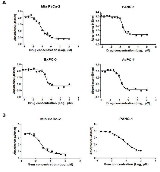
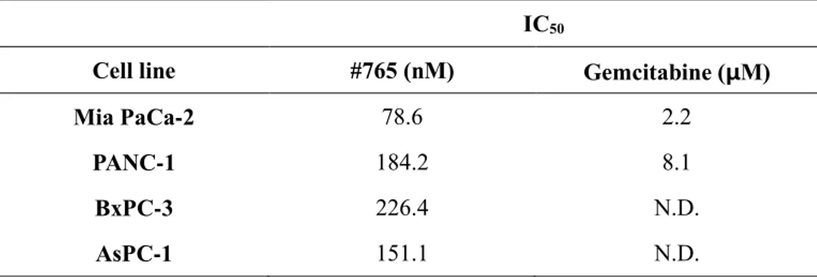
To investigate the effect of #765 on cell proliferation against human pancreatic cancer cell lines (Mia PaCa-2, PANC-1, BxPC-3, and AsPC-1), cell proliferation was measured by CCK-8 assay. As shown in Figure 1A, #765 suppressed proliferation of all four human pancreatic cancer cell lines in a dose-dependent manner. The half-maximal inhibitory concentrations (IC50) were shown in Table 1.
Most notably, the IC50 values of #765 in pancreatic cancer cell lines are nanomolar
range (78-226 nM). Gemcitabine is commonly used as a first line drug in pancreatic cancer, so the anti-proliferative effect of gemcitabine in Mia PaCa-2 and PANC-1 cell was measured to compare with #765 (Figure 1B). As shown in Table 1, #765 shows more potent inhibitory effect on cell proliferation than gemcitabine in Mia PaCa-2 and PANC-1.
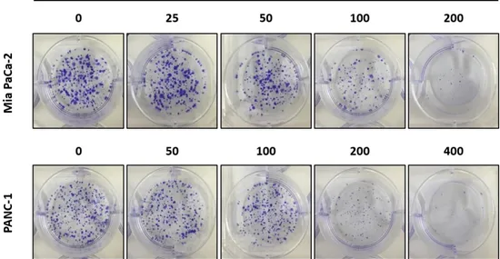
In addition, #765 significantly inhibited colony formation of Mia PaCa-2 and PANC-1 cells in a dose-dependent manner supporting its anti-proliferative effect on human pancreatic cancer cells (Figure 2).
17
Figure 1. Effect of #765 and gemcitabine on pancreatic cancer cell proliferation.
(A) Four pancreatic cancer cell lines (Mia PaCa-2, PANC-1, BxPC-3, and AsPC-1) were treated with various concentrations of #765 for 72 hours. (B) Mia PaCa-2 and PANC-1 cells were treated with various concentrations of gemcitabine for 72 hours. Cell proliferation was measured by Cell Counting Kit-8 (CCK-8). DMSO was used as a solvent control.
18
Table 1. Half-maximal inhibitory concentration (IC50) of #765 and gemcitabine
in pancreatic cancer cell lines
IC50
Cell line #765 (nM) Gemcitabine (μM)
Mia PaCa-2 78.6 2.2
PANC-1 184.2 8.1
BxPC-3 226.4 N.D.
AsPC-1 151.1 N.D.
IC50 is the concentration that results in a 50% decrease in the number of cells
19
Figure 2. Effect of #765 on colony formation of pancreatic cancer cells. Mia
PaCa-2 and PANC-1 cells were treated with the indicated concentrations of #765 for 48 hours. Subsequently, culture media containing #765 was changed with fresh media. After 9 days incubation, colonies were stained with crystal violet. DMSO was used as a solvent control.
20
2. #765 induces mitotic arrest by regulating cell cycle-related proteins in
human pancreatic cancer cells
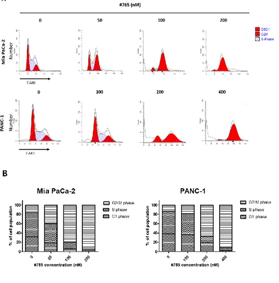
To investigate whether #765-induced inhibition of cell proliferation was caused by arresting the cell cycle, the effect of #765 on cell cycle in pancreatic cancer cells was examined by flow cytometry. As shown in Figure 3A and Figure 3B, #765 significantly led to accumulation of cells at G2/M phase and decrease of cell
population at G1 and S phase in both Mia PaCa-2 and PANC-1 cells. Also, treatment
of #765 with higher concentration caused a clear increase of cell population at subG1, indicating that #765 induced cell death. Since #765 induced G2/M arrest in
human pancreatic cancer cells, the expression of cell cycle regulatory proteins was evaluated by western blotting. Paclitaxel was used as a positive control of causing mitotic arrest. It was reported that dephosphorylation of Cdc2 at Tyr15 is a key regulatory step in activating Cdc2/cyclinB1 complex and further promotes G2/M
transition.29 As shown in Figure 4A, expression of phospho-Cdc2 (Tyr15) was
significantly decreased in both Mia PaCa-2 and PANC-1 cells treated with both #765 and paclitaxel. Expression of cyclin B1 increased in response to the treatment of #765 in PANC-1 cells but not in Mia PaCa-2 cells. In addition, expression of phospho-Histone H3 (Ser10) was clearly elevated in both Mia PaCa-2 and PANC-1 cells treated with #765 (Figure 4B). The increased expression of phospho-Histone H3 (Ser10) in #765-treated PANC-1 cells was also confirmed by
21
immunofluorescence analysis (Figure 4C). All these data suggest that #765 induced activation of Cdk1/cyclin B1 complex and upregulation of phospho-Histone H3 (Ser10), thus leading to mitotic arrest.
22
Figure 3. Analysis of cell cycle in #765-treated pancreatic cancer cells. (A) Mia
PaCa-2 and PANC-1 cells were treated with the indicated concentrations of #765 for 24 hours. #765-treated cells were stained with 7-AAD and analyzed by flow cytometry. FACS data was analyzed using ModFit software. (B) Graphical representation of cell cycle analysis using GraphPad Prism. DMSO was used as a solvent control.
24
Figure 4. Analysis of cell cycle-related protein expression in #765-treated pancreatic cancer cells. Mia PaCa-2 and PANC-1 cells were treated with the
indicated concentrations of #765 or paclitaxel (10 μM) for 24 hours. Expression of cell cycle regulatory proteins, including (A) p-Cdc2 (Tyr15), Cdc2, cyclin B1, and (B) p-HH3 (Ser10), was determined by western blotting. (C) PANC-1 cells were treated with the indicated concentrations of #765 for 24 hours. Cells were analyzed by immunofluorescence using p-HH3 (Ser10) antibodies (red) and DAPI (blue). Cells were visualized using confocal microscopy. DMSO was used as a solvent control and β-actin was used as a loading control. PTX, paclitaxel. p-HH3(Ser10), phospho-Histone H3 (Ser10)
25
3. #765 promotes tubulin depolymerization
Many anti-cancer drugs including paclitaxel and nocodazole induce mitotic arrest by disrupting microtubule dynamics. These agents disrupt microtubule dynamics by triggering polymerization or depolymerization of the tubulins which play a key role in mitosis, further resulting in mitotic arrest.30 Therefore, to
elucidate the underlying mechanism of #765-induced mitotic arrest, the effect of #765 on microtubule dynamics was examined using in vitro cell-free tubulin polymerization assay. As shown in Figure 5A, similar to microtubule-depolymerizing agent nocodazole, #765 significantly inhibited tubulin polymerization compared to the DMSO control. Further, immunofluorescence analysis was used to evaluate the effect of #765 on microtubule network organization in PANC-1. As shown in Figure 5B, #765-treated PANC-1 cells exhibited disruption of the microtubule cytoskeleton, similar to nocodazole-treated cells. These results suggest that #765 triggers depolymerization of tubulin, and this might lead to mitotic arrest of human pancreatic cancer cells.
27
Figure 5. Effect of #765 on microtubule dynamics. (A) Tubulin proteins (>99%
purity) were suspended in Tubulin polymerization buffer in the presence or absence of #765 (400 nM, 2 μM, or 4 μM), paclitaxel (10 μM), or nocodazole (10 μM). Polymerization of microtubules was determined by measuring absorbance at 340 nm over 60 minutes at 37℃ using a spectrophotometer. (B) PANC-1 cells were treated with paclitaxel (50 nM), nocodazole (100 ng/ml), and #765 (200 nM) for 18 hours. Cells were analyzed by immunofluorescence using α-tubulin antibodies (green) and DAPI (blue). Cells were visualized using confocal microscopy. DMSO was used as a solvent control while paclitaxel and nocodazole were used as positive controls for tubulin polymerization and depolymerization, respectively.
28
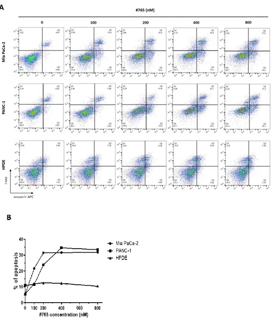
4. #765 induces apoptosis of human pancreatic cancer cells
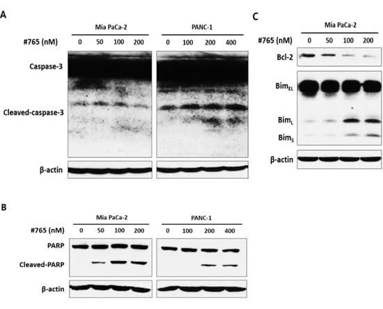
According to the cell cycle analysis data by flow cytometry (Figure 3), accumulation of cell population at subG1 was observed when treated with #765, indicating that #765 induced cell death. Thus, flow cytometry using Annexin-Ⅴ/7-AAD was performed to clarify whether apoptosis is induced by #765. As shown in Figure 6, cell population of both early and late apoptosis was significantly increased in a dose-dependent manner in #765-treated Mia PaCa-2 and PANC-1 cells. Treatment of #765 induced little apoptosis in HPDE. These results indicate that #765 has selectivity for pancreatic cancer cells without showing significant cytotoxicity for normal cells. Next, since #765 induced apoptosis, expression of proteins related to apoptosis was evaluated by western blotting. As shown in Figure 7A and Figure 7B, expression of cleaved-caspase-3 and cleaved-PARP was markedly increased when treated with #765 in both Mia PaCa-2 and PANC-1 cells. In addition, anti-apoptotic protein Bcl-2 was decreased and pro-apoptotic protein Bim was increased (Figure 7C). These data suggest that #765 induces apoptosis via cleavage of caspase-3 and PARP in Mia PaCa-2 and PANC-1 cells.
29
Figure 6. Effect of #765 on apoptosis in pancreatic cancer cells and HPDE. (A)
Mia PaCa-2, PANC-1, and HPDE cells were treated with the indicated concentrations of #765 for 48 hours. Cells treated with #765 were stained with
30
APC-conjugated Annexin-Ⅴ/7-AAD and analyzed by FACS. FACS data was analyzed using FlowJo. (B) Graphical representation of the percentage of cells in apoptosis using GraphPad Prism. DMSO was used as a solvent control. Data are representative of three independent experiments.
31
Figure 7. Analysis of apoptosis-related protein expression in #765-treated pancreatic cancer cells. Expression of apoptosis-related proteins, including (A)
caspase-3, (B) PARP, (C) Bcl-2, and Bim, was determined in Mia PaCa-2 and PANC-1 cells. Cells were treated with the indicated concentrations of #765 for 48 hours. DMSO was used as a solvent control and β-actin was used as a loading control.
32
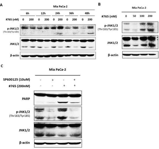
5. Activation of JNK is associated with #765-induced apoptosis
It was reported that microtubule-interfering agents activate JNK signaling and subsequently promotes apoptosis.22 Since JNK is involved in apoptosis21 and #765
caused tubulin depolymerization (Figure 5), the role of JNK pathway in the action of #765-induced apoptosis was investigated. First, whether #765 induced activation of JNK was examined. As shown in Figure 8A and Figure 8B, the expression of phosphorylated-JNK (Thr183/Tyr185) was increased time- and dose-dependently in Mia PaCa-2 cells. The activation was observed 24 hours after #765 treatment, and it lasted for 48 hours. To determine whether the activation of JNK was required for the #765-induced apoptosis, a JNK-specific inhibitor SP600125 was used. As shown in Figure 8C, #765-induced cleavage of PARP was clearly reversed when cells were treated with SP600125. These results suggest that JNK pathway might be involved in #765-induced apoptosis.
33
Figure 8. Effect of JNK on #765-induced apoptosis in Mia PaCa-2 cell. (A) Mia
PaCa-2 cells were treated with #765 at 200 nM for the indicated times and expression of p-JNK (Thr183/Tyr185) and JNK was determined. (B) Mia PaCa-2 cells were treated with the indicated concentrations of #765 for 48 hours and expression of p-JNK (Thr183/Tyr185) and JNK was determined. (C) Mia PaCa-2 cells were treated with SP600125 (10 μM) for 2 hours prior to the treatment of #765 (200 nM) for 48 hours. Expression of p-JNK (Thr183/Tyr185), JNK, and PARP was determined. DMSO was used as a solvent control and β-actin was use d as a loading control.
34
IV. DISCUSSION
Hallmarks of cancer are characterized with six concepts; sustaining proliferative signaling, evading growth suppressors, resisting cell death, enabling replicative immortality, inducing angiogenesis, and activating invasion and metastasis.31 These traits of cancer cells culminate in uncontrolled cell proliferation.
In normal cells, mechanism to maintain the balance between proliferation, growth arrest, and apoptosis exists. By this orchestrated mechanism, normal cells can cause cell cycle arrest when there is something wrong giving time to repair the damage and to maintain genomic integrity, or they can eventually induce apoptosis to prevent malignant progression if problems are not resolved successfully. However, defects in this mechanism may lead to the development of cancer.32
Several distinct anti-cancer drugs ultimately have goal in eliminating cancer cells or holding their growth. Among these anti-cancer drugs, anti-mitotic agents have been used to treat various types of cancers and shown to have effective results in the clinic for decades.12 Therefore, the search for new effective drugs which
induce mitotic arrest and apoptosis may be a promising approach for development of anti-cancer drug.
Based on this study, #765 shows anti-proliferative effect in four human pancreatic cancer cell lines (Figure 1) and inhibits cells to form colony in Mia
35 PaCa-2 and PANC-1 cells (Figure 2).
In the process of G2/M transition, activity of Cdk1/cyclin B1 complex is
important for M phase entry. Cdk1, also called as Cdc2, can be phosphorylated at Thr161, Thr14, and Tyr15. It was reported that key regulatory step in the G2/M
transition is the dephosphorylation of Cdk1 at Tyr15. Reduction in the levels of inhibitory phospho-Cdk1 (Tyr15) promotes the binding of activated phospho-Cdk1 (Thr161) to cyclin B1 resulting in cell cycle progression to mitosis.29 Histone H3, a
nuclear core histone protein of DNA chromatin, has an important role in chromosome condensation and cell cycle progression during mitosis after phosphorylation at Ser10 by Aurora kinase. Thus, phosphorylated Histone H3 at Ser10 could be used as a specific mitosis marker.33 Several studies have
demonstrated that microtubule dynamics disturbing agents have an effect on causing mitotic arrest.34,35 In this study, it was shown that #765 induces downregulation of
phospho-cdc2 (Tyr15) and upregulation of phospho-Histone H3 (Ser10) in Mia PaCa-2 and PANC-1 cell, indicating that #765 causes mitotic arrest (Figure 4). In addition, #765 promotes the disruption of microtubule dynamics by depolymerizing tubulins (Figure 5), and this might lead to mitotic arrest.
Several studies reported that prolonged mitotic arrest leads to apoptosis.18,19
Intrinsic pathway of apoptosis is controlled by tight regulation of pro- and anti-apoptotic proteins of Bcl-2 family. For example, anti-anti-apoptotic protein Bcl-2
36
inhibits pro-apoptotic protein Bax and Bak, which results in breakdown of mitochondrial outer membrane integrity followed by release of cytochrome c into cytosol which triggers the activation of caspase.36 Pro-apoptotic protein Bim, which
has three alternatively spliced isoforms including BimEL, BimL, and Bims, has role
in linking between cytoskeleton and the apoptotic machinery. BimEL and BimL is
sequestered in microtubule cytoskeleton by binding to dynein motor protein. Bims
does not interact with microtubule, yet still have ability to binding with Bcl-2, thus acts as a constitutive death inducer. Following an apoptotic stimulus, the release of BimEL and BimL from microtubules allows their interaction with Bcl-2 leading to
activation of Bax followed by apoptosis.37 In this study, #765 significantly increases
cell populations in early- and late-apoptosis in Mia PaCa-2 and PANC-1 cells. Interestingly, #765 shows less cytotoxicity in normal pancreas cell HPDE compared to pancreatic cancer cell lines, augmenting #765’s therapeutic potential as an anti-pancreatic cancer drug. In addition, #765 induced downregulation of Bcl-2 and upregulation of cleaved caspase-3, cleaved PARP, BimL, and Bims, supporting that
#765 induces apoptosis. The increased expression of Bim in response to the treatment of #765 also implies a possibility that microtubule is also involved in the action of #765.
JNK has a role in apoptosis as well as cell proliferation and differentiation. Furthermore, activation of JNK is involved in microtubule-interfering agent-induced apoptosis.21,22 In this study, #765 caused activation of JNK and JNK
37
inhibitor SP600125 reversed induced cleavage of PARP, indicating that #765-induced apoptosis is partially mediated by JNK pathway.
There is yet to be defined relationship between #765-induced disruption of microtubule dynamics and JNK activation. It can be inferred that activation of JNK is triggered by #765-induced tubulin depolymerization and mitotic arrest, considering that expression of phospho-JNK1/2 (Thr183/Tyr185) was started to be upregulated at 24 hours after #765 treatment (Figure 8A), when tubulin depolymerization and mitotic arrest was already occurred (Figure 3 and Figure 5). Moreover, among the downstream substrates of JNK, there are members of Bcl-2 family that control intrinsic apoptosis, and 14-3-3, which controls the fate of many apoptosis-regulatory proteins.38 Previous studies have reported that activation of
JNK plays an important role in mitotic arrest, leading to inhibition of cancer development and apoptosis.39,40 Therefore, it can be assumed that #765-induced
tubulin depolymerization induces mitotic arrest and simultaneously activates the JNK pathway, thus leading to apoptosis of pancreatic cancer cells.
38
V. CONCLUSION
In the present study, I examined therapeutic potential of #765 as an anti-pancreatic cancer drug in human anti-pancreatic cancer cells. #765 shows remarkable cytotoxicity with nanomolar IC50 value in Mia PaCa-2, PANC-1, BxPC-3, and
AsPC-1 cells. In addition, #765 exhibits more potent anti-proliferative activity than gemcitabine which is commonly used for treatment of pancreatic cancer showing 27- to 43-fold lower IC50 value compared to gemcitabine. Here, the molecular action
mechanism of #765 is elucidated. #765 triggers tubulin depolymerization leading to mitotic arrest. Furthermore, #765 induces apoptosis in human pancreatic cancer cells, which is associated with the activation of JNK pathway. These findings provide evidence that #765 could be a promising therapeutic agent against pancreatic cancer.
39
REFERENCES
1. Siegel RL, Miller KD, Jemal A. Cancer Statistics, 2017. CA Cancer J Clin 2017;67:7-30.
2. Oberstein PE, Olive KP. Pancreatic cancer: why is it so hard to treat? Therap Adv Gastroenterol 2013;6:321-37.
3. Stathis A, Moore MJ. Advanced pancreatic carcinoma: current treatment and future challenges. Nat Rev Clin Oncol 2010;7:163-72.
4. Burris HA, 3rd, Moore MJ, Andersen J, Green MR, Rothenberg ML, Modiano MR, et al. Improvements in survival and clinical benefit with gemcitabine as first-line therapy for patients with advanced pancreas cancer: a randomized trial. J Clin Oncol 1997;15:2403-13.
5. Moore MJ, Goldstein D, Hamm J, Figer A, Hecht JR, Gallinger S, et al. Erlotinib plus gemcitabine compared with gemcitabine alone in patients with advanced pancreatic cancer: a phase III trial of the National Cancer Institute of Canada Clinical Trials Group. J Clin Oncol 2007;25:1960-6. 6. Conroy T, Desseigne F, Ychou M, Bouche O, Guimbaud R, Becouarn Y, et
al. FOLFIRINOX versus gemcitabine for metastatic pancreatic cancer. N Engl J Med 2011;364:1817-25.
7. Von Hoff DD, Goldstein D, Renschler MF. Albumin-bound paclitaxel plus gemcitabine in pancreatic cancer. N Engl J Med 2014;370:479-80.
8. Binenbaum Y, Na'ara S, Gil Z. Gemcitabine resistance in pancreatic ductal adenocarcinoma. Drug Resist Updat 2015;23:55-68.
9. Kleeff J, Korc M, Apte M, La Vecchia C, Johnson CD, Biankin AV, et al. Pancreatic cancer. Nat Rev Dis Primers 2016;2:16022.
10. Waters AM, Der CJ. KRAS: The Critical Driver and Therapeutic Target for Pancreatic Cancer. Cold Spring Harb Perspect Med 2018;8.
11. Cicenas J, Kvederaviciute K, Meskinyte I, Meskinyte-Kausiliene E, Skeberdyte A, Cicenas J. KRAS, TP53, CDKN2A, SMAD4, BRCA1, and BRCA2 Mutations in Pancreatic Cancer. Cancers (Basel) 2017;9.
40
12. Jackson JR, Patrick DR, Dar MM, Huang PS. Targeted anti-mitotic therapies: can we improve on tubulin agents? Nat Rev Cancer 2007;7:107-17.
13. Chen MC, Kuo YC, Hsu CM, Chen YL, Shen CC, Teng CM, et al. The apoptotic mechanisms of MT-6, a mitotic arrest inducer, in human ovarian cancer cells. Sci Rep 2017;7:46149.
14. Sakurikar N, Eichhorn JM, Alford SE, Chambers TC. Identification of a mitotic death signature in cancer cell lines. Cancer Lett 2014;343:232-8. 15. Smits VA, Medema RH. Checking out the G(2)/M transition. Biochim
Biophys Acta 2001;1519:1-12.
16. Musacchio A. The Molecular Biology of Spindle Assembly Checkpoint Signaling Dynamics. Curr Biol 2015;25:R1002-18.
17. Mollinedo F, Gajate C. Microtubules, microtubule-interfering agents and apoptosis. Apoptosis 2003;8:413-50.
18. Topham CH, Taylor SS. Mitosis and apoptosis: how is the balance set? Curr Opin Cell Biol 2013;25:780-5.
19. Haschka M, Karbon G, Fava LL, Villunger A. Perturbing mitosis for anti-cancer therapy: is cell death the only answer? EMBO Rep 2018;19.
20. Elmore S. Apoptosis: a review of programmed cell death. Toxicol Pathol 2007;35:495-516.
21. Dhanasekaran DN, Reddy EP. JNK-signaling: A multiplexing hub in programmed cell death. Genes Cancer 2017;8:682-94.
22. Wang TH, Wang HS, Ichijo H, Giannakakou P, Foster JS, Fojo T, et al. Microtubule-interfering agents activate c-Jun N-terminal kinase/stress-activated protein kinase through both Ras and apoptosis signal-regulating kinase pathways. J Biol Chem 1998;273:4928-36.
23. Blagosklonny MV, Giannakakou P, el-Deiry WS, Kingston DG, Higgs PI, Neckers L, et al. Raf-1/bcl-2 phosphorylation: a step from microtubule damage to cell death. Cancer Res 1997;57:130-5.
41 Cancer Res 1997;57:229-33.
25. Samaj J, Baluska F, Hirt H. From signal to cell polarity: mitogen-activated protein kinases as sensors and effectors of cytoskeleton dynamicity. J Exp Bot 2004;55:189-98.
26. Franken NA, Rodermond HM, Stap J, Haveman J, van Bree C. Clonogenic assay of cells in vitro. Nat Protoc 2006;1:2315-9.
27. Nunez R. DNA measurement and cell cycle analysis by flow cytometry. Curr Issues Mol Biol 2001;3:67-70.
28. Adan A, Alizada G, Kiraz Y, Baran Y, Nalbant A. Flow cytometry: basic principles and applications. Crit Rev Biotechnol 2017;37:163-76.
29. Stark GR, Taylor WR. Control of the G2/M transition. Mol Biotechnol 2006;32:227-48.
30. Singh P, Rathinasamy K, Mohan R, Panda D. Microtubule assembly dynamics: an attractive target for anticancer drugs. IUBMB Life 2008;60:368-75.
31. Hanahan D, Weinberg RA. Hallmarks of cancer: the next generation. Cell 2011;144:646-74.
32. Swanton C. Cell-cycle targeted therapies. Lancet Oncol 2004;5:27-36. 33. Hans F, Dimitrov S. Histone H3 phosphorylation and cell division.
Oncogene 2001;20:3021-7.
34. Mitchison T, Kirschner M. Dynamic instability of microtubule growth. Nature 1984;312:237-42.
35. Desai A, Mitchison TJ. Microtubule polymerization dynamics. Annu Rev Cell Dev Biol 1997;13:83-117.
36. Taylor RC, Cullen SP, Martin SJ. Apoptosis: controlled demolition at the cellular level. Nat Rev Mol Cell Biol 2008;9:231-41.
37. Puthalakath H, Huang DC, O'Reilly LA, King SM, Strasser A. The proapoptotic activity of the Bcl-2 family member Bim is regulated by interaction with the dynein motor complex. Mol Cell 1999;3:287-96. 38. Bogoyevitch MA, Kobe B. Uses for JNK: the many and varied substrates
42
of the c-Jun N-terminal kinases. Microbiol Mol Biol Rev 2006;70:1061-95. 39. Davis RJ. Signal transduction by the JNK group of MAP kinases. Cell
2000;103:239-52.
40. Tournier C, Hess P, Yang DD, Xu J, Turner TK, Nimnual A, et al. Requirement of JNK for stress-induced activation of the cytochrome c-mediated death pathway. Science 2000;288:870-4.
43
ABSTRACT (IN KOREAN)
새로운 췌장암 치료제의 개발 및 작용 기전 규명 <지도교수 이재면> 연세대학교 대학원 의과학과 김청비 췌장암은 가장 치명적인 질병 중에 하나로서, 5년 생존율이 8% 이하이며 발병률과 사망률이 거의 일치한다. 췌장암을 치료하기 위해 효 과적인 항암제를 개발하려는 많은 노력이 있음에도 불구하고, 췌장암 치 료제는 여전히 한정적인 실정이다. 따라서, 효과적인 췌장암 치료제의 개발이 시급한 상황이다. 본 연구에서는 새로운 물질인 #765의 치료 효 과를 인간 췌장암 세포주에서 확인하고자 했다. #765는 나노몰 (nM) 수 준의 농도에서 췌장암 세포의 증식을 억제하는 효과를 보였다. #765는 췌장암 치료에 사용되고 있는 젬시타빈(gemcitabine) 보다 더욱 효과적 으로 췌장암 세포의 증식을 억제하였다. 또한, #765는 췌장암 세포의 콜
44 로니(colony) 형성을 억제하였다. 더욱이, #765를 처리하였을 때, 췌장 암 세포에서 튜불린 중합해체(tubulin depolymerization)가 일어남으로 써 마이크로튜불 동역학(microtubule dynamics)이 저해되었고, 유사분열 중단(mitotic arrest)이 일어났다. #765는 유사분열 특이적 표지로 알려 진 히스톤 H3 의 10번째 세린 잔기(Ser10)에 인산화를 일으켰고, G2/M 이행에 중요한 역할을 하는 것으로 알려진 Cdc2의 15번째 타이로신 잔기 (Tyr15)에 탈인산화를 일으켰다. 이외에도, #765에 의해 췌장암 세포에 서 카스파아제-3(caspase-3)의 활성과 다중(에이디피-리보스)당중합효소 (poly-(ADP-ribose) polymerase; PARP)의 불활성을 유도하여 세포사멸이 일어났다. #765가 JNK를 활성화 시켰고, JNK 억제제인 SP600125 를 처리 하면 #765에 의해서 증가한 cleaved-PARP의 발현이 다시 감소하였다. 이 러한 결과들을 종합해보면, #765는 인간 췌장암 세포에서 튜불린 동역학 를 저해시킴으로써 유사세포분열 중단을 일으키고, JNK를 매개로하여 세 포사멸을 일으킨다는 것을 알 수 있다. 따라서, #765가 새로운 췌장암 치료제로 쓰일 수 있을 것으로 기대된다. 핵심되는 말 : 췌장암, 유사세포분열 중단, 튜불린 중합해체, 세포사멸, JNK