R E S E A R C H A R T I C L E
Open Access
Classification of ginseng berry (Panax ginseng C.A.
MEYER) extract using
1
H NMR spectroscopy and its
inhibition of lipid accumulation in 3 T3-L1 cells
Seung Ok Yang
1, Hae Ran Park
2, Eun Suk Sohn
3, Sang Won Lee
2, Hyung Don Kim
2, Young Chang Kim
2,
Kee Hong Kim
2, Sae Won Na
4, Hyung-Kyoon Choi
5, Mariadhas Valan Arasu
6and Young Ock Kim
2*Abstract
Background: Panax ginseng is a famous traditional medicine in Korea for its beneficial effect on obesity, cardiac and liver associated diseases. The aim of this study was to investigate the metabolite in Panax ginseng (P. ginseng, Aralicaceae) berries depending on the ripen stages and evaluate its potential inhibition on adipocyte differentiation in 3 T3-L1 cells. Methods: Different ripening stage samples of P. ginseng berry were analyzed through global metabolite profiling by NMR spectroscopy. Lipid accumulation in the cells was analyzed by Oil Red O staining.
Results: The PLS-DA clearly distinguished P. ginseng berry extract (PGBE) according to the partial ripe (PR), ripe(R) and fully ripe (FR) stage. Lipid accumulation of PGBE was examined by measuring triglyceride content and Oil-Red O staining. These results suggested that the FR stage of PGBE decrease in lipid accumulation during adipocyte differentiation and the amount of threonine, asparagine, fumarate, tyraine, tyrosine, and phenylalanine increased with longer ripening of ginseng berries.
Conclusion: Metabolite profiling of P. ginseng was identified by1H NMR spectra. P. ginseng extract efficiently inhibits adipogenesis in 3 T3-L1 adipocytes concluded that the P. ginseng has the antiobesity properties.
Keywords: Panax ginseng berry, Obesity, Metabolite profiling, Maturation Background
Obesity is a risk factor for major metabolic disease in-cluding type 2 diabetes, atherosclerosis, hyperlipidemia and hypertension and multifactorial syndrome in human [1,2]. It is an abnormal condition in which the lipids are accumulated in adipose tissues and various kinds of adipokine [3]. Panax ginseng is a perennial plant that has been used as a tonic and for the treatment of various diseases [4-8]. P. ginseng root is normally harvested between the fourth and sixth year of growth. The multiple active constituents such as ginsenosides, polysaccharides, peptides, polyacetylenic alcohols and fatty acids are identi-fied in the P. ginseng root [9]. On the other hand, P. ginseng berry easily be harvested several times after the third year of growth [10]. Quan et al. [11] reported that
ginsenoside Re (groups, namely protopanaxatriol-type saponin from ginseng) lowers blood glucose and lipid in high-fat diet fed mice. However, effects on adipocyte differentiation in 3 T3-L1 cells on PGBE have not yet been reported. The chemical composition and biological activities of the P. ginseng berry may differ according to the maturation stage. We have looked for lipid accumu-lation in adipocyte inhibitory plants using PGBE as an in vitro assay system. During the course of screening, the water extract of P. ginseng berry was significantly inhibited this activity. The metabolic profiling can be useful for quantifying a group of related compounds. There are few previous studies about profiling metabolic compounds of ginseng by using NMR [12,13]. However, no study reported in differences of the metabolic compounds among different maturation stages in ginseng fruits. The aim of this study was to classify the ginseng berry (Panax ginseng) extract using1H NMR spectroscopy and evaluates its inhibition of lipid accumulation in 3 T3-L1 cells.
* Correspondence:[email protected]
2
Department of Medicinal Crop Research NIHHS RDA Eumsung, Chungbuk 369-873, Republic of Korea
Full list of author information is available at the end of the article
© 2014 Yang et al.; licensee BioMed Central Ltd. This is an Open Access article distributed under the terms of the Creative Commons Attribution License (http://creativecommons.org/licenses/by/4.0), which permits unrestricted use, distribution, and reproduction in any medium, provided the original work is properly credited. The Creative Commons Public Domain Dedication waiver (http://creativecommons.org/publicdomain/zero/1.0/) applies to the data made available in this article, unless otherwise stated.
Methods
Plant material
Three steps berries of five year-old P. ginseng were obtained from a local farm in Eumseong province (GPS N 36° 56’ 34”, E 127° 45’ 14”), Republic of Korea. The collected plants were identified by the botanist in Ginseng Research Institiute, Daegu, South Korea. The collected samples were stored in the Medicinal Crop Research Insti-tute, NIHHS, RDA, with voucher number MCRI-241. Three periods were June 8th, 2012 (12ea), June 18th, 2012 (10ea), and July 16th, 2012 (8ea), respectively. The collected P. ginsengs were classified into three major categories according to their stage of maturation: ripe (R), and fully ripe (FR). The PGBE was freeze-dried and then stored at −70°C before analysis. Voucher specimens were de-posited at the Department of Medicinal Crop Research, Rural Development Administration in Republic of Korea (RDAPGBE 201201–201230).
Sample preparation for1H NMR
PGBE were extracted by adding 1 mL of 100% D2O to
30 mg of powdered P. ginseng berries in a micro tube, vortexed for 1 min, and sonicated for 5 min. The materials were then centrifuged at 14000 × rpm for 10 min. KH2PO4
was added as a buffering agent to 100 mL of D2O
contain-ing 0.05% 3-(trimethylsilyl)-propionic-2,2,3,3-d4 acid, sodium salt (TSP) as an internal standard for D2O. The
pH of the D2O used for NMR measurements was adjusted
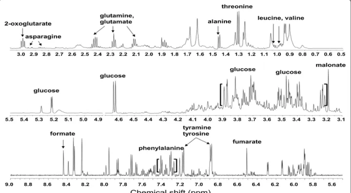
Figure 1 Representative total1H NMR spectra (600 MHz) of the P. ginseng berry extract analyzed with direct D
2O extraction.
Table 1 Assignment of1H NMR spectral peaks obtained
from P. ginseng berry extract analyzed by using D2O
solvent. S: singlet, d: doublet, t: triplet, m: multiplet, dd: doublet of doublet
No Compounds Chemical shift (δ), Peak multiplicity, J value (Hz) 1 Leucine 0.95 (d, J = 6.0) 2 Valine 0.99 (d, J = 5.4), 1.03 (d, J = 7.1) 3 Threonine 1.32(d, J = 6.9) 4 Alanine 1.47 (d, J = 7.3) 5 Glutamate 2.10-2.16(m), 2.29 (t, J = 7.4, 2.48-2.48 (m) 6 Glutamine 2.10-2.16 (m), 2.40-2.48 (m) 7 Asparagine 2.85 (dd, J = 16.9, 7.7), 2.94 (dd, J = 16.9, 4.2) 8 2-Oxoglutaric acid 3.00 (t, J = 7.2) 9 Malonate 3.19 (s) 10 Glucose 3.21-3.25(m), 3.36-3.43 (m), 3.43-3.54 (m), 3.67-3.82 (m), 3.89 (dd, J = 11.9, 1.6), 4.63 (d, J = 8.0), 5.22 (d, J = 3.8) 11 Fumarate 6.51 (s) 12 Tyramine, 6.89 (d, J = 8.5), 7.18 (d, J = 8.3) 13 Tyrosine 6.89 (d, J = 8.5), 7.18 (d, J = 8.3) 14 Phenylalanine 7.31 (d, J = 7.8), 7.36 (t, J = 7.3), 7.41 (t, J = 7.6) 15 Formate 8.44 (s)
to 6.0 by careful addition of 1 N NaOD and then trans-ferred to a 5 mm NMR tube.
Data reduction and processing
MestReNova (version 6.0.4) was used to obtain the NMR spectra, which were all automatically binning using Chenomx (version 5.1) software. The spectral1H NMR region from δ = 0.56 to δ = 10.00 was segmented into regions with widths of 0.04 ppm, giving 232 integrated regions in each NMR spectrum.
Cell culture
3 T3-L1 preadipocyte purchased from ATCC were cultured in 24 well plate at a density of 3×10⁴cells/well. In DMEM containing 10% FBS, 2 mM glutamine, 20 mM Hepes, 50U/ml penicillin, and 50 mg/ml streptomycin
sulfate. After 100% confluency, cells were cultured with differentiation medium (DMEM with 10% FBS, 0.5 mM IBMX, 2 mM DEX and 1.7 mM INS). After 48 h of stimu-lation, cells were cultured in DMEM supplemented with 10% FBS with/without PGBE and changed every two days up to 8 days.
Oil Red O staining
Lipid accumulation of PGBE was examined by measur-ing triglyceride content usmeasur-ing Oil-Red O stainmeasur-ing. For Oil Red O staining, cells were washed gently with PBS twice, fixed with 3.7% fresh formaldehyde in PBS for 1 h at room temperature and stained with filtered Oil Red O solution (60% isopropanol and 40% water) for at least 1 h. After staining of lipid droplets with Red, the Oil Red O staining solution was removed and the plates
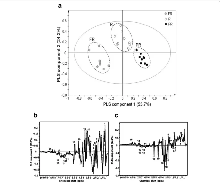
Figure 2 PLS-DA-derived score plot (a) and loading plot (PLS component 1: b and PLS component 2: c) of P. ginseng berry extract analyzed with D2O direct extraction. (green circle symbol): PR, (blue circle symbol): R, (red circle symbol): FR . 1: leucine, 2:valine, 3: threonine,
4: alanine, 5: glutamate, 6: glutamine, 7: asparagines, 8: 2-oxoglutaric acid, 9: malonate, 10: glucose, 11: fumarate, 12: tyramine, 13: tyrosine, 14: phenylalanine, 15: formate.
were rinsed with water and dried. Images were collected on an Olympus microscope (Tokyo, Japan). Finally, the dye retained in the cells was eluted with isopropanol and quantified by measuring the optical absorbance at 500 nm.
Multivariate statistics analysis
Principal component analysis (PCA) was performed using mean Pareto-scaled data obtained from aqueous solvent system. Then, partial least squares-discriminant analysis (PLS-DA) was also performed, which can yield a clearer differentiation of each class and enable a less complicated investigation of marker compounds
Statistical analysis
Unless otherwise specified, all data are expressed as the mean ± standard error (SE) from triplicate experiments. One-way ANOVA (Scheffe test or student t test) was used for multiple comparisons using the Statistical Package for the Social Sciences (SPSS) program (version 16.0) (SPSS,
Inc., Chicago, IL, USA). Values of p < 0.05 were considered statistically significant.
Results and discussion
Figure 1 shows a representative NMR spectrum of the D2O extracts from the PGBE at the mature stage. As
described in Table 1, the signals were assigned based on comparisons with the database of the Chenomx NMR software suite: amino acid such as leucine, valine, threo-nine, alathreo-nine, glutamate, glutamine, asparagines, malonate, tyramine, tyrosine, and phenylalanine; organic acid such as 2-oxoglutarate, fumarate, and formate; sugars such as glucose.
PCA could clearly separate the maturation of ginseng berries based on the score plots (data not shown). PLS-DA was applied to the1H NMR spectral data of P. ginseng berries according to three different maturation periods. The different maturation of the ginseng fruit samples were clearly distinguished based upon the PLS-DA-derived score plots (Figure 2a). Depending on the maturation of
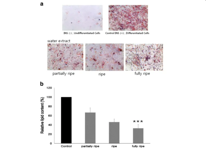
Figure 3 Effects P. ginseng berry extract on Oil Red O staining in cultured 3 T3-L1 adipocytes. 3 T3-L1 cells were treated with 100μg/ml of P. ginseng berry extract. (a) Effects of P. ginseng berry extract on fat droplet formation in 3 T3-L1 cells. It was stained with Oil Red O dye and examined with a light microscope; (b) Relative lipid content by quantification method of Oil Red O staining. Data are presented as average ± SD (n =5). * indicates *** p <0.001 compared with control (expressed by t-test).
the sample, the positive direction was shifted to the nega-tive direction of PC1. Loading plot analysis was performed to investigate the contributing metabolites for separating each PGBE from the score plots. As shown in Figure 2b, the levels of leucine, valine, alanine, glutamate, glutamine, 2-oxoglutarate, malonate, and glucose were higher in the PR stage samples than the FR berries. In addition, threonin, asparagines, fumarate, tyramine, tyrosine, and phenylalan-ine were higher in the FR berries than the PR ones. The levels of leucine, valine, threonin, alanine, asparagines, 2-oxoglutarate, malonate, phenylalanine, and formate were higher in the ripening fruits (Figure 2c) than other stages.
3 T3-L1 cells of the adipocyte morphology increase the synthesis and accumulation of triglycerides and acquire the signet ring appearance of adipose cells [14]. 3 T3-L1 cells are extensively used to study adipogenesis [15]. For this reason, we chose these cells for research to identity the feasibility of PGBE as a possible new anti-obesity herbal agent. This observation was further supported with the quantitative analysis of neutral lipid content by meas-uring the absorbance at 500 nm. 3 T3-L1 cells were treated with various maturation stage of PGBE. After 8 days, intracellular lipid accumulation was examined with Oil red O staining for lipid droplets as an indicator of the degree of adipogenesis. Figure 3a showed that the cell size was bigger and the intracellular fat drops were comparatively more in 3 T3-L1 cells. And comparatively adipogenesis and cell viability were decreased 100ug/ml treated group. In addition, the lipid accumulation rate was significantly reduced with PGBE treatment compared with the control; the decrease was maturation stage dependent (p <0.001); accumulation was about 67%, 46%, and 33% at PR, R, and FR by aqueous extraction concentration in Figure 3b.
In previous, many studies have reported the anti-obesity effects in various medicinal plants, such as Nigella sativa [16], Camellia sinensis [17], Hibiscus sabdariffa [18], Psyl-lium fibre [19], and Lycium barbarum [20]. Dey et al. [21] demonstrated the anti-obesity effect in Asian ginseng berry extract, and Attele et al. [22] also showed the anti-hyperglycemic effect in ginsenoside Re. According to Kim et al. [23], the group of people whom the black soybean peptide had been taken showed the decreased body mass and fat. The scientific study shows that natural products contain a large variety of components that possess lipid inhibition activity. Especially, a variety of herbs from plants have been used as traditional natural medicines for cure many kinds of diseases or restore to health. In particular, various oriental medicinal herbs are reported to have biological activity [24,25].
In this study, it was confirmed that the amount of phenylalanine is higher in FR stage of P. ginseng berries, thus expecting to lower the obesity. Further in vivo research and clinical trials are still need to clarify the efficacy, safety,
and precise molecular mechanisms of the anti-obesity effects of PGBE.
Conclusion
In conclusion, this is the first study regarding metabolic profiling of PGBE using D2O solvent. Moreover,
multi-parameter pattern recognition analysis established 1H NMR spectra of PGBE. And PGBE efficiently inhibits adipogenesis in 3 T3-L1 adipocytes as indicated by sig-nificant reduction lipid accumulation. It is predicted that P. ginseng berry extract may apparently inhibit the adipogenic differentiation and lipid accumulation in the cells through the activation of various adipogenic regulatory genes such as peroxisome proliferator-activated receptor (PPARγ) and CCAAT element binding protein (C/EBP-α). Further the mechanism underlying the anti-adipogenic activity of P. ginseng berry extract has to be studied in the future.
Competing interests
The authors declare that they have no competing interests.
Authors’ contributions
YCK, HKC and MVA designed all the experiments, analyzed data and wrote the paper. SOK, HRP and ESS carried out the main experiments. HDK, YCK, KHK and SWN provided valuable suggestions for this study and helped to draft the manuscript. All authors read and approved the final manuscript. Acknowledgments
This work was performed with the support of the Agenda Program (PJ008568), Rural Development Administration, Republic of Korea. Author details
1
Dapartment of food science and engineering, Ewha Womans University, 11-1 Daehyun-dong, Seodaemun-gu, Seoul 120-750, Republic of Korea.
2
Department of Medicinal Crop Research NIHHS RDA Eumsung, Chungbuk 369-873, Republic of Korea.3Department of Applied Biochemistry, College of
Biomedical & Health Science, Konkuk University, Chungju 380-701, South Korea.4College of Veterinary Medicine, ChonBuk National University,
Jeonju-si, South Korea.5College of pharmacy, Chung-Ang University, Seoul 156-756, Republic of Korea.6Department of Botany and Microbiology,
Addiriyah Chair for Environmental Studies, College of Science, King Saud University, P. O. Box 2455, Riyadh 11451, Saudi Arabia.
Received: 25 August 2014 Accepted: 13 November 2014 Published: 24 November 2014
References
1. Leonhardt M, Hrupka B, Langhans W: New approaches in the pharmacological treatment of obesity. Eur J Nutr 1999, 38:1–13. 2. Monteiro R, Azevedo I: Chronic inflammation in obesity and the
metabolic syndrome. Medi Infla 2010, 2010:1–10.
3. Zalilah MS, Khor GL, Mirnalini K, Norimah AK, Ang M: Dietary intake, physical activity and energy expenditure of Malaysian adolescents. Sin Med J 2006, 47:491.498.
4. Block KI, Mead MN: Immune system effects of echinacea, ginseng, and astragalus: A review. Int Can Ther 2003, 2:247–267.
5. Nah SY, Kim DH, Rhim H: Ginsenosides: Are any of them candidates for drugs acting on the central nervous system? CNS Drug Rev 2007, 13:381–404. 6. Xie JT, Mchendale S, Yuan CS: Ginseng and diabetes. Am J Chin Med 2005,
33:397–404.
7. Yun TK: Experimental and epidemiologic evidence on non-organ specific cancer preventive effect of Korean red ginseng and identification of active compounds. Mut Res 2003, 523–524:63–74.
8. Kim S, Kim T, Ahn K, Park WK, Nah SY, Rhim H: Ginsenoside Rg3 antagonizes NMDA receptors through a glycine modulatory site in rat cultured hippocampal neurons. Biochem Biophy Res Comm 2004, 323:416–424. 9. Lee Florence C: Facts About Ginseng: The Elixir of Life. In Elizabeth, NJ,
USA: Hollym International Corporation; 1992.
10. Kim YK, Yoo DS, Xu H, Park NI, Kim HH, Choi JE, Park SU: Ginsenoside content of berries and roots of three typical Korean ginseng (Panax ginseng) cultivars. Nat Pro Comm 2009, 4:903–906.
11. Quan HY, Yuan HD, Jung MS, Ko SK, Park YG, Chung SH: Ginsenoside Re lowers blood glucose and lipid levels via activation of AMP-activated protein kinase in HepG2 cells and high-fat diet fed mice. Int J Mol Med 2012, 29:73–80.
12. Yang SY, Kim HK, Lefeber AWM, Erkelens C, Angelova N, Choi YH, Verpoorte R: Application of two dimensional nuclear magnetic resonance spectroscopy to quality control of ginseng commercial products. Planta Med 2006, 72:364–369.
13. Lee EJ, Shaykhutdinov R, Weljie AM, Vogel HJ, Facchini PJ, Park SU, Kim YK, Yang TJ: Quality assessment of ginseng by 1H NMR metabolite fingerprinting and profiling analysis. J Agri Food Chem 2009, 57:7513–7522. 14. Green H, Kehinde O: An established preadipose cell line and its
differentiation in culture. II. Factors affecting the adipose conversion. Cell 1975, 5:19–27.
15. Atanasov AG, Wang JN, Gu SP, Bu J, Kramer MP, Baumgartner L, Fakhrudin N, Ladurner A, Malainer C, Vuorinen A, Noha SM, Schwaiger S, Rollinger JM, Schuster D, Stuppner H, Dirsch VM, Heiss EH: Honokiol: A non-adipogenic PPARγ agonist from nature. Biochim Biophys Acta 1830, 2013:4813–4819. 16. Datau EA, Wardhana Surachmanto EE, Pandelaki K, Langi JAF: Efficacy of
Nigella sativa on serum free testosterone and metabolic disturbances in central obese male. Acta Med Indones 2010, 42:130–134.
17. Basu A, Du M, Sanchez K, Leyva MJ, Betts NM, Blevins S, Wu M, Aston CE, Lyons TJ: Green tea minimally affects biomarkers of inflammation in obese subjects with metabolic syndrome. Nutr 2011, 27:206–213. 18. Gurrola-Díaz CM, García-López PM, Sánchez-Enríquez S, Troyo-Sanromán R,
Andrade-González I, Gómez-Leyva JF: Effects of Hibiscus sabdariffa extract powder and preventive treatment [diet] on the lipid profiles of patients with metabolic syndrome [MeSy]. Phytomed 2010, 17:500–505. 19. Pal S, Khossousi A, Binns C, Dhaliwal S, Ellis V: The effect of a fibre
supplement compared to a healthy diet on body composition, lipids, glucose, insulin and other metabolic syndrome risk factors in overweight and obese individuals. Bri J Nutr 2011, 105:90–100.
20. Amagase H, Nance DM: Lycium barbarum increases caloric expenditure and decreases waist circumference in healthy overweight men and women: pilot study. J Am Coll Nutr 2011, 30:304–309.
21. Dey L, Zhang L, Yuan CS: Letter to the editor: anti-diabetic and anti-obese effects of ginseng berry extract: comparison between intraperitoneal and oral administrations. Am J Chin Med 2002, 30:645–647.
22. Attele AS, Zhou YP, Xie JT, Wu JA, Zhang L, Dey L, Pugh W, Rue PA, Polonsky KS, Yuan CS: Antidiabetic effects of Panax ginseng berry extract and the identification of an effective component. Diabetes 2002, 51:1851–1858. 23. Kim MJ, Yang HJ, Kim JH, Ahn CW, Lee JH, Kim KS, Kwon DY: Obesity-related
metabolomic analysis of human subjects in black soybean peptide intervention study by ultraperformance liquid chromatography and quadrupole-time-of-flight mass spectrometry. J Obesity 2013, 2013:1–11. 24. Kim YS, Lee YM, Kim H, Kim J, Jang DK, Kim JH, Kim JS: Anti-obesity effect
of morus bombycis root extract: anti-lipase activity and lipolytic effect. J Ethnopharmacol 2010, 130:621–624.
25. Yun JW: Possible anti-obesity therapeutics from nature-a review. Phytochem 2010, 71:1625–1641.
doi:10.1186/1472-6882-14-455
Cite this article as: Yang et al.: Classification of ginseng berry (Panax ginseng C.A. MEYER) extract using1H NMR spectroscopy and its inhibition
of lipid accumulation in 3 T3-L1 cells. BMC Complementary and Alternative Medicine 2014 14:455.
Submit your next manuscript to BioMed Central and take full advantage of:
• Convenient online submission
• Thorough peer review
• No space constraints or color figure charges
• Immediate publication on acceptance
• Inclusion in PubMed, CAS, Scopus and Google Scholar
• Research which is freely available for redistribution
Submit your manuscript at www.biomedcentral.com/submit