INTRODUCTION
Urinary tract stones are one of the most prevalent urological disorders. It has been estimated that up to 12% of the population will suffer from urinary stones during
Ureteral stenting can be a negative predictor
for successful outcome following shock wave
lithotripsy in patients with ureteral stones
Dong Hyuk Kang
1, Kang Su Cho
2, Won Sik Ham
1, Doo Yong Chung
1, Jong Kyou Kwon
3, Young Deuk Choi
1,
Joo Yong Lee
11Department of Urology, Severance Hospital, Urological Science Institute, Yonsei University College of Medicine, Seoul, 2Department of Urology, Gangnam Severance Hospital, Urological Science Institute, Yonsei University College of Medicine, Seoul, 3Department of Urology, Severance Check-Up, Yonsei University Health System, Seoul, Korea
Purpose: To evaluate ureteral stenting as a negative predictive factor influencing ureteral stone clearance and to estimate the probability of one-session success in shock wave lithotripsy (SWL) patients with a ureteral stone.
Materials and Methods: We retrospectively reviewed the medical records of 1,651 patients who underwent their first SWL. Among these patients, 680 had a ureteral stone measuring 4–20 mm and were thus eligible for our study. The 57 patients who underwent ureteral stenting during SWL were identified. Maximal stone length (MSL), mean stone density (MSD), skin-to-stone distance (SSD), and stone heterogeneity index (SHI) were determined by pre-SWL noncontrast computed tomography.
Results: After propensity score matching, 399 patients were extracted from the total patient cohort. There were no significant dif-ferences between stenting and stentless groups after matching, except for a higher one-session success rate in the stentless group (78.6% vs. 49.1%, p=0.026). In multivariate analysis, shorter MSL, lower MSD, higher SHI, and absence of a stent were positive pre-dictors for one-session success in patients who underwent SWL. Using cutoff values of MSL and MSD obtained from receiver opera-tor curve analysis, in patients with a lower MSD (≤784 HU), the success rate was lower in those with a stent (61.1%) than in those without (83.5%) (p=0.001). However, in patients with a higher MSL (>10 mm), the success rate was lower in those with a stent (23.6%) than in those without (52.2%) (p=0.002).
Conclusions: Ureteral stenting during SWL was a negative predictor of one-session success in patients with a ureteral stone. Keywords: Lithotripsy; Stents; Treatment outcome; Ureter; Urinary calculi
This is an Open Access article distributed under the terms of the Creative Commons Attribution Non-Commercial License (http://creativecommons.org/licenses/by-nc/4.0) which permits unrestricted non-commercial use, distribution, and reproduction in any medium, provided the original work is properly cited.
Received: 20 June, 2016 • Accepted: 8 September, 2016
Corresponding Author: Joo Yong Lee
Department of Urology, Severance Hospital, Urological Science Institute, Yonsei University College of Medicine, 50-1 Yonsei-ro, Seodaemun-gu, Seoul 03722, Korea TEL: +82-2-2228-2320, FAX: +82-2-312-2538, E-mail: joouro@yuhs.ac
ⓒ The Korean Urological Association, 2016
their lifetime, and recurrence rates approach 50% [1]. Several treatment methods exist, including observation (awaiting spontaneous passage), shock wave lithotripsy (SWL), retrograde endoscopic procedures, and percutaneous nephrolithotomy. SWL is a safe, effective, noninvasive, and
www.icurology.org
Investig Clin Urol 2016;57:408-416. https://doi.org/10.4111/icu.2016.57.6.408 pISSN 2466-0493 • eISSN 2466-054X
well-established treatment modality, which is now the first-choice treatment for most upper urinary tract stones [2].
Becoming stone-free after SWL does not occur imme-diately; instead, the stones are pulverized during the procedure, then spontaneously passed through the urinary tract. Thus, the time course of stone clearance varies considerably. In most cases, fragmented particles of calculi pass uneventfully through the urinary tract after SWL, but fragments sometimes obstruct the ureter, causing post-SWL complications such as acute renal colic, hydronephrosis, acute kidney injury, or urinary tract infection [3]. Particularly for larger calculi, a number of stone fragments may become impacted in the ureter, forming an obstructing column of sand known as steinstrasse. According to the European Association of Urology Urolithiasis Guidelines, ureteral stenting reduces the risk of renal colic and obstruction [4], and many physicians consider inserting ureteral stents before SWL to create an artificial chamber with an impro-ved stone-fluid interface for better fragmentation during SWL and to reduce the risk of obstruction [5]. Accordingly, several studies have been performed to determine whether routine pre-SWL ureteral stenting is helpful in preventing obstructive complications, but the issue remains somewhat controversial [6-8].
Preventing post-SWL complication is surely important, but the ultimate goal of SWL treatment is to establish a stone-free status. Similar to the issue of ureteral stenting and SWL complications, the effects of SWL on stone-free rates (SFRs) are also controversial [7,9,10]. Several recent studies have demonstrated that ureteral stenting reduces the SFR following SWL [11-13], but the significance of this finding is debated. Thus, the current study was conducted to evaluate the effects of ureteral stenting and stone characteristics on ureteral stone clearance and to estimate the probability of one-session success in SWL patients with ureteral calculi according to whether they underwent ureteral stenting or exhibited various other factors.
MATERIALS AND METHODS
1. Patient cohort
Medical records were obtained from a database of patients (n=1,651) who underwent an initial session of SWL between November 2005 and September 2014 at Severance Hospital, Seoul, Korea. The study inclusion criteria were a single, 4–20 mm, radiopaque calculus located within the ureter on plain-film X-rays, presenting within 1 month prior to SWL treatment, and without evidence of stone migration. Patients with bilateral ureteral stones, urinary tract
congenital anomalies, or a single kidney, as well as those who received prophylactic medical expulsion therapy, were excluded from the analysis. This left 680 patients eligible for analysis.
2. Good clinical practice protocols
The study was performed in accordance with all applicable laws and regulations, good clinical practices, and the ethical principles described in the Declaration of Helsinki. The Institutional Review Board of Severance Hospital approved this study protocol (approval number: 4-2015-1052). The study was exempt from requiring the participants’ written informed consent because of its retrospective design and because the patients’ records and information were anonymized and de-identified prior to analysis.
3. Extracorporeal SWL
SWL was performed using an electroconductive lithotripter (EDAP Sonolith Praktis, Technomed, Lyon, France) until 2011. Beginning in 2012, this was replaced by an electromagnetic generative lithotriptor (Dornier Compact Delta II lithotripter, Dornier Medtech, Wessling, Germany). All ESL procedures were conducted under fluoroscopic guidance. The number of shock waves per SWL session varied from 2,500 to 4,000, at a rate of 60–90 shock waves per minute. We prematurely terminated the session if the stone became difficult to visualize during the session. The launch intensity was conducted when the focal peak pressure ranged from 16 to 55 MPa, as determined by the pain reported by the patients while SWL was being performed.
4. Demographic data and stone characteristics on
noncontrast computed tomography
A detailed history of the ureteral stone was obtained, including the number of past stone events, history of pain onset, and stone characteristics. The stone characteristics included the location, maximal stone length (MSL), stone heterogeneity index (SHI), skin-to-stone distance (SSD), and mean stone density (MSD). The SSD was measured in the axial plane, 45o from the vertical axis [14]. The MSL was
the longest stone length measured in three dimensions on noncontrast computed tomography (NCCT) images. We used the GE Centricity system (GE Healthcare Bio-Sciences Corp., Piscataway, NJ, USA) during the measurement procedure. The MSD was measured using bone windows on the magnified, axial NCCT image of the stone in the maximal diameter, in which the elliptical region of
interest incorporated the largest cross-sectional area of the stone without including adjacent soft tissue [15]. The SHI was defined as the standard deviation of the Hounsfield units (HUs) in the same region of interest by Lee et al. [16]. Complication rate and each variables including post-SWL complication were also obtained. Successful post-SWL treatment of the ureteric calculus was defined as the patient being rendered stone-free or asymptomatic with clinically insignificant residual fragments ≤3 mm in maximal diameter 2 weeks after a single SWL treatment (as measured by simple X-ray) [2] and not requiring additional treatment within a 3-month follow-up period.
5. Statistical analysis
Data are presented as mean±standard deviation. After total cohort analyses, propensity score matching was performed to further elucidate the characteristics of our patients with ureteral stones. Stenting cases were 1:6 matched with the closest-propensity stentless cases. Propensity scores were then calculated using a multivariable
logistic regression model with a binomial method based on age and MSL (2 factors that demonstrated significant differences between the stenting and stentless groups in the total cohort) [17]. Propensity score matching can improve matching of patients, thereby forming a better comparator group. It is a balancing score, wherein the conditional distribution of the pretreatment characteristics given the propensity score is the same for the case and control groups [18]. The propensity score is most commonly estimated via an observational study involving patient and other background characteristics and using a multivariate logistic regression model.
Statistical comparisons of patient demographic continuous variables were performed using either a Student or Welch’s two-sample t-test. Categorical variables were compared using Pearson chi-square test with Yates' continuity correction. Univariate and multivariate logistic regression analysis using a binomial method were performed to identify factors significantly associated with one-session success. Optimal cutoff values for symptom severity were identified from
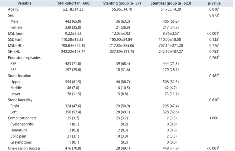
Table 1. Demographic data and success rate comparisons between stenting and stentless groups for the total cohort
Variable Total cohort (n=680) Stenting group (n=57) Stentless group (n=623) p-value
Age (y) 52.18±14.33 56.86±14.10 51.75±14.29 0.010a Sex 0.873b Male 442 (65.0) 36 (63.2) 406 (65.2) Female 238 (35.0) 21 (36.8) 217 (34.8) MSL (mm) 9.22±3.92 12.03±6.02 8.96±3.57 <0.001a SSD (cm) 110.50±19.22 105.90±24.84 110.00±18.58 0.135a MSD (HU) 708.04±272.19 717.86±285.06 707.14±271.20 0.776a SHI (HU) 242.22±108.47 237.89±121.75 242.62±107.27 0.753a
Prior stone episodes 0.763b
FSF 483 (71.0) 39 (68.4) 444 (71.3) RSF 197 (29.0) 18 (31.6) 179 (28.7) Stone location 0.482b Upper 554 (81.5) 46 (80.7) 508 (81.5) Middle 48 (7.0) 6 (10.5) 42 (6.7) Lower 78 (11.5) 5 (8.8) 73 (11.7) Stone laterality 0.610b Right 324 (47.6) 29 (50.9) 295 (47.4) Left 356 (52.4) 28 (49.1) 328 (52.6) Complication rate 25 (3.7) 23 (3.7) 2 (3.5) 1.000 Pyelonephritis 1 (0.1) 1 (0.2) 0 (0.0) Hematuria 2 (0.3) 2 (0.3) 0 (0.0) Colic pain 21 (3.1) 19 (3.0) 2 (3.5) GI symptoms 1 (0.1) 1 (0.2) 0 (0.0) One-session success 476 (70.0) 28 (49.1) 448 (71.9) <0.001b
Values are presented as mean±standard deviation or number (%).
MSL, maximal stone length; SSD, skin-to-stone distance; MSD, mean stone density; SHI, stone heterogeneity index; FSF, first-time stone formers; RSF, recurrent stone formers; HU, Hounsfield units.
the receiver operator characteristic (ROC) curves using Youden methods. Statistical analyses were performed using R software (ver. 3.0.3, R Foundation for Statistical Computing, Vienna, Austria; http://www.r-project.org) and its OptimalCutpoints package for optimal cutoff value.
RESULTS
Table 1 lists the baseline characteristics of the 680 patients who underwent primary SWL for a single ureteral calculus. The overall incidence of stenting during SWL for ureteral calculi was 8.3% (n=57). Comparisons between the stenting group and stentless groups based on patient and stone NCCT characteristics demonstrated that patient age and stone MSL were significantly different between the 2 groups. Stenting patients had a significantly longer MSL (12.03±6.02 mm in the stenting group, 8.96±3.57 mm in the stentless group, p<0.001). There were no significant differences between groups for SSD (105.90±24.84 cm in the stenting group, 110.00±18.58 cm in the stentless group, p=0.135), MSD (717.86±285.06 HU in the stenting group, 707.14±271.20 HU in the stentless group, p=0.776), and SHI (237.89±121.75 HU in the stenting group, 242.62±107.27 in the stentless group, p=0.753). The number of previous stone
episodes, stone location, and stone laterality demonstrated no differences between groups. Complication rate and each variables including post-SWL complication did not show significant differences between 2 groups. One-session success was significantly lower in the stenting group: 28 cases (49.1%) in the stenting group and 448 cases (71.9%) in the stentless group (p<0.001) (Table 1).
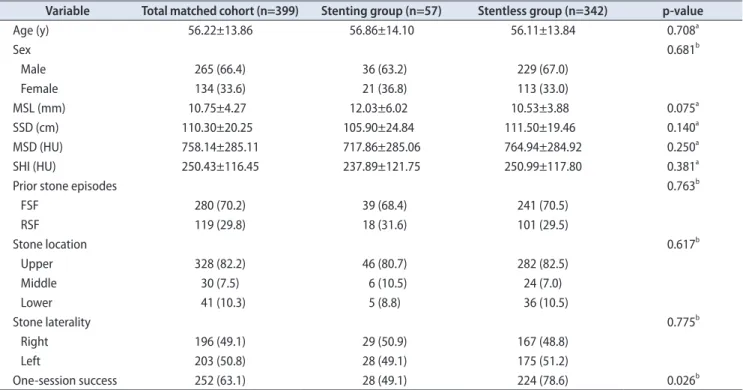
After stenting and stentless cases were 1:6 propensity-matched, the one-session success rate of the stentless group (224 cases, 78.6%) was higher than that of the stenting group (28 cases, 49.1%) (p=0.026) (Table 2).
The univariate logistic regression models revealed the following predictive factors of one-session success following SWL for ureteral stones: shorter MSL (odds ratio [OR], 0.832; 95% confidence interval [CI], 0.792–0.872; p<0.001), lower MSD (OR, 0.997; 95% CI, 0.996–0.998; p<0.001), higher SHI (OR, 1.003; 95% CI, 1.001–1.005; p<0.001) and absence of a stent (OR, 0.377; 95% CI, 0.217–0.653; p<0.001). Multivariate analyses also demonstrated that a shorter MSL, lower MSD, higher SHI, and stentless cases were independent predictors of one-session success after SWL for ureteral calculi (Table 3).
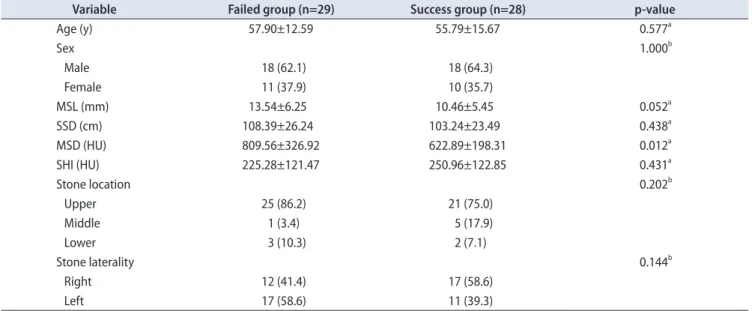
In stenting cases, one-session failed and success groups were divided for subgroup analyses. Between failed and success groups in stenting cases, MSL and MSD
Table 2. Demographic data and success rate comparisons between stenting and stentless groups for the propensity-matched cohort
Variable Total matched cohort (n=399) Stenting group (n=57) Stentless group (n=342) p-value
Age (y) 56.22±13.86 56.86±14.10 56.11±13.84 0.708a Sex 0.681b Male 265 (66.4) 36 (63.2) 229 (67.0) Female 134 (33.6) 21 (36.8) 113 (33.0) MSL (mm) 10.75±4.27 12.03±6.02 10.53±3.88 0.075a SSD (cm) 110.30±20.25 105.90±24.84 111.50±19.46 0.140a MSD (HU) 758.14±285.11 717.86±285.06 764.94±284.92 0.250a SHI (HU) 250.43±116.45 237.89±121.75 250.99±117.80 0.381a
Prior stone episodes 0.763b
FSF 280 (70.2) 39 (68.4) 241 (70.5) RSF 119 (29.8) 18 (31.6) 101 (29.5) Stone location 0.617b Upper 328 (82.2) 46 (80.7) 282 (82.5) Middle 30 (7.5) 6 (10.5) 24 (7.0) Lower 41 (10.3) 5 (8.8) 36 (10.5) Stone laterality 0.775b Right 196 (49.1) 29 (50.9) 167 (48.8) Left 203 (50.8) 28 (49.1) 175 (51.2) One-session success 252 (63.1) 28 (49.1) 224 (78.6) 0.026b
Values are presented as mean±standard deviation or number (%).
MSL, maximal stone length; SSD, skin-to-stone distance; MSD, mean stone density; SHI, stone heterogeneity index; FSF, first-time stone formers; RSF, recurrent stone formers; HU, Hounsfield units.
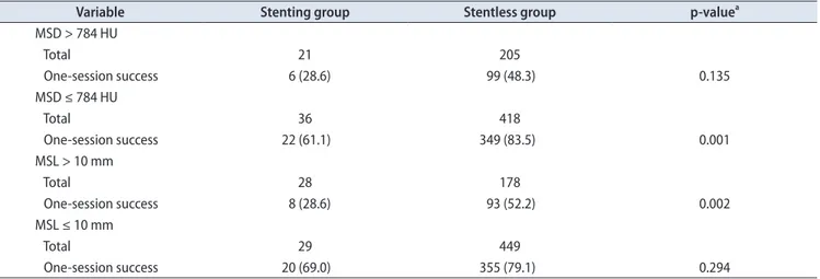
demonstrated the significant differences (Table 4). For one-session success rates, the area under the curve (AUC) of ROC curves was 0.689 (95% CI, 0.635–0.742) for MSL and 0.686 (95% CI, 0.632–0.740) for MSD. The cutoff values for MSL and MSD were 10.0 mm and 784 HU, respectively. As shown in Table 5, the number of patients with one-session success
status was higher in the stentless group than in the stenting group for patients with an MSL >10 mm (p=0.002) or an MSD ≤784 HU (p=0.001).
Table 3. Univariate and multivariate logistic regression models for predictive factors of one-session success following shock wave lithotripsy for ureteral stones
Parameter Odds ratio 95% CI p-value
Univariate Age 1.006 0.995–1.018 0.275 Male sex 0.793 0.557–1.122 0.195 MSL 0.832 0.792–0.872 <0.001 MSD 0.997 0.996–0.998 <0.001 SSD 1.001 0.992–1.010 0.799 SHI 1.003 1.001–1.005 <0.001
Recurrent stone formers 1.152 0.802–1.670 0.452
Stone location (%) Upper Reference Middle 0.683 0.373–1.281 0.222 Lower 0.868 0.527–1.466 0.588 Laterality, right 1.204 0.866–1.674 0.269 Stenting 0.377 0.217–0.653 <0.001 Multivariate MSL 0.901 0.854–0.948 <0.001 MSD 0.996 0.994–0.997 <0.001 SHI 1.010 1.007–1.012 <0.001 Stenting 0.432 0.217–0.863 0.017
CI, confidence interval; MSL, maximal stone length; MSD, mean stone density; SSD, skin-to-stone distance; SHI, stone heterogeneity index.
Table 4. Demographic and factor comparisons between failed and success groups in stenting patients
Variable Failed group (n=29) Success group (n=28) p-value
Age (y) 57.90±12.59 55.79±15.67 0.577a Sex 1.000b Male 18 (62.1) 18 (64.3) Female 11 (37.9) 10 (35.7) MSL (mm) 13.54±6.25 10.46±5.45 0.052a SSD (cm) 108.39±26.24 103.24±23.49 0.438a MSD (HU) 809.56±326.92 622.89±198.31 0.012a SHI (HU) 225.28±121.47 250.96±122.85 0.431a Stone location 0.202b Upper 25 (86.2) 21 (75.0) Middle 1 (3.4) 5 (17.9) Lower 3 (10.3) 2 (7.1) Stone laterality 0.144b Right 12 (41.4) 17 (58.6) Left 17 (58.6) 11 (39.3)
Values are presented as mean±standard deviation or number (%).
MSL, maximal stone length; SSD, skin-to-stone distance; MSD, mean stone density; SHI, stone heterogeneity index; HU, Hounsfield units. a:Based on Student or Welch's two-sample t-tests. b:Based on Pearson chi-square tests with Yates' continuity correction.
DISCUSSION
Since the introduction of ureteroscopy, SWL, and percutaneous nephrolithotomy, researchers have expended much effort to determine the factors associated with high success and low complication rates in the treatment of urinary stone disease. For SWL, factors reported to influence success and complication rates include the stone size, composition, density, and location; total number and frequency of shock waves; operators’ experience; and type of lithotripter. Pre-SWL ureteral stenting has also been proposed as an important way to reduce complications following SWL.
A primary rationale for performing ureteral stenting is to prevent complications associated with ureteral obstruction as stone fragments pass down the ureter. Several previous studies investigated the efficacy of ureteral stent in preventing these complications. Mohayuddin et al. [19] reported that steinstrasse and fever were not affected by whether or not a ureteral stent was used, but ureteral colic was significantly lower in their ureteral stenting group. In a prospective randomized clinical trial, the ureteral stenting group exhibited lower rates of hospital readmission and Emergency Department visits, and 13% of the stentless group but only 2% of the stenting group had steinstrasse formation [10]. In a recent meta-analysis, the authors noted that the steinstrasse rate was significantly lower in the stenting group, whereas fever, urinary tract infection, pain, and auxiliary treatment did not differ between groups [7]. From these results about post-SWL complications, the effects of ureteral stent for passage of fragments during SWL seem to be controversial.
Until now, reducing complications following SWL has been one of the most important goals of SWL for urinary stone management, although achieving stone-free status is the ultimate objective. Unfortunately, few reports have indicated that ureteral stents increase stone-free status or success rates following SWL. Several previous studies demonstrated that ureteral stents do not improve SWL success or SFR [9,10,19-21]. Rather, recent studies found that ureteral stents negatively affect SWL success or SFR. For example, Pettenati et al. [12] demonstrated that the presence of a ureteral stent negatively affects the efficacy of SWL in treating lumbar ureteral stones These authors found that the success rate with stenting was significant lower than the rate without stenting in patients with stones larger than 8 mm. Ozkan et al. [13] also reported that SFR was significantly higher in their stentless group than in their stenting group. Furthermore, in 2 randomized controlled trials, ureteral stenting was found to reduce the SFR [11], and the absence of an indwelling ureteral stent was an independent predictor of success [22].
Our results were similar to those of previous studies, supporting the negative effects of ureteral stenting on SFR. Although MSL, which can affect SFR, differed significantly between the stenting and stentless groups in our total cohort, the presence of a ureteral stent was the only factor that differed significantly between groups after 1:6 propensity score matching. The negative effects of stenting may be explained by 2 theories, which are not mutually exclusive. First, in order to have a maximal effect, shock waves must impinge on a stone surrounded by liquid. The ureteral stent may absorb some of the energy created by the shock waves, thus reducing their effect on the stones [9].
Table 5. Comparison of stone-free status in stenting and stentless group according to optimal cutoff value for MSD and MSL
Variable Stenting group Stentless group p-valuea
MSD > 784 HU Total 21 205 One-session success 6 (28.6) 99 (48.3) 0.135 MSD ≤ 784 HU Total 36 418 One-session success 22 (61.1) 349 (83.5) 0.001 MSL > 10 mm Total 28 178 One-session success 8 (28.6) 93 (52.2) 0.002 MSL ≤ 10 mm Total 29 449 One-session success 20 (69.0) 355 (79.1) 0.294
Values are presented as number or number (%).
MSD, mean stone density; MSL, maximal stone length; HU, Hounsfield units. a:Pearson chi-square test with Yates' continuity correction.
Second, the presence of a ureteral stent may cause ureteral edema, thus interrupting the passage of stone fragments [23-25]. The impact would be greater with ureteral stones than renal stones because the area of contact between the stone and stent would be larger and ureteral edema would have a greater effect on ureteral stones. Very few studies have distinguished between renal stones and ureteral stones.
Based on our findings and results of previous studies, pre-SWL ureteral stenting does not appear to have definite advantages in terms of SFR or complications, compared to in situ SWL. In addition, the decision to proceed with ureteral stenting requires much caution because it is a relatively invasive procedure. Furthermore, we should also consider the possibility of stent-related voiding symptoms, such as bladder irritation symptoms and flank pain or discomfort [26,27]. In their randomized control study, Ghoneim et al. [9] noted that microscopic hematuria, pyuria, dysuria, and suprapubic pain were significantly more common in patients with a ureteral stent than in those without. Another study, by El-Assmy et al. [20], of patients with ureteral stones 2 cm or less causing moderate or severe obstruction also showed that the rates of post-SWL morbidities related to ureteral stents (such as suprapubic pain, gross or microscopic hematuria, pyuria, and positive urine cultures) were significantly higher in their ureteral stenting group. Thus, we recommend against routine pre-SWL ureteral stenting; instead, ureteral stents should be reserved for special indications, such as complicated urinary tract infection or severe pain.
A unique aspect of our study is that we calculated the AUC and cutoff values for MSL and MSD, and then analyzed the SWL success rates according to the presence of a stent in subgroups based on these cutoff values. Since NCCT was introduced in the management of urinary tract stone disease, MSL and MSD have been widely recognized as predictors of SWL success rate [15,28-30]. In our multivariate logistic regression analysis, a shorter MSL, lower MSD, and absence of a stent were positive predictors for one-session success in patients who underwent SWL, which is consistent with the results of previous studies. The AUCs for MSL and MSD were high (0.689 and 0.686, respectively), compared to the AUCs for other factors. The cutoff values for MSL and MSD were 10.0 mm and 784 HU, respectively. When we analyzed the success rate of subgroups based on these cutoff values, the results for MSL were interestingly contrary to those observed for MSD. In patients with an MSL>10 mm, the success rate was significantly lower in the stenting group, but in those with an MSL≤10 mm, there was no difference in success rate between the 2 groups. These findings are similar to those of Pettenati et al. [12], in
which SWL success was lower in the stenting group only with larger stones, defined as a size >8 mm. The differential effects of MSL may be explained to some degree by the theories mentioned above. Larger stones have a wider area of contact between the stone and stent, which presumably leads to greater energy absorption by the stent, thus increasing the likelihood of impaired stone fragmentation. By contrast, the success rate was higher in the stentless group than in the stent group in patients with a lower MSD (≤784 HU), representing a more fragile stone. If a stone is fragile, the stent can play a more significant role as an interference factor, thus promoting treatment failure.
Our results thus suggest that when physicians are deciding whether to perform ureteral stent insertion before SWL, they may consider MSL and MSD as factors influencing SWL failure (although further study is required to more definitively address this issue). In patients with a ureteral stone with a low MSD and large size, the decision to perform pre-SWL stenting should be based on symptoms and renal function of the patient. In addition, when physicians are trying to choose between treatment options, including retrograde ureteroscopic surgery and SWL, our overall higher one-session success rates with stones exhibiting a lower MSD and lower MSL (including both the stenting and stentless groups) suggest that it may be more appropriate to perform surgery in patients with stones with a high MSD and large volume.
This study has some inherent limitations because of its retrospective design, which may have introduced sampling bias; however, we used a relatively large cohort of patients undergoing SWL for ureteric stones. In addition, to overcome this type of limitation and elucidate the impact of MSL and MSD on SWL outcomes more clearly, we limited the study to include subjects who had only ureteric stones. With renal stones, anatomical considerations including the location of calyx and renal pelvic stones or the infundibulopelvic angle can be another source of bias. Furthermore, we could not analyze the reasons for stenting due to retrospective design. This may work as a significant bias because renal function or hydronephrosis grade which acts main causes of prestenting can be impact factors of SWL success rate. The 2 different lithotripsy machines may be a source of bias, but there were no statistical differences between the 2 time periods (data not shown). Despite these limitations, the study has certain strengths, including our focus on the results of SFR as a more important goal than complication rates and our analysis of one-session success rates following SWL according to stone characteristics (MSL and MSD) in relation to the presence of a stent. In the future, large
prospective studies are needed to confirm our observations on the negative effect of ureteral stenting on SFR.
CONCLUSIONS
Ureteral stenting during SWL was a negative predictive factor for one-session success in patients with a single ureteral stone. Furthermore, in patients with stones that exhibited a lower MSD and higher MSL, ureteral stenting negatively influenced one-session outcomes, compared to SWL without stenting.
CONFLICTS OF INTEREST
The authors have nothing to disclose.ACKNOWLEDGMENTS
This study was supported by a faculty research grant from the Yonsei University College of Medicine for 2014 (6-2014-0156). This work was received Korean National Academic Award in the 2016 The Annual Meeting of Korean Endourological Society, Daegu, June 10-11, 2016.
REFERENCES
1. Teichman JM. Clinical practice: acute renal colic from ureteral calculus. N Engl J Med 2004;350:684-93.
2. Seitz C, Tanovic E, Kikic Z, Memarsadeghi M, Fajkovic H. Rap-id extracorporeal shock wave lithotripsy for proximal ureteral calculi in colic versus noncolic patients. Eur Urol 2007;52:1223-7.
3. Salem S, Mehrsai A, Zartab H, Shahdadi N, Pourmand G. Complications and outcomes following extracorporeal shock wave lithotripsy: a prospective study of 3,241 patients. Urol Res 2010;38:135-42.
4. Türk C, Petřík A, Sarica K, Seitz C, Skolarikos A, Straub M, et al. EAU Guidelines on diagnosis and conservative manage-ment of urolithiasis. Eur Urol 2016;69:468-74.
5. Morgentaler A, Bridge SS, Dretler SP. Management of the im-pacted ureteral calculus. J Urol 1990;143:263-6.
6. Kirkali Z, Esen AA, Akan G. Place of double-J stents in extra-corporeal shock wave lithotripsy. Eur Urol 1993;23:460-2. 7. Shen P, Jiang M, Yang J, Li X, Li Y, Wei W, et al. Use of
ure-teral stent in extracorporeal shock wave lithotripsy for upper urinary calculi: a systematic review and meta-analysis. J Urol 2011;186:1328-35.
8. Musa AA. Use of double-J stents prior to extracorporeal shock wave lithotripsy is not beneficial: results of a prospective
ran-domized study. Int Urol Nephrol 2008;40:19-22.
9. Ghoneim IA, El-Ghoneimy MN, El-Naggar AE, Hammoud KM, El-Gammal MY, Morsi AA. Extracorporeal shock wave lithotripsy in impacted upper ureteral stones: a prospective randomized comparison between stented and non-stented techniques. Urology 2010;75:45-50.
10. Chandhoke PS, Barqawi AZ, Wernecke C, Chee-Awai RA. A randomized outcomes trial of ureteral stents for extracorporeal shock wave lithotripsy of solitary kidney or proximal ureteral stones. J Urol 2002;167:1981-3.
11. Sfoungaristos S, Polimeros N, Kavouras A, Perimenis P. Stent-ing or not prior to extracorporeal shockwave lithotripsy for ureteral stones? Results of a prospective randomized study. Int Urol Nephrol 2012;44:731-7.
12. Pettenati C, El Fegoun AB, Hupertan V, Dominique S, Ravery V. Double J stent reduces the efficacy of extracorporeal shock wave lithotripsy in the treatment of lumbar ureteral stones. Cent European J Urol 2013;66:309-13.
13. Ozkan B, Dogan C, Can GE, Tansu N, Erozencı A, Onal B. Does ureteral stenting matter for stone size? A retrospectıve analyses of 1361 extracorporeal shock wave lithotripsy pa-tients. Cent European J Urol 2015;68:358-64.
14. Cho KS, Jung HD, Ham WS, Chung DY, Kang YJ, Jang WS, et al. Optimal skin-to-stone distance is a positive predictor for successful outcomes in upper ureter calculi following extra-corporeal shock wave lithotripsy: a bayesian model averaging approach. PLoS One 2015;10:e0144912.
15. Chung DY, Cho KS, Lee DH, Han JH, Kang DH, Jung HD, et al. Impact of colic pain as a significant factor for predicting the stone free rate of one-session shock wave lithotripsy for treat-ing ureter stones: a Bayesian logistic regression model analysis. PLoS One 2015;10:e0123800.
16. Lee JY, Kim JH, Kang DH, Chung DY, Lee DH, Do Jung H, et al. Stone heterogeneity index as the standard deviation of Hounsfield units: a novel predictor for shock-wave lithotripsy outcomes in ureter calculi. Sci Rep 2016;6:23988.
17. Rubin DB, Thomas N. Matching using estimated propensity scores: relating theory to practice. Biometrics 1996;52:249-64. 18. Lee JY, Diaz RR, Cho KS, Yu HS, Chung JS, Ham WS, et al.
Lymphocele after extraperitoneal robot-assisted radical prosta-tectomy: a propensity score-matching study. Int J Urol 2013;20: 1169-76.
19. Mohayuddin N, Malik HA, Hussain M, Tipu SA, Shehzad A, Hashmi A, et al. The outcome of extracorporeal shockwave lithotripsy for renal pelvic stone with and without JJ stent--a comparative study. J Pak Med Assoc 2009;59:143-6.
20. El-Assmy A, El-Nahas AR, Sheir KZ. Is pre-shock wave litho-tripsy stenting necessary for ureteral stones with moderate or severe hydronephrosis? J Urol 2006;176:2059-62.
21. Mustafa M, Ali-El-Dein B. Stenting in extracorporeal shock-wave lithotripsy; may enhance the passage of the fragments! J Pak Med Assoc 2009;59:141-3.
22. Nguyen DP, Hnilicka S, Kiss B, Seiler R, Thalmann GN, Roth B. Optimization of extracorporeal shock wave lithotripsy delivery rates achieves excellent outcomes for ureteral stones: results of a prospective randomized trial. J Urol 2015;194:418-23. 23. Abdel-Khalek M, Sheir K, Elsobky E, Showkey S, Kenawy M.
Prognostic factors for extracorporeal shock-wave lithotripsy of ureteric stones: a multivariate analysis study. Scand J Urol Nephrol 2003;37:413-8.
24. Singh I, Gupta NP, Hemal AK, Dogra PN, Ansari MS, Seth A, et al. Impact of power index, hydroureteronephrosis, stone size, and composition on the efficacy of in situ boosted ESWL for primary proximal ureteral calculi. Urology 2001;58:16-22. 25. Joshi HB, Obadeyi OO, Rao PN. A comparative analysis of
nephrostomy, JJ stent and urgent in situ extracorporeal shock wave lithotripsy for obstructing ureteric stones. BJU Int 1999;84:264-9.
26. Kwon JK, Cho KS, Oh CK, Kang DH, Lee H, Ham WS, et al. The beneficial effect of alpha-blockers for ureteral stent-related discomfort: systematic review and network meta-analysis for alfuzosin versus tamsulosin versus placebo. BMC Urol 2015;15: 55.
27. Abt D, Mordasini L, Warzinek E, Schmid HP, Haile SR, Engeler DS, et al. Is intravesical stent position a predictor of associated morbidity? Korean J Urol 2015;56:370-8.
28. Perks AE, Gotto G, Teichman JM. Shock wave lithotripsy cor-relates with stone density on preoperative computerized to-mography. J Urol 2007;178(3 Pt 1):912-5.
29. El-Nahas AR, El-Assmy AM, Mansour O, Sheir KZ. A pro-spective multivariate analysis of factors predicting stone disin-tegration by extracorporeal shock wave lithotripsy: the value of high-resolution noncontrast computed tomography. Eur Urol 2007;51:1688-93.
30. Pareek G, Armenakas NA, Fracchia JA. Hounsfield units on computerized tomography predict stone-free rates after extra-corporeal shock wave lithotripsy. J Urol 2003;169:1679-81.