저작자표시-비영리-동일조건변경허락 2.0 대한민국 이용자는 아래의 조건을 따르는 경우에 한하여 자유롭게 l 이 저작물을 복제, 배포, 전송, 전시, 공연 및 방송할 수 있습니다. l 이차적 저작물을 작성할 수 있습니다. 다음과 같은 조건을 따라야 합니다: l 귀하는, 이 저작물의 재이용이나 배포의 경우, 이 저작물에 적용된 이용허락조건 을 명확하게 나타내어야 합니다. l 저작권자로부터 별도의 허가를 받으면 이러한 조건들은 적용되지 않습니다. 저작권법에 따른 이용자의 권리는 위의 내용에 의하여 영향을 받지 않습니다. 이것은 이용허락규약(Legal Code)을 이해하기 쉽게 요약한 것입니다. Disclaimer 저작자표시. 귀하는 원저작자를 표시하여야 합니다. 비영리. 귀하는 이 저작물을 영리 목적으로 이용할 수 없습니다. 동일조건변경허락. 귀하가 이 저작물을 개작, 변형 또는 가공했을 경우 에는, 이 저작물과 동일한 이용허락조건하에서만 배포할 수 있습니다.
The Relationship Between Volumetric Plaque
Components and Classical Cardiovascular Risk
Factors and the Metabolic Syndrome
by
Mingri Zheng
Major in Medicine
Department of Medical Sciences
The Graduate School, Ajou University
The Relationship Between Volumetric Plaque Components
and Classical Cardiovascular Risk Factors and the
Metabolic Syndrome:
A Three-Vessel Coronary
Artery Virtual
Histology-Intravascular Ultrasound Analysis
by
Mingri Zheng
A Dissertation Submitted to The Graduate School of
Ajou University in Partial Fulfillment of the Requirements
for the Degree of Ph. D. in Medicine
Supervised by
Seung-Jea Tahk, M.D., Ph.D.
Major in Medicine
Department of Medical Sciences
The Graduate School, Ajou University
This certifies that the dissertation
of Mingri Zheng is approved.
SUPERVISORY COMMITTEE
Seung-Jea Tahk
Myeong-Ho Yoon
So-Yeon Choi
In-Ho Chae
Hong-seak Lim
The Graduate School, Ajou University
June 23rd, 2011
Acknowledgement
First and foremost, I would like to express my deepest gratitude to my
supervisor, Mr. Seung-Jea Tahk, a respectable, responsible and resourceful
scholar who has offered me valuable guidance in the academic studies.
I owe a special debt of gratitude to Professor So-Yeon Choi who provided
me with valuable suggestion in every stage of writing of this thesis.
I’m also extremely grateful to many people whose kindness and patient
guidance and invaluable suggestions are indispensable to the completion of
this thesis.
I should finally like to express my gratitude to my family, especially to
my wife who have always been helping me out of difficulties and supporting
without a word of complaint.
- ABSTRACT -
The Relationship Between Volumetric Plaque Components and
Classical Cardiovascular Risk Factors and the Metabolic
Syndrome: A Three-Vessel Coronary Artery Virtual
Histology-Intravascular Ultrasound Analysis
Objectives: The aim of this study was to analyze volumetric plaque composition of the coronary arterial tree according to the classical cardiovascular risk factors and metabolic syndrome (MS) by using virtual histology-intravascular ultrasound (VH-IVUS).
Background: It remains unclear how the cardiovascular risk factors correlate with the histological components of coronary plaques.
Methods: “Whole vessel” VH-IVUS analysis was performed in 189 vessels of 63 patients. The components of atherosclerotic plaques were classified as fibrous, fibrofatty, necrotic core (NC), and dense calcium. Quantitative assessment of these plaque components and the
presence of VH-IVUS-derived thin-cap fibroatheroma (VH-TCFA) in the coronary arterial trees were compared to cardiovascular risk factors.
Results: There was a significantly larger mean plaque+media (P+M) burden in patients with diabetes mellitus (DM) (47±5 vs. 39±7% in non-DM patients, p<0.001) and MS (47±4 vs. 39±7% in non-MS patients, p<0.001). DM patients had a significantly larger %NC (17.8±5.6 vs. 12.5±6.1, p=0.003) compared to non-DM patients; and MS patients had a significantly larger %NC (17.3±5.8 vs. 12.8±6.2, p=0.016) as compared to non-MS patients. Finally, VH-TCFAs were more frequent in DM patients (3.4±2.0 vs. 2.1±1.7 in non-DM patients,
p=0.016) and in MS patients (4.1±2.1 vs. 1.9±1.4 in non-MS patients, p=0.001).
Conclusions: Three-vessel VH-IVUS analysis showed that DM and MS patients had a larger P+M burden, larger amount of NC, and more frequent VH-TCFAs in coronary arterial trees compared to patients without DM or MS implying greater plaque vulnerability in DM and MS patients.
Key Words: virtual histology intravascular ultrasound; cardiovascular risk factors; coronary artery disease; imaging; plaque vulnerability
TABLE OF CONTENTS
ABSTRACT ··· ⅰ TABLE OF CONTENTS ··· ⅲ LIST OF FIGURES ··· ⅴ LIST OF TABLES ··· ⅵ ABBREVIATIONS ··· ⅶ I. INTRODUCTION ··· 1II. MATERIALS AND METHODS ··· 3
A. MATERIALS ··· 3
1. Study population ··· 3
2. Definition of the risk factors ··· 3
B. METHODS ··· 4
3. IVUS imaging and analysis ··· 4
4. Statistical analysis ··· 6
III. RESULTS ··· 8
A. The baseline clinical and VH-IVUS characteristics ··· 8
B. IVUS and VH-IVUS findings according to the classical risk factors and the metabolic risk factors associated to metabolic syndrome ··· 11
VH-TCFAs ··· 20 IV. DISCUSSION ···22 V. STUDY LIMITATION ··· 27 VI. CONCLUTION ··· 29 REFERENCES ··· 30 국문요약 ··· 36
LIST OF FIGURES
Fig. 1. VH-TCFAs in regard to being with or without risk factors.··· 19 Fig. 2. The correlation between the plaque components and the metabolic syndrome
LIST OF TABLES
Table. 1. Baseline data for conventional cardiovascular risk factors and metabolic
risk factors associated with MS (n=63) ··· 9 Table. 2. Baseline lesion characteristics in the VH-IVUS analysis (n=63) ··· 10 Table. 3. Volume indices of VH-IVUS quantitative findings in regard to being with
or without cardiovascular risk factors (n=63) ··· 13 Table. 4. The mean percentages of plaque burden and VH-IVUS components and the
VH-TCFAs according to each risk factor associated with metabolic syndrome (n=63) ··· 16 Table. 5. Multivariate predictors of the plaque burden, the % necrotic core and the
ABBREVIATIONS
DC = dense calcium DM = diabetes mellitus FF = fibrofatty
FI = fibrous
IVUS= intravascular ultrasound
LAD= left anterior descending coronary artery LCX= left circumflex coronary artery
MS = metabolic syndrome
MDCT = multi-detector computed tomography NC = necrotic core
P+B = plaque and media RCA= right coronary artery
VH-IVUS = virtual histology-intravascular ultrasound
I. INTRODUCTION
According to autopsy data, the fate of atherosclerosis is related to the composition of the plaque(Davies et al,1993; Felton et al,1997). Recognizing the histological characteristics of coronary plaques using an in vivo diagnostic modality may be a key to a lesion-specific treatment strategy of patients with coronary artery disease(Komiyama et al, 2000). Tomographic images of intravascular ultrasound (IVUS) are widely used for assessing the morphological characteristics of vessels(Nissen et al, 1991; von Birgelen et al, 1997). however, conventional gray-scale IVUS has significant limitations for identifying specific plaque components(Yock and Linker, 1991; DiMario C et al, 1998). Spectral analysis of the radiofrequency ultrasound backscatter signals known as virtual histology-IVUS (VH-IVUS) allows identification of four different components of atherosclerotic plaques: fibrous tissue (FI), fibro-fatty plaque (FF), dense calcium (DC), and necrotic core (NC)(Rodriguez-Granillo et al, 2005; Nair et al, 2007). While the relationship between cardiovascular risk factors and the risk of coronary events is well established, it remains unclear how these risk factors correlate with the histological components of coronary artery plaques in vivo. To the best of our knowledge, no three-vessel volumetric VH-IVUS study has yet reported the relationship between plaque components and cardiovascular risk factors. The aim of the current study was to evaluate plaque composition in the coronary arterial tree using volumetric three-vessel VH-IVUS according to classical cardiovascular risk factors as well
II. MATERIALS AND METHODS
A. MATERIALS 1. Study population
Between September 2006 and August 2008, a preintervention three-vessel VH-IVUS examination was successfully performed in 63 consecutive patients who were diagnosed with ischemic heart disease for the first time at a single medical center. On the diagnostic coronary angiography, there were one or more native coronary arteries with ≥30% luminal stenosis by visual estimation. The primary aim of this prospective study was to determine the clinical correlation of the VH-IVUS parameters in a consecutive, non-selected population of patients. Written informed consent was obtained from all patients. The patients with chronic total occlusion, severe vessel tortiousity, extensively calcified lesions, severe left main coronary artery disease(diameter stenosis ≥50%) and haemodynamic instability were excluded in this study.
2. Definition of the risk factors
Old age was defined as ≥ 65years, hypercholesterolemia was defined as total cholesterol level ≥ 220 mg/dl, smoking was defined as current smoking, a family history of coronary artery disease was defined as premature of coronary artery disease in a first-degree relative(in a male<55 years; in a female <65 years); hypertension was defined as a systolic
blood pressure ≥140 mm Hg or a diastolic blood pressure ≥90 mm Hg, or the use of any antihypertensive drug, diabetes mellitus(DM) was defined as a confirmed diagnosis or using antidiabetic medication(insulin or oral hypoglycemic) at entry into the study. The definition of the metabolic syndrome met the Adult Treatment Panel III(ATP III) of the National Cholesterol Education Program. The waist circumference criteria was replaced by >90 cm for males and >80 cm for females.
B. METHODS
3. IVUS imaging and analysis
The VH-IVUS studies were performed with a phased-array, 20 MHz, 2.9F IVUS
catheter(Eagle Eye, Volcano Corporation, Rancho Cordova, CA, USA). After intracoronary administration of 0.2 mg nitroglycerin, the transducer was introduced up to the distal coronary bed of the each major epicardial artery in coronary tree. Using motorized pull-back system(0.5 mm/s), the imaging was performed back to aorta-ostial junction. During pull-back, the grayscale IVUS was recorded and the raw radiofrequency data was captured at the top of R wave for the reconstruction of color-coded map by VH data recorder(Volcano Corporation).
Manual contour revision of the lumen and the media-adventitia interface was performed by an experienced analyst. Volumetric VH-IVUS analysis was performed from distal start-point to respective ostium, and the volumetric data was generated with using pcVH 2.1
analyzing three vessels in each patient, and the volume index was calculated as the total plaque volume / total vessel length analyzed. The mean plaque burden(PB) was calculated as the total plaque volume/total vessel volume×100. The VH-IVUS analysis classified the color-coded tissue as green(FI), yellow-green(FF), red(NC), and white(DC). Each plaque component was measured in every recorded frame and it was expressed as the volume index and the percentages of the total plaque volume. VH-IVUS-derived thin-cap fibroatheroma (VH-TCFA) was defined as a lesion that fulfilled the following criteria in at least three consecutive frames: 1) necrotic cores ≥10% with directly attaching to the lumen and 2) ≥40% PB(Rodriguez-Granillo et al, 2005). It was reported that identifying of 2 separate plaques in the same artery required a ≥5 mm reference segment between them; if not, they were considered to be part of one long lesion(Alberti et al, 2009). Referring to these criteria, 2 separate VH-TCFAs required a reference segment ≥5 mm between them. Two experienced observers, who were unaware of the patients’ clinical histories, evaluated the VH-TCFA in consensus. Volumetric VH-IVUS analyses of all the plaques of the three vessels were compared to the cardiovascular risk factors.
4. Statistical analysis
Statistical analysis was performed with SPSS software, version 13.0(Chicago, IL, USA). The categorical data is expressed as numbers or the frequencies of occurrence. The continuous data is reported as means ± SDs. Comparisons were performed with using two-tailed unpaired Student’s t tests. The correlation between two parameters was assessed by
linear regression analysis. Multivariate analysis used multiple linear regression analysis and the variables with a p value <0.15 on univariate analysis were included. A p value <0.05 was considered statistically significant
.
III. RESULTS
A. The baseline clinical and VH-IVUS characteristics
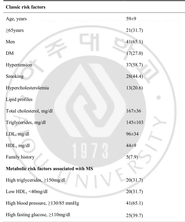
Baseline clinical characteristics are listed in Table 1. The study subjects had a mean age of 59 years, 31.7% of the patients were ≥65years, 65.1% were male, 27.0% had a history of DM, 58.7% had a history of hypertension, 44.4% had a history of smoking, 20.6% had a history of hypercholesterolemia, 7.9% had a family history of coronary artery disease, 25.4% had MS. Forty nine patients(77.8%) were diagnosed with acute coronary syndrome in this population. The analyzed vessel length was 56.1±17.4mm for the LAD, 51.9±19.0mm for the LCX, and 74.2±18.8mm for the RCA(Table 2). The number of VH-TCFAs was 1.0±0.8 for the LAD, 0.6±0.7 for the LCX and 0.8±1.0 for the RCA.
Table 1. Baseline data for conventional cardiovascular risk factors and metabolic risk factors associated with MS(n=63).
Classic risk factors
Age, years 59±9 ≥65years 21(31.7) Men 41(65.1) DM 17(27.0) Hypertension 37(58.7) Smoking 28(44.4) Hypercholesterolemia 13(20.6) Lipid profiles Total cholesterol, mg/dl 167±36 Triglycerides, mg/dl 145±103 LDL, mg/dl 96±34 HDL, mg/dl 44±9 Family history 5(7.9)
Metabolic risk factors associated with MS High triglycerides, ≥150mg/dl 20(31.7)
Low HDL, <40mg/dl 20(31.7) High blood pressure, ≥130/85 mmHg 41(65.1) High fasting glucose, ≥110mg/dl 25(39.7)
Abdominal obesity, WC ≥90cm in male or ≥80cm in female 23(36.5) MS scores 0 3(4.8) 1 16(25.4) 2 28(44.4) 3 10(15.9) 4 3(4.8) 5 3(4.8)
Values are given as n(%) or mean ± standard deviation.
DM = diabetes mellitus; LDL = low-density lipoprotein; HDL = high-density lipoprotein; MS = metabolic syndrome; WC = waist circumference.
Table 2. Baseline lesion characteristics in the VH-IVUS analysis(n=63) No. of vessels with >60% plaque burden* (n=63)
0 15(23.8)
1 16(25.4)
2 18(28.6)
3 14(22.6)
Mean analyzed vessel length, mm
Total length per patient 182.6±45.4
LAD 56.1±17.4
LCX 51.9±19.0
RCA 74.2±18.8
VH-TCFA, n
Total number per patient 2.5±1.9
LAD 1.0±0.8
LCX 0.6±0.7
RCA 0.8±1.0
Values are given as n(%) or mean ± standard deviation.
* Number of vessels with >60% plaque burden was defined based on maximum plaque burden in each vessel. LAD = left anterior descending coronary artery; LCX = left circumflex coronary artery; RCA = right coronary artery; VH-IVUS = virtual histology-intravascular ultrasound; VH-TCFA = virtual histology-histology-intravascular ultrasound-derived thin-cap fibroatheroma.
B. IVUS and VH-IVUS findings according to classical risk factors and risk factors associated to the metabolic syndrome.
The quantitative volumetric VH-IVUS findings with regard to the presence or absence of each cardiovascular risk factor are shown in Table 3. Total analyzed vessel length was not different regardless of any risk factor(data not shown). Patients with DM had larger absolute measures of NC(p=0.032) than those without DM; and patients with MS had larger normalized plaque mass(p<0.001) and larger absolute measures of NC(p=0.008) and DC(p=0.032) than patients without MS. Mean P+M burden was significantly correlated with increasing patient age(r=0.292, p<0.05). In addition, patients with DM had significantly larger mean P+M burden(47±5% vs. 39±7%, p<0.001) compared to non-DM patients; and MS patients had significantly larger mean P+M burden(47±4% vs. 39±7%, p<0.001) compared to the non-MS patients. Mean P+M burden did not correlate with any other risk factors(data not shown).
Patients with DM had significantly larger values of mean %NC(17.8±5.6% vs. 12.5±6.1%, p=0.003) and mean %DC(10.7±5.4% vs. 7.7±5.3%, p=0.032). MS patients also had significantly larger mean %NC(17.3±5.8% vs. 12.8±6.2%, p=0.016) and mean %DC(10.6±4.6% vs. 7.4±5.5%, p=0.052) as compared to non-MS patients.
Table 3. Volume indices of VH-IVUS quantitative findings in regard to being with or without cardiovascularrisk factors(n=63).
P+M volume index (mm3/mm) FI volume index (mm3/mm) FF volume index (mm3/mm) NC volume index (mm3/mm) DC volume index (mm3/mm) ≥65 years 6.4[5.5-7.2] 1.9[1.3-2.3] 0.4[0.2-0.6] 0.4[0.3-0.6] 0.2[0.1-0.3] <65 years 5.6[4.7-7.2] 1.6[0.9-2.7] 0.3[0.2-0.4] 0.3[0.2-0.5] 0.2[0.1-0.3] p value 0.205 0.937 0.773 0.261 0.504 pc value 1.000 1.000 1.000 1.000 1.000 Man 6.1[5.1-7.2] 1.7[1.1-2.4] 0.3[0.2-0.5] 0.4[0.2-0.5] 0.2[0.1-0.3] Woman 6.3[4.8-7.1] 1.8[0.7-2.6] 0.3[0.3-0.5] 0.3[0.2-0.4] 0.1[0.1-0.3] p value 0.858 0.476 0.519 0.542 0.819 pc value 1.000 1.000 1.000 1.000 1.000 Diabetes 6.7[5.6-8.1] 1.9[1.5-2.6] 0.3[0.2-0.5] 0.5[0.4-0.9] 0.3[0.2-0.4] No diabetes 5.7[4.8-7.0] 1.7[1.0-2.5] 0.3[0.2-0.5] 0.3[0.2-0.4] 0.1[0.1-0.2] p value 0.037 0.736 0.565 0.004 0.024 pc value 0.296 1.000 1.000 0.032 0.192 Hypertension 6.7[5.1-7.8] 1.9[1.0-2.7] 0.3[0.2-0.6] 0.4[0.2-0.6] 0.2[0.1-0.4] No hypertension 5.6[4.9-6.7] 1.6[1.0-2.1] 0.2[0.1-0.5] 0.3[0.2-0.4] 0.1[0.1-0.2]
pc value 0.512 1.000 1.000 1.000 0.432 Smoker 5.7[4.9-7.4] 1.6[1.1-2.7] 0.3[0.2-0.5] 0.4[0.2-0.5] 0.2[0.1-0.3] Non-Smoker 6.4[5.2-7.1] 1.9[1.0-2.4] 0.3[0.2-0.5] 0.3[0.2-0.5] 0.2[0.1-0.4] p value 0.833 0.427 0.721 0.791 0.236 pc value 1.000 1.000 1.000 1.000 1.000 Hypercholester olemia 6.7[4.9-7.9] 2.1[1.1-2.9] 0.4[0.2-0.5] 0.4[0.2-0.5] 0.2[0.1-0.2] Normal cholesterol 6.1[4.99-7.1] 1.8[1.0-2.4] 0.3[0.2-0.5] 0.3[0.2-0.5] 0.2[0.1-0.3] p value 0.374 0.724 0.749 0.436 0.519 pc value 1.000 1.000 1.000 1.000 1.000 Familial CAD 7.0[7.0-7.1] 2.2[1.7-2.9] 0.6[0.5-1.3] 0.3[0.2-0.5] 0.2[0.1-0.2] No family history of CAD 5.9[4.9-7.2] 1.8[1.0-2.5] 0.3[0.2-0.5] 0.3[0.2-0.5] 0.2[0.1-0.3] p value 0.170 0.633 0.115 0.750 0.932 pc value 1.000 1.000 0.920 1.000 1.000 Metabolic syndrome* 7.5[6.9-8.3] 2.6[1.9-2.9] 0.4[0.3-0.5] 0.7[0.4-0.9] 0.4[0.2-0.6] No metabolic 5.5[4.8-6.7] 1.5[0.9-2.1] 0.3[0.2-0.5] 0.3[0.2-0.4] 0.1[0.1-0.2]
syndrome
p value <0.001 0.120 0.605 0.001 0.004
pc value <0.001 0.960 1.000 0.008 0.032
Values are given as median [lowest and highest quartile].
pc : A p value corrected by Bonferroni correction.
CAD = coronary artery disease; DC = dense calcium; FF = fibrofatty; FI = fibrous; NC = necrotic core; P+M = plaque and media; VH-IVUS = virtual histology-intravascular ultrasound.
*including 8 patients with both diabetes and metabolic syndrome.
The higher triglyceride and higher glucose levels were related to a larger mean plaque burden; and the lower HDL level was related to a larger mean %NC. The presence of a VH-TCFA was more frequent in patients with a higher TG level or a higher blood sugar level(Table 4).
Table 4. The mean percentages of plaque burden and IVUS components and the VH-TCFAs according to each risk factor associated with metabolic syndrome(n=63).
Mean P+M burden (%) Mean %FI (%) Mean %FF (%) Mean %NC (%) Mean %DC (%) VH-TCFAs (n) Triglyceride <150mg/dl 39.7±7.6 63.1±7.2 15.5±8.2 13.0±6.4 7.8±5.6 1.9±1.4 Triglyceride ≥150mg/dl 46.1±6.3 62.0±7.1 13.2±6.8 15.4±6.3 9.2±5.0 3.7±2.1 p value 0.002 0.593 0.273 0.242 0.327 0.002 pc value 0.010 1.000 1.000 1.000 1.000 0.010 BP <130/80mmHg 40.8±8.5 65.1±7.7 15.5±7.1 12.6±6.3 6.5±4.5 2.0±1.5 BP ≥130/80mmHg 42.2±7.4 61.5±6.6 14.4±8.2 14.7±6.4 9.1±5.8 2.7±2.0 p value 0.506 0.060 0.613 0.216 0.074 0.266 pc value 1.000 0.300 1.000 1.000 0.370 1.000 Fasting blood sugar <110mg/dl 39.5±7.7 63.6±7.5 16.5±8.4 12.4±6.1 7.4±5.3 1.8±1.5 Fasting blood 45.0±6.7 61.5±6.6 12.3±6.1 16.3±6.3 9.6±5.4 3.4±2.0
sugar ≥110mg.dl p value 0.006 0.274 0.026 0.017 0.118 0.002 pc value 0.030 1.000 0.130 0.085 0.590 0.010 HDL>40mg/dl 40.9±7.4 64.3±6.4 16.4±7.8 12.1±5.4 7.0±4.6 2.3±1.7 HDL≤40mg/dl 43.4±8.5 59.3±7.7 11.4±6.9 18.0±6.6 11.0±6.3 2.9±2.1 p value 0.252 0.009 0.017 0.000 0.006 0.330 pc value 1.000 0.045 0.085 < 0.001 0.030 1.000 Absence of abdominal obesity 40.9±7.0 61.7±7.8 13.8±8.0 15.3±6.6 9.0±5.8 2.6±2.2 Abdominal obesity 43.1±9.0 64.5±5.5 16.6±7.3 11.6±5.4 7.0±4.5 2.4±1.7 p value 0.282 0.147 0.170 0.027 0.167 0.960 pc value 1.000 0.735 0.850 0.135 0.835 1.000
Values are given as mean ± standard deviation.
pc : A p value corrected by Bonferroni correction.
BP = blood pressure; HDL = high density lipoprotein; TG = triglycerides; VH-TCFA = virtual histology-intravascular ultrasound-derived thin-cap fibroatheroma.
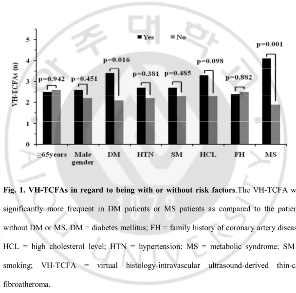
VH-TCFAs were significantly more frequent in DM patients than non-DM patients(3.4±2.0 vs. 2.1±1.7, p=0.016). VH-TCFAs were significantly more frequent in MS patients than non-MS patients(4.1±2.1 vs. 1.9±1.4, p=0.001) (Fig.1).
Fig. 1. VH-TCFAs in regard to being with or without risk factors.The VH-TCFA was significantly more frequent in DM patients or MS patients as compared to the patients without DM or MS. DM = diabetes mellitus; FH = family history of coronary artery disease; HCL = high cholesterol level; HTN = hypertension; MS = metabolic syndrome; SM = smoking; VH-TCFA = virtual histology-intravascular ultrasound-derived thin-cap fibroatheroma.
The mean P+M burden, % NC and the number of VH-TCFAs were significantly correlated with increasing MS scores (Fig. 2).
Fig. 2. The correlation between the plaque components and the metabolic syndrome scores.Results showing the positive correlations between the MS scores and the plaque burden(A), the % necrotic core(B) and virtual histology-intravascular ultrasound -derived thin-cap fibroatheroma(C). MS = metabolic syndrome; VH-TCFA = virtual histology-intravascular ultrasound-derived thin-cap fibroatheroma.
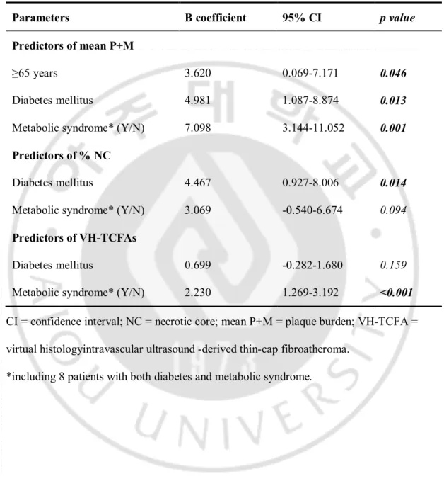
C. Multivariate analysis of the risk factors for the plaque burden, the %NC and the VH-TCFAs
In the multivariate analysis age ≥65 years, DM, and MS were independent predictors of the mean P+M burden; and DM was also the independent predictor of the %NC. MS was the only independent predictor for the presence of VH-TCFAs(Table 5).
Table 5. Multivariate predictors of the plaque burden, the % necrotic core and the VH-TCFAs(n=63).
Parameters B coefficient 95% CI p value
Predictors of mean P+M
≥65 years 3.620 0.069-7.171 0.046
Diabetes mellitus 4.981 1.087-8.874 0.013
Metabolic syndrome* (Y/N) 7.098 3.144-11.052 0.001
Predictors of % NC
Diabetes mellitus 4.467 0.927-8.006 0.014
Metabolic syndrome* (Y/N) 3.069 -0.540-6.674 0.094
Predictors of VH-TCFAs
Diabetes mellitus 0.699 -0.282-1.680 0.159
Metabolic syndrome* (Y/N) 2.230 1.269-3.192 <0.001
CI = confidence interval; NC = necrotic core; mean P+M = plaque burden; VH-TCFA = virtual histologyintravascular ultrasound -derived thin-cap fibroatheroma.
IV. DISCUSSION
To the best of our knowledge, this is the first clinical study to analyze the relationship between cardiovascular risk factors and three-vessel plaque components using volumetric VH-IVUS. The major findings of this study were that DM or the metabolic syndrome was associated with a larger P+M burden and a higher %NC than the absence of DM or the metabolic syndrome, VH-TCFAs were significantly more frequent in DM or metabolic syndrome patients as compared to patients without DM or metabolic syndrome, and among the metabolic syndrome components, a high blood sugar level was related to larger P+M burden and NC and more frequent VH-TCFA(Davies et al, 1993; Felton et al, 1997; Komiyama et al, 2000). In addition, the mean P+M burden, % NC and the number of VH-TCFAs were well correlated with increasing metabolic syndrome scores. Previous pathological studies have shown the relationship between lesion instability and the size of the NCs or the presence of TCFAs. One study showed that ruptured plaques had the largest NCs, followed by TCFA, plaque erosion, and fibrocalcific plaques(Virmani et al, 2000). Atheromatous plaques may be rendered unstable by increases in their size, increases in intraand extracellular lipid accumulation, and development of intraplaque hemorrhage(Rao et al, 2005). As the size of a NC within a TCFA enlarges, the TCFA may become more likely to rupture(Cheruvu et al, 2007). The relation between cardiovascular risk factors and
epidemiological approach(Dawber et al, 1951; Jousilahti et al, 1999). Recently, investigators have used non-invasive modalities - including B-mode ultrasonography, magnetic resonance imaging, and multidetector computed tomography(MDCT)-or invasive imaging modalities-including IVUS or VH-IVUS-to study the correlation between the cardiovascular risk factors and the morphological characteristics of atherosclerotic plaques(Tonstad et al, 1996; Wasserman et al, 2005; Taniguchi et al, 2004; Kaneda et al, 2008). Among them, IVUS provides high-resolution tomographic visualization and quantifies atherosclerotic plaque area and volume and plaque burden. However, other recent studies have suggested that gray-scale IVUS has limited value for the identification of specific plaque components(Yock and Linker, 1990; DiMario et al, 1998) that lead to the development of VH-IVUS(Moore et al, 1998; Nasu et al, 2006; Rodriguez-Granillo et al, 2006; Prasad et al, 2008). Previous IVUS studies had reported that male gender and DM are strong independent predictors of the atherosclerotic burden in coronary disease patients, either at severely narrowed segments or at mildly narrowed segments(Nicholls et al, 2006; Kaneda et al, 2008). Some authors also showed that in culprit lesions, the plaque burden was significantly associated with age, male gender, and DM(Kahlon et al, 2006). Using MDCT, DM patients had more coronary segments with atherosclerosis per patient(Zeina et al, 2008). An IVUS study showed that subjects with MS had a significantly high percent plaque volume and as well as more frequent eccentricity, calcification, and a lipid pool like image than subjects without MS(Hitsumoto et al, 2007). In a MDCT study, MS was independently associated with the
presence and extent of both calcified and noncalcified coronary atherosclerotic plaques(Butler et al, 2008).
Several VH-IVUS studies have demonstrated for the relationship between risk factors and plaque composition. The proportion of NC and DC increased with increasing age, and more advanced calcified lesions were observed in men than women(Qian et al, 2009; Pundziute et al, 2010). Study from global VH registry, patients with diabetes and hypertension had an increased proportion of NC and DC, and HDL-cholesterol level negatively correlated with FF and NC. Also, greater amounts of NC were associated with diabetes, hypertension, MI, and low HDL-C(Philipp et al, 2010). However, previous studies analyzed plaque composition at target lesions or non-obstructive lesion not from entire coronary arterial tree. Compared with these previous studies, the present study extends the association between cardiovascular risk factors(age, DM, and MS) and plaque burden and plaque composition, especially as it affects the entire coronary tree. Therefore, analysis of the whole plaque burden as performed in the current study is more representative than focal plaque analysis for assessing risk factors and the prognosis of atrisk patients.
Until now, few studies have depicted the pathohistological relation between NC or TCFA and DM or MS. Pathologically, the NC size was positively correlated with the diabetic status, independent of other risk factors(Burke et al, 2004). Another study showed that MS was independently correlated with the percentage of lipid volume at the non-target
found patients with DM or MS had more NCs and more frequent VH-TCFAs than patients without DM or MS. NC or TCFA has currently become the surrogate for assessing the vulnerability of plaque. The strong relations of the P+M burden, the %NC and the number of VH-TCFAs with increased MS scores suggest that the plaque vulnerability might be increasing with the severity of MS.
V. STUDY LIMITATIONS
This study was a single-center study, and the findings of this study were based on a small patient population. These patients are not typical of patients presenting to catheterization laboratories in the United States, and the results thus may not fully apply to a US patient population. While pathologic correlations of VH-IVUS vs ex vivo coronary arteries and directional coronary atherectomy specimens have been published(Nasu et al, 2006; Nair et al, 2007), a recent comparison of VH-IVUS vs a model of porcine atherosclerosis found no correlation in the assessment of necrotic core(Thim et al, 2010), and there are limited data on the reproducibility of IVUS or the ability of IVUS to predict future events. VH-IVUS is limited in analyzing small vessels, including distal vessels or those typical of diabetic patients. On one hand, the system imposes at least a 300 micron thick gray “media” obscuring small amounts of plaque; on the other hand, there are limitations in imaging near the 20MHz transducer used during VH analysis. The VH-IVUS differences between patients with vs without DM and between patients with vs without MS were small, may not be clinically meaningful or predictive of future events, and must be studied prospectively in larger patient populations. We imaged only the proximal 56.1±17.4mm of the LAD, 51.9±19.0mm of the LCX, and 74.2±18.8mm of the RCA; thus, while earlier studies suggested that vulnerable plaques are mostly proximal, the recent PROSPECT study
thought. This study contains a great number of independent t-tests comparisons raising the specter of Type I error.
VI. CONCLUSION
Three-vessel VH-IVUS volumetric analysis showed that there are more NCs and TCFAs and a greater plaque burden in DM and MS patients. This implies higher plaque vulnerability in these groups.
REFERENCES
1. Alberti KG, Eckel RH, Grundy SM, et al. Harmonizing the metabolic syndrome: a joint interim statement of the International Diabetes Federation Task Force on Epidemiology and Prevention; National Heart, Lung, and Blood Institute; American Heart Association; World Heart Federation; International Atherosclerosis Society; and International Association for the Study of Obesity. Circulation. 120(16):1640-1645, 2009
2. Amano T, Matsubara T, Uetani T, et al. Impact of metabolic syndrome on tissue characteristics of angiographically mild to moderate coronary lesions. Integrated backscatter intravascular ultrasound study. J Am Coll Cardiol. 49:1149–1156, 2007 3. Burke AP, Kolodgie FD, Zieske A, et al. Morphologic findings of coronary
atherosclerotic plaques in diabetics: a postmortem study. Arterioscler Thromb Vasc Biol. 24:1266-1271, 2004
4. Butler J, Mooyaart EA, Dannemann N, et al. Relation of the metabolic syndrome to quantity of coronary atherosclerotic plaque. Am J Cardiol. 101:1127–1130, 2008
5. Cheruvu PK, Finn AV, Gardner C, et al. Frequency and distribution of thin-cap fibroatheroma and ruptured plaques in human coronary arteries. A pathologic study. J
Am Coll Cardiol. 50:940–949, 2007
6. Davies MJ, Richardson PD, Woolf N, et al. Risk of thrombosis in human atherosclerotic plaques: role of extracellular lipid, macrophage, and smooth muscle cell content. Br
Heart J. 69:377-381, 1993
7. Dawber TR, Meadors GF, Moore FE Jr. Epidemiological approaches to heart disease: the Framingham study. Am J Public Health. 41: 279–281, 1951
8. DiMario C, Gorge G, Peters R, et al. Clinical application and image interpretation in intracoronary ultrasound. Eur Heart J.19:207–229, 1998
9. Felton CV, Crook D, Davies MJ, et al. Relation of plaque lipid composition and morphology to the stability of human aortic plaques. Arterioscler Thromb Vasc Biol. 17:1337-1345, 1997
10. García-García HM, Mintz GS, Lerman A, et al. Tissue characterisation using intravascular radiofrequency data analysis: recommendations for acquisition, analysis, interpretation and reporting. EuroIntervention. 5(2):177-189, 2009
11. Hitsumoto T, Takahashi M, Lizuka T, et al. Relationship between metabolic syndrome and early stage coronary atherosclerosis. J Atheroscler Thromb. 14:294-302, 2007 12. Jousilahti P, Vartiainen E, Tuomilehto J, et al. Sex, age, cardiovascular risk factors, and
coronary heart disease: a prospective follow-up study of 14 786 middle-aged men and women in Finland. Circulation. 99:1165-1172, 1999
13. Kahlon JP, Torey J, Nordstrom CK, et al. The impact of coronary artery disease risk factors on intravascular ultrasound-derived morphologic indices of human coronaries.
Echocardiography. 23:308-311, 2006
14. Kaneda H, Kataoka T, Ako J, et al. Coronary risk factors and coronary atheroma burden at severely narrowing segments. Int J Cardiol. 124: 124–126, 2008
15. Komiyama N, Berry GJ, Kolz ML, et al. Tissue characterization of atherosclerotic plaques by intravascular ultrasound radiofrequency signal analysis: an in vitro study of human coronary arteries. Am Heart J. 140:565–574, 2000
ultrasound study with histological and radiological validation. Heart. 79:459–467, 1998 17. Nair A, Margolis MP, Kuban BD, Vince DG. Automated coronary plaque
characterization with intravascular ultrasound backscatter: ex vivo validation. Euro
Intervention. 3:113-120, 2007
18. Nasu K, Tsuchikane E, Katoh O, et al. Accuracy of in vivo coronary plaque morphology assessment. A validation study of in vivo virtual histology compared with in vitro histopathology. J Am Coll Cardiol. 47:2405–2412, 2006
19. Nicholls SJ, Tuzcu EM, Crowe T, et al. Relationship between cardiovascular risk factors and atherosclerotic disease burden measured by intravascular ultrasound. J Am Coll
Cardiol. 47:1967–1975, 2006
20. Nissen SE, Gurley JC, Grines CL, et al. Intravascular ultrasound assessment of lumen size and wall morphology in normal subjects and patients with coronary artery disease.
Circulation. 84:1087–1099, 1991
21. Philipp S, Böse D, Wijns W, et al. Do systemic risk factors impact invasive findings from virtual histology? Insights from the international virtual histology registry. Eur
Heart J. 31(2):196-202, 2010
22. Prasad A, Cipher DJ, Prasad A, et al. Reproducibility of intravascular ultrasound virtual histology analysis. Cardiovasc Revasc Med. 9:71–77, 2008
23. Pundziute G, Schuijf JD, van Velzen JE, et al. Assessment with multi-slice computed tomography and gray-scale and virtual histology intravascular ultrasound of gender-specific differences in extent and composition of coronary atherosclerotic plaques in relation to age. Am J Cardiol. 105(4):480-486, 2010
histology-intravascular ultrasound imaging plaque characterization (from the global Virtual Histology Intravascular Ultrasound [VH-IVUS] registry). Am J Cardiol. 103(9):1210-1214, 2009
25. Rao DS, Goldin JG, Fishbein MC. Determinants of plaque instability in atherosclerotic vascular disease. Cardiovascular Pathology. 14:285– 293, 2005
26. Rodriguez-Granillo GA, Garcia-Garcia HM, Mc Fadden EP, et al. In vivo intravascular ultrasound-derived thin-cap fibroatheroma detection using ultrasound radiofrequency data analysis. J Am Coll Cardiol. 46:2038–2042, 2005
27. Rodriguez-Granillo GA, Vaina S, He´ctor M. et al. Reproducibility of intravascular ultrasound radiofrequency data analysis: implications for the design of longitudinal studies. Int J Cardiovasc Imaging, 22:621–631, 2006
28. Taniguchi H, Momiyama Y, Fayad ZA, et al. In vivo magnetic resonance evaluation of associations between aortic atherosclerosis and both risk factors and coronary artery disease in patients referred for coronary angiography. Am Heart J. 148:137–143, 2004 29. Thim T, Hagensen MK, Wallace-Bradley D, Granada JF, Kaluza GL, Drouet L, Paaske
WP, Bøtker HE, Falk E. Unreliable assessment of necrotic core by virtual histology intravascular ultrasound in porcine coronary artery disease. Circ Cardiovasc Imaging. 3(4):384-391, 2010
30. Tonstad S, Joakimsen O, Stensland-Bugge E, et al. Risk factors related to carotid intimamedia thickness and plaque in children with familial hypercholesterolemia and control subjects. Arterioscler Thromb Vasc Biol. 16:984–991, 1996
Arterioscler Thromb Vasc Biol. 20:1262-1275, 2000
32. von Birgelen C, de Vrey EA, Mintz GS, et al. ECG-gated three dimensional intravascular ultrasound: feasibility and reproducibility of the automated analysis of coronary lumen and atherosclerotic plaque dimensions in humans. Circulation. 96:2944 –2952, 1997 33. Wasserman BA, Sharrett AR, Lai s, et al. Risk factor associations with the presence of a
lipid core in carotid plaque of asymptomatic individuals using high-resolution MRI. The multi-ethnic study of atherosclerosis (MESA). Stroke. 39:329-335, 2008
34. Yock PG, Linker DT. Intravascular ultrasound looking below the surface of vascular disease. Circulation. 81:1715-1718, 1990
35. Zeina AR, Odeh M, Rosenschein U, et al. Coronary artery disease among asymptomatic diabetic and nondiabetic patients undergoing coronary computed tomography angiography. Coron Artery Dis. 19:37–41, 2008
- 국문요약 -
관상동맥 경화반 구성 요소의 용적 전형적인
심혈관 위험 요인과 대사 증후군과의 관계
아주대학교 대학원 의학과 정 명 일 (지도교수:탁 승 제) 배경: 현재까지 전체관상동맥 죽상경화반의 조직적성분과 심혈관계 위험인자와의 관계는 명확하지 않다. 본 연구의 목적은 관상동맥 세분지의 죽상경화반의 성분을 VH-IVUS(가상 조직학적 혈관내 초음파)로 분석하고 그 결과를 심혈관계 위험인자와 대사증후군 (MS)으로 비교한다. 대상 및 방법: 63명 환자의 189개혈관죽상경화반을 VH-IVUS로 용적적분석을 했다. 동맥경화 죽상반의 성분은 아래와 같이 나누웠다. 섬유,섬유지방,괴사 (NC) 및 밀집한 칼슘로 나눈다. 그리고 죽상경화반의 성분의 각자와 VH-IVUS-derived thin-cap fibroatheroma (VH-TCFA)을 심혈관위험인자와의 상호관계을 분석했다.DM환자군이 비DM환자군에 비하여 %NC (17.8±5.6 vs. 12.5±6.1, p=0.003) 더 많다. MS환자군도 마찬가지였다(17.3±5.8 vs. 12.8±6.2, p=0.016). 그리고 VH-TCFA은 DM환자군(3.4±2.0 vs. 2.1±1.7, p=0.016)과 MS 환자군(4.1±2.1 vs. 1.9±1.4, p=0.001)에서 더 많다. 결론: 관상동맥 용적적 세혈관 VH-IVUS분석에서 DM와 MS환자군의 PB, NCs와 VH-TCFAs이 많다. 이는 이런한 환자들이 죽상경화반의 취약성을 말한다. 핵심되는 말: 가상 조직학적 혈관내 초음파 (VH-IVUS), 심혈관계 위험인자, 관상동맥질환, 화상 진찰; 죽상경화반의 취약성.