135
-Vol. 11, No. 2(December), 2014 The Journal of Medicine and Life Science
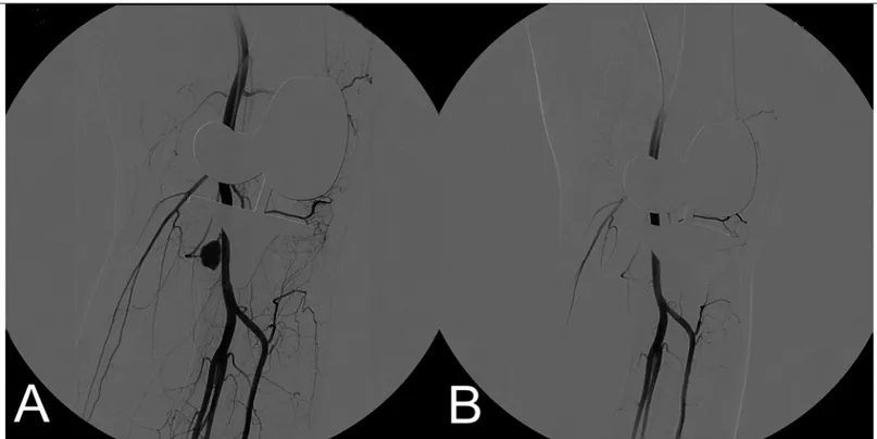
슬관절 전치환술 후에 발생하는 혈관손상은 0.03~0.2% 로 드 물게 발생하나 사지에 심각한 손상을 일으킬 수 있는 잠재적인 합병증을 야기 할 수 있다1-4). 이러한 문제들은 심각한 경우 사망 에도 이를 수 있는데 이는 오진 또는 치료가 늦어질 경우 그 확 률이 증가한다1-4). 슬와동맥 (popliteal artery)의 손상은 삼출, 잡 음 그리고 비박동성 종괴 등의 증상을 나타낸다. 슬와동맥 및 슬 와동맥의 분지인 슬동맥 (genicular artery)의 손상은 가성 동맥 류의 형태로도 나타난다. 가성동맥류의 기원, 크기, 가성동맥류 입구 (pseudoaneurysm neck)의 진로, 그리고 원위부 가지 (distal runoff)에 대한 평가는 치료 전 평가에 있어서 매우 중요 하다. 슬관절 주변의 가성동맥류의 치료에는 보존적 치료, 색전 술, 혈관내 그물망 삽입, 또는 수술적 치료 등이 있다4). 저자들은 수술적 절개 없이 경피적 색전술을 이용하여 슬관절 인공관절 치 환술 이후 발생한 슬동맥의 가성동맥류를 치료한 1개의 증례에 대해 보고하고자 한다. 2009년 6월, 67세 남자가 좌측 무릎 통증을 주소로 내원하였 다. 술 전 평가에서 우측 슬관절는 14도의 외반변형 및 5~130도 의 슬관절가동범위를 보였다. 그는 좌측 슬관절에 대하여 퇴행성 골관절염 진단 하에 좌측 슬관절 전치환술을 시행받았다. 접합제 (Cement)를 사용하기 전 총 56분간 지혈대 (Tourniquet)를 유지 하였다. 수술 부위를 봉합하기 전 지혈대의 공기를 뺀 후, 지혈을 완료 하였다. 환자는 심부정맥혈전증 예방을 위하여 enoxaparin 을 투여 받았으며 외상력은 없었다. 수술 후 2일째까지 배액관을 통해 배출된 총량은 1L 로 측정 되었으며 배액관은 술 후 2일째 제거되었다. 배액관을 제거 한 뒤 환자는 좌측 하지에 중등도의 통증 및 부종이 발견 되었으며 원위부 맥박은 정맥 폐쇄 증상 없 이 잘 촉진 되었다. 수술 후 3일 째 환자는 좌측 하지의 심한 통 증을 호소 하였으며 좌측 슬관절의 부종이 증가하기 시작하였다. 혈관 손상 의심하에 즉시 혈관 조영술을 실시 하였고 외측슬하동 맥 (lateral inferior genicular artery)의 가성동맥류를 확인 하였 다(Fig. 1A.). 외측슬하동맥의 가성동맥류를 0.014 inch micro coils 과 histoacryl 1cc, lipiodol 0.5 cc 를 혼합하여 색전술을 시행 하였다(Fig. 1B.). 색전술 시행 후 환자의 좌측 슬관절에 대 한 통증 및 부종이 완화되었으며, 수술 후 17일째 추가적인 합병 증 없이 퇴원하였다. 이후 외래 통하여 경과 관찰하였으며, 수술 후 2년째 슬관절가동범위는 5~130도로 측정 되었으며 가성동맥 류의 재출혈은 발견되지 않았다.
인공 슬관절 전치환술 후 발생한 외측슬하동맥의 손상에 대한 치료 : 증례 보고
김상림, 박용근
제주대학교 의학전문대학원 제주대학교병원 정형외과학교실(Received October 16, 2014; Revised October 23, 2014; Accepted October 30, 2014)
Therapeutic Embolization for Postoperative Hemorrhage of Lateral Inferior Genicular
Artery after Total Knee Arthroplasty : A Case Report
Sang-Rim Kim, Yong-Geun Park
Department of Orthopedic Surgery, Jeju National University hospital, Jeju National University School of Medicine, Jeju, Korea We describe a case of 67-year-old man who had a hemorrhage of the lateral inferior genicular artery after total knee arthroplasty. The authors encountered rare case of a pseudoaneurysm that was occurred due to the genicular artery injury during the knee arthroplasty operation. The pseudoaneurysm was treated successfully without the need for surgical intervention using transcatheter embolization. No recurrence of the hemarthrosis was observed in the patient after a follow-up of 2 years. (J Med Life Sci 2014;11(2):135-137)
Key Words
: Knee, Pseudoaneurysm, Embolization, Arthroplasty, Replacement서 론
Correspondence to : Yong-Geun Park
Department of Orthopedic Surgery, Jeju National University School of Medicine, Aran 13gil 15, Jeju-si, Jeju Special Self-governing Province, Republic of Korea, 690-767
E-mail : [email protected]
Abstract
증 례
본 논문은 2014년도 제주대학교 학술진흥연구비 지원사업의 지원을 받아 이루어졌음.
동맥류는 구조에 따라 진성동맥류 또는 가성동맥류로 분류된 다. 진성동맥류는 통상적인 혈관보다 2배 이상 확장이 되며 혈관 벽의 모든 층의 확장을 특징으로 가지며 가성동맥류는 섬유낭으 로 이루어진 박동성 종괴를 특징으로 한다5). 인공관절치환술 이 후 가성동맥류의 발생률은 낮으며 연구에 의하면 0.03% 의 확률 로 발생할 수 있다고 한다21). 가성동맥류의 원인은 견인으로 인한 천공, 혈관 손상, 시멘트에 의한 열손상, 또는 압박띠로 인한 것 으로 설명되고 있다6). 슬와부 가성동맥류는 대개 수술용 진동톱 (oscillating saw)과 함께 근위 경골 혹은 후방 대퇴과의 절제 및 후방 관절낭의 유리술 시행 동안 발생하기도 하며, 이들 혈관 손 상으로 인한 합병증은 절단을 포함한 심각한 재앙을 가져올 수 있다2,4,7). 슬동맥의 가성동맥류는 내측 및 외측구의 유리술 동안 에 흔히 발생하는 것으로 되어 있으며, 이들 혈관의 손상은 지속 적인 소량의 혈 배출, 혈종 형성, 혹은 미미한 혈색소 변화를 나 타낸다8-10). 위의 증례의 경우 혈관 손상에 의한 출혈로 갑작스런 활력증후 변화, 극심한 통증 및 순환계/신경학적 이상 소견보다 는 혈종 형성, 미미한 혈색소의 변화 및 관절운동의 제한이 주로 나타난 점으로 보아 슬와동맥의 손상보다는 그 분지 동맥의 손상 으로 판단하였었다. 외상성 혈관손상 합병증이 있는 경우 조기 진단은 차후 발생 할 수 있는 심각한 혈관 손상을 막는데 매우 중요하다. 만약 환 자가 수술 후 슬관절에 심한 부종을 동반한 통증을 호소 할 경우 박동성 종괴의 유무, 종괴의 크기 변화, 배액량의 변화를 유심히 살펴야 하며 동맥 손상 조기 진단을 위한 계획, 그리고 치료를 시행 하여야 한다. 이전에는 침습적인 검사 또는 도플러 초음파 (Doppler ultrasonography)가 혈관 손상을 조기 발견하는데 도 음이 된다고 보고하였다7,11). 슬동맥과 같은 작은 동맥의 출혈의 경우 도플러 초음파 또는 CT angiography로 출혈부위를 명확히 찾지 못할 가능성이 있고, 수술적 절개를 통하여 치료를 할 경우 그 출혈부위를 성공적으로 찾는다는 보장도 없으며 절개에 따른 추가적인 합병증도 발생할 수 있다. 본 저자들은 환자의 출혈양 상이 슬와동맥의 손상보다는 슬동맥의 손상을 먼저 의심하고, 진 단적 목적과 치료적 목적을 함께하는 방법으로 혈관조영술을 선 택하게 되었고 성공적으로 출혈부위를 찾아 지혈할 수 있었다. 인공슬관절 치환술 이후 발생한 외상성 가성동맥류를 치료 하는 데 있어서 선택적 색전술이 치료결과가 좋다는 일부 보고들이 있 다1,4-6,8-10,12). 슬관절 전치환술 후 급격히 진행하는 통증 및 종창이 있고 출 혈이 지속된다고 의심되는 경우 동맥 합병증의 발생 가능성을 의 심하고 조기에 진단 및 치료가 필요하며, 가능한 혈관조영술로 정확한 출혈지점을 확인하는 것이 중요하고, 슬동맥의 손상의 경 우 색전술을 시행하여 불필요한 수술적 치료를 피하고 치료할 수 있는 방법이라 생각한다.
고 찰
Sang-Rim Kim, Yong-Geun Park
136
-Figure 1. Arteriogram showing an aneurysm of the inferior lateral genicular artery at the knee joint level (A). Arteriogram
showing no filling of pseudoaneurysm after glue embolization (Histoacryl 1cc + Lipiodol 0.5cc mixture) (B). 11-2호 논문-최종진짜_8호제주대학교논문-수정3 15. 01. 12 오전 11:27 페이지 136
참고문헌
Therapeutic Embolization for Postoperative Hemorrhage of Lateral Inferior Genicular Artery after Total Knee Arthroplasty : A Case Report
137 -1) Barriga A, Valenti Nin JR, Delgado C, Bilbao JJ.
Therapeutic embolisation for postoperative haemorrhage after total arthroplasty of the hip and knee. The Journal of bone and joint surgery British volume 2001;83:90-2. 2) Calligaro KD, Dougherty MJ, Ryan S, Booth RE. Acute
arterial complications associated with total hip and knee arthroplasty. Journal of vascular surgery 2003;38: 1170-7.
3) Hartford JM, Kwolek C, Circle B. Popliteal pseudoaneurysm after total knee arthroplasty: MRI of the vascular anatomy. Orthopedics 2002;25:187-9. 4) Ibrahim M, Booth RE, Jr., Clark TW. Embolization of
traumatic pseudoaneurysms after total knee arthroplasty. The Journal of arthroplasty 2004;19:123-8.
5) Pai VS. Traumatic aneurysm of the inferior lateral geniculate artery after total knee replacement. The Journal of arthroplasty 1999;14:633-4.
6) Saini P, Meena S, Malhotra R, Gamanagatti S, Kumar V, Jain V. Pseudoaneurysm of the superior lateral genicular artery: case report of a rare complication after total knee arthroplasty. Patient safety in surgery 2013;7:15. 7) Karkos CD, Thomson GJ, D'Souza SP, Prasad V. False
aneurysm of the popliteal artery: a rare complication of
total knee replacement. Knee surgery, sports traumatology, arthroscopy : official journal of the ESSKA 2000;8:53-5.
8) Kirschner S, Konrad T, Weil EJ, Buhler M. [False aneurysm of the lateral superior genicular artery. A complication after the implantation of a knee prosthesis]. Der Orthopade 2004;33:841-5.
9) Moran M, Hodgkinson J, Tait W. False aneurysm of the superior lateral geniculate artery following Total Knee Replacement. The Knee 2002;9:349-51.
10) Noorpuri BS, Maxwell-Armstrong CA, Lamerton AJ. Pseudo-aneurysm of a geniculate collateral artery complicating total knee replacement. European journal of vascular and endovascular surgery : the official journal of the European Society for Vascular Surgery 1999;18:534-5.
11) Karkos CD. Re: B.S.W. Noorpuri et al.: Pseudo-aneurysm of a geniculate collateral artery complicating total knee replacement. European journal of vascular and endovascular surgery : the official journal of the European Society for Vascular Surgery 2000;19:559-60. 12) Law KY, Cheung KW, Chiu KH, Antonio GE.
Pseudoaneurysm of the geniculate artery following total knee arthroplasty: a report of two cases. Journal of orthopaedic surgery 2007;15:386-9.