저작자표시-비영리-변경금지 2.0 대한민국 이용자는 아래의 조건을 따르는 경우에 한하여 자유롭게 l 이 저작물을 복제, 배포, 전송, 전시, 공연 및 방송할 수 있습니다. 다음과 같은 조건을 따라야 합니다: l 귀하는, 이 저작물의 재이용이나 배포의 경우, 이 저작물에 적용된 이용허락조건 을 명확하게 나타내어야 합니다. l 저작권자로부터 별도의 허가를 받으면 이러한 조건들은 적용되지 않습니다. 저작권법에 따른 이용자의 권리는 위의 내용에 의하여 영향을 받지 않습니다. 이것은 이용허락규약(Legal Code)을 이해하기 쉽게 요약한 것입니다. Disclaimer 저작자표시. 귀하는 원저작자를 표시하여야 합니다. 비영리. 귀하는 이 저작물을 영리 목적으로 이용할 수 없습니다. 변경금지. 귀하는 이 저작물을 개작, 변형 또는 가공할 수 없습니다.
Fully Covered, Retrievable Self-Expanding Metal
Stent(Niti-S) in Palliation of Malignant Esophageal
Obstruction
by
Sung Jun Choi
Major in Medicine
Department of Medical Sciences
The Graduate School, Ajou University
Fully Covered, Retrievable Self-Expanding Metal
Stent(Niti-S) in Palliation of Malignant Esophageal
Obstruction
by
Sung Jun Choi
A Dissertation Submitted to The Graduate School of Ajou University
in Partial Fulfillment of the Requirements for the Degree of
Master of Medical Sciences
Supervised by
Jin Hong Kim, M.D., Ph.D.
Major in Medicine
Department of Medical Sciences
The Graduate School, Ajou University
This certifies that the dissertation
of Sung Jun is approved.
SUPERVISORY COMMITTEE
Jin Hong Kim
Sung Won Cho
Ki Myung Lee
The Graduate School, Ajou University
December, 21st, 2009
i
-Abstract
-
Fully Covered, Retrievable Self-Expanding Metal Stent(Niti-S) in
Palliation of Malignant Esophageal Obstruction
Background: The covered self expanding metal stent (SEMS) has become the main
treatment option of malignant esophageal obstruction. However, the fully covered SEMS has not been as popular as the partially covered one for the fear of migration in spite of much advantage. So, we performed a prospective study to evaluate clinical efficacy of the fully covered SEMS.
Methods: Between October 1998 and February 2009, 100 consecutive patients with
malignant esophageal obstruction who were treated with the fully covered Niti-S stent (Niti-S, Taewoong Medical, Seoul, Korea) were included. Data collected contained technical success rate of deployment and retrieval, dysphagia score changes, success rate of esophagorespiratory fistula (ERF) resolution, survival, stent patency and complications. We also conducted analyses concerning the associations between results and variables.
Result: Technical success rate of stent deployment was 100%. Dysphagia score was
improved significantly from a mean of 3.1±0.8 to 1.3±0.7 (p=0.000) and 13 of 14 patients(14/100,14%) with ERF were resolved successfully with the initial stent(13/14, 92.9%). Median survival and stent patency was 74(51~97) and 54(47~67) days respectively. Recurrent dysphagia occurred in 19 patients (19/100,19%) with tumor ingrowth (2/100,2%), tumor overgrowth(7/100,7%), stent migration(6/100,6%), and food impaction(4/100,4%). Among other complications, early(≤7days) complications were chest pain (12/100, 12%), regurgitation (2/100 2%), tracheal compression (1/100, 1%) and late(>7days) ones were bleeding (2/100, 2%), persistent chest pain (2/100, 2%), and GERD (7/100, 7%). Reintervention had to be done in 19 patients (19/100,19%). Among them, endoscopic stent retrieval and replacement was done in 17 patients (17%) with 100%
ii
success rate and additional stents were inserted for the remaining two patients. There was no stent related mortality or 30 day mortality. In our analyses, there was no significant variable associated with clinical outcomes and complications.
Conclusions: The fully covered Niti-S metal stent has proved its effectiveness in
palliation of malignant dysphagia and safety of endoscopic retrieval with a comparably low migration rate maintaining its good advantage of lower incidence of tumor ingrowth and overgrowth.
iii
TABLE OF CONTENTS
ABSTRACT ··· ⅰ
TABLE OF CONTENTS ··· iii
LIST OF FIGURES ··· iv
LIST OF TABLES ··· v
Ⅰ. INTRODUCTION ··· 1
Ⅱ. PATIENTS AND METHODS ··· 3
A. Study design ··· 3
B. Patients ··· 4
C. Stent and stent insertion ··· 4
D. Statistics ··· 4
Ⅲ. RESULTS ··· 6
Ⅳ. DISCUSSION ··· 13
REFERENCES ··· 18
iv
LIST OF FIGURES
Fig. 1. Survival curve ---12
v
LIST OF TABLES
Table. 1. Dysphagia grade ---3
Table. 2. General characteristics ---7
Table. 3. Clinical outcomes ---9
Table. 4. Recurrent dysphagia related complications ---11
1
I. Introduction
Malignant dysphagia is defined as swallowing difficulty caused by malignant cancer and this condition may result from direct cancer involvement of esophagus or extrinsic tumor compression narrowing the esophageal lumen and may be complicated by ERF(Esophago-Respiratory Fistula). Adenocarcinoma and squamous cell carcinoma of esophagus and gastric cardia are the most frequent causes of malignant dysphagia and often inoperable at presentation. So it is common that there is no curative treatment modality (Kubba et al 2000; Enzinger et al 2003). In addition to lung cancer, the second most common cause of malignant
dysphagia (Kassis et al 1998; Baltayiannis et al 2006), various kinds of tumors may cause this condition mainly by extrinsic compression of the esophagus.(Pohl et al 2005;Sobel et al 2005;Salis et al 1998). ERF may be developed by tumor invasion to esophagus and lung at the same time or loss of tumor tissue after treatment. The effectiveness of the covered SEMS for malignant dysphagia and ERF has been already documented (Nelson et al 1997; Saxon et al 1997; Kosarek et al 1996). Laser ablation and photodynamic therapy were also investigated as treatment modalities of malignant dysphagia but stents have proved their superiority to them in randomized trials (Adam et al 1997; Dallal et al 2001). Although there are some evidence that stents are more effective for palliation of dysphagia caused by intrinsic obstruction than extrinsic compression, stents have also been proved to be effective for palliation of dysphagia caused by extrinsic tumor compression (Bethge et al 1998).
Among currently available stents, the most popular type is the covered SEMS(Self Expandable Metal Stent). Previously used rigid plastic stents have become replaced by the SEMS because of the high risk of complication and the difficulty of deployment as documented in some comparative studies(Maroju et al 2006; Kynrim et al 1993; Sanyika et al 1999; O’Donnell et al 2002; De Palma et al 1996; Siersema et al 1998). The SEMS is divided into two groups as covered and uncovered ones according to whether it has covering material or not. The first generation SEMS was uncovered and stent obstruction by cell growth of tumor and regenerative tissue was the main problem. The covered SEMS, the next generation decreased the frequency of tumor ingrowth and repeated intervention in spite of increased risk of migration (Rozanes et al 2002; Ell et al 1994; Saranovic et al 2005; Vakil et
2
al 2001). This tendency was clearly demonstrated in a recent meta-analysis (Yakoub et al 2008) and the covered SEMS rather than uncovered has become more favorable in palliation of malignant dysphagia and ERF. However, the main stream of covered stents have been partially covered ones for the fear of increase in migration rate and tumor or tissue ingrowth through the bare portion of partially covered stent is still the main cause of recurrent dysphagia after stent placement. The fully covered Niti-S stent has been invented to overcome this problem without big increase in migration rate. Flange shaped ends was applied instead of bare portion of partially covered SEMS to preclude stent migration. Until now, there has been no English literature about the fully covered SEMS except for a recent report about only retrieval outcomes of a fully covered SEMS (Eloubeidi et al 2009) in medical searching engines like Medline or Pubmed.
In this study, we tried to evaluate the clinical outcomes including retrieval and complications of fully covered Niti-S stents in palliation of malignant esophageal obstruction.
3
II. PATIENTS AND METHODS
A. Study design
Between October 1998 and February 2009, 100 consecutive patients with malignant esophageal obstruction were involved in this study and fully covered Niti-S stents were used for the treatment. Patients were prospectively followed up after stent placement by scheduled out-patient department visiting or telephone calls, weekly for first month, every month for the next 5 months and then every 3 month until death. Patients were evaluated about dysphagia before and 4weeks after stent placement using the following dysphagia grade system: 0=normal swallowing, 1= able to swallow some but not all solid, 2= able to swallow semisolid, 3= able to swallow fluid only, 4= unable to swallow fluid and they were also evaluated about recurrent dysphagia related complications, early complications (<7days), and late complications(≥ 7days). Patients with ERF were evaluated at the moment of procedure and 4 weeks later by using water soluble contrast study under the endoscopic view. We defined survival as the period from the initial stent placement to death of patients and stent patency as interval days between initial procedures and additional interventions (eg, stent removal and reinsertion, stent in stent, tracheal stent, …etc.) or the period until expire day after stent placement in the case of patients without complication. All information including dysphagia grade and complications was recorded in a planned case record form.
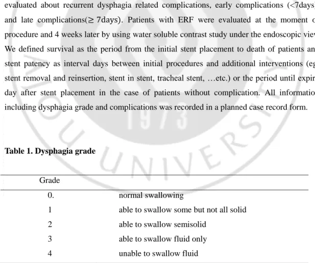
Table 1. Dysphagia grade
Grade 0. 1 2 3 4 normal swallowing
able to swallow some but not all solid able to swallow semisolid
able to swallow fluid only unable to swallow fluid
4 B. Patients
Patients with esophageal obstruction caused by primary and secondary malignant esophageal cancers with or without ERF who need palliative stent placement(dysphagia grade>3) were eligible. Patients who got chemotherapy and radiation therapy and patients with competent esophageal lumen including EGJ after getting operations like subtotal gastrectomy or lung surgery were included but patients who had undergone operations causing changes in normal esophageal lumen like esophagectomy or total gastrectomy were excluded. Written informed consents were obtained from all patients before study enrollment and this study was approved by Institutional Review Board of Ajou University Hospital. (05-166, post facto approval)
C. Stents and stent insertion
The fully covered Niti-S stent (Taewoong Medical ,Seoul, Korea) consists of braided nickel alloy wire and covering membrane. At an early stage of this stent, the covering membrane was made of polyurethane (PU) but it’s been replaced by biochemically more stable Polytetrafluoroethylene (PTFE) since 2003. This fully covered SEMS has dog-bone shaped flanges at both ends to prevent migration and has nylon drawstring at the proximal end which makes it easy to remove stent with minimal trauma by means of collapsing the proximal flared opening when grasping the string. There are 7 kinds of stents available according to length from 6 cm to 18cm. Diameters of body and flanges, compressed in 16Fr delivery system are 18mm and 26mm. All procedures were held in consciously sedative state induced by midazolam under fluoroscopic guidance. Stricture locations were marked by attaching small metals to the patients ‘skin and 2~4cm longer stents than the lengths of stricture were chosen to cover the lesions completely.
D. Statistics
Basic characteristics of patients and clinical outcomes were analyzed as proportions with 95% confidence interval or using the mean(standard deviation) and median(range) according to whether variables were categorical or continuous.
5
Comparisons of complication rates among patients’ subgroups according to categorical variables were made by using Chi-square test or the Fisher exact test as appropriate. Linear associations among categorical variables were analyzed by Mantel-Haenzel test. Logistic regression was used to find out continuous variables related with major complications and make up multivariate models for major complication rates.
Overall survival and stent patency were estimated by using Kaplan-Meier and log-rank test was used for comparisons between subgroups.
6
III. RESULTS
A. General characteristics
Total 100 patients with 66±10 years mean age were included and their general characteristics are summarized in table.1.
Male and female patients were 88 and 12 respectively(M/F 88/12, 87.1%/12.9%) and the origins of tumors were as followings ; esophageal cancer(63/100, 63%), gastric cardial cancer(24/100, 24%), lung cancer(10/100,10%), NHL(1/100,1%), hepatoma(1/100,1%), and CUPS(cancer of unknown primary site)(1/100,1%). Histologic types of esophageal cancers were squamous cell carcinoma(61/63, 96.8%), small cell carcinoma(1/63,1.6%), and adenocarcinoma(1/63,1.6%). Gastric cardial cancers were all adenocarcinoma and lung cancers included squamous cell carcinoma(9/10) and small cell carcinoma(1/10). The distribution of tumor location was as followings; middle esophagus(42/100, 42%), EGJ(26/100, 26%), lower(19/100, 19%) and upper esophagus(13/100, 13%). ERF was complicated in 14 of 100 patients (14/100, 14%) and all of them got palliative chemotherapy or radiation therapy before stent placement.
7 Table 2. General characteristics
Total(100)
Age 66±10
Sex(M/F) 88/12
Tumor origin & histology
Esophageal Ca 63(63%) Squamous cell ca 61 Adenocarcinoma 1 Small cell ca 1 Stomach Ca 24(24%) Adenocarcinoma 24 Lung Ca 10(10%) Squamous cell ca 9 Small cell ca 1 Others(NHL,hepatoma, CUPS) 3(3%) Tumor location Esophagus upper 13(13%) middle 42(42%) lower 19(19%) EGJ/cardia 26(26%) Stricture length(mean,cm) 5.6±2.6 Stenting reasons Malignant obstruction 86(86%) Obstruction+ERF 14(14%) Adjuvant Therapy No 63(63%) CTx 10(10%) RTx 13(13%) CTx+RTx 14(14%)
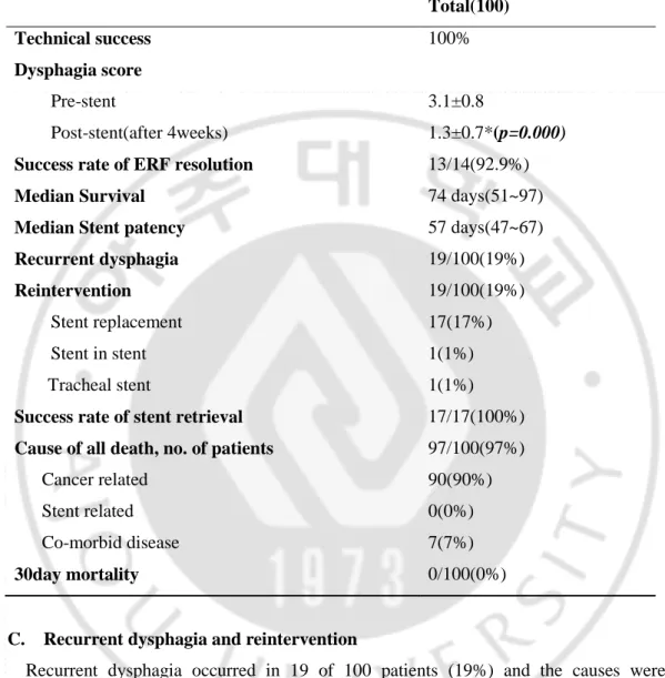
8 B. Clinical outcomes
Clinical outcomes are summarized in table 2. Technical success rate of stent placement was 100%. There was no procedure related complication like perforation, bleeding, or aspiration pneumonia. All patients experienced improvement of dysphagia by mean score change from 3.1(able to swallow fluid only) to 1.3(able to swallow some but not all solid). 13 of 14 patients with ERF were completely resolved with the initial stent placement (92.9%)
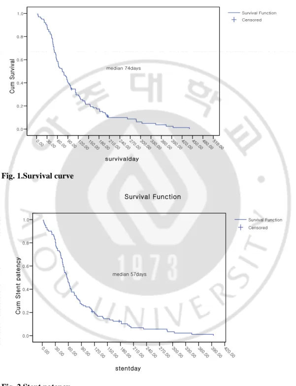
Median survival of total patients was 74days (51~97days) and subgroup analyses showed that female, younger age than 70, and lung cancer origin were independent predictors of poor survival with statistical significance (p=0.0196,p=0.0143,p=0.0224).
Median stent patency of total Niti-S stents was 57 days(47~67days). We also conducted subgroup analyses of independent variables and the results showed no significant factor associated with stent patency including stricture site or prior adjuvant therapy. In subgroup analyses according to stricture site, Median stent patency of EGJ lesions was not worse than other lesions’one (EGJ 59 days, upper 42days, middle 55days, lower 64days, p=0.2941).
90 patients died from cancer progression (90/100, 90%) and seven patients(7/100, 7%) died from co-morbid disease(3 acute coronary events, 3 COPD aggravations, and 1 hepatic coma). 3 patients were still alive at the end point of study (2009-2-28). But, there was no stent related mortality or 30 day-mortality.
9
Table 3. clinical outcomes (mean follow up: 94±82days)
Total(100)
Technical success 100%
Dysphagia score
Pre-stent 3.1±0.8
Post-stent(after 4weeks) 1.3±0.7*(p=0.000)
Success rate of ERF resolution 13/14(92.9%)
Median Survival 74 days(51~97)
Median Stent patency 57 days(47~67)
Recurrent dysphagia 19/100(19%) Reintervention 19/100(19%) Stent replacement Stent in stent 17(17%) 1(1%) Tracheal stent
Success rate of stent retrieval
1(1%) 17/17(100%)
Cause of all death, no. of patients 97/100(97%)
Cancer related 90(90%)
Stent related 0(0%)
Co-morbid disease 7(7%)
30day mortality 0/100(0%)
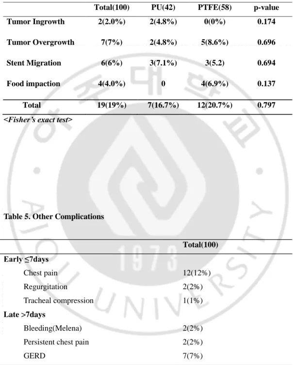
C. Recurrent dysphagia and reintervention
Recurrent dysphagia occurred in 19 of 100 patients (19%) and the causes were as followings; tissue ingrowth(2/100, 2%), overgrowth(7/100, 7%), stent migration (6/100, 6%), food impaction (4/100, 4.0%). There was no significant factor related with occurrence of these complications in univariate and multivariate analyses including stricture site, covering material, age, sex, adjuvant therapy, stricture length, histology, and tumor origin.
Tumor ingrowth only occurred in PU (polyurethane) covered stent group but there was no statistical significance in ingrowth rate between PU and PTFE group(p=0.170), Meanwhile,
10
any variables didn’t affect the rate of stent migration including stricture sites (upper 7.7%,middle 2.3%, lower 15.8%, EGJ 3.8% p=0.205).
Reintervention had to be done in 19 of 100 patients (19%). Among them, 17 (17%) patients with overgrowth(7/17), tumor ingrowth(2/17), stent migration(6/17), and persistent chest pain(2/17) got stent replacement with 100% success rate and there was no retrieval related complication. Longer Niti-S stents were used for the patients with overgrowth. As for patients with stent migration, longer Niti-s stents (3/6), modified Choo’s stents(2/6), and double layered Niti-s stent(1/6) were used. Tumor ingrowth only occurred in 2 patients with PU stent just before 2003 when PTFE stents started to be available and the old stents were replaced by PTFE stents. Patients who underwent stent replacement due to persistent chest pain possessed their stents in upper esophagus and revealed marginal ulcers above the proximal ends of stents on endoscopic views. So, 1 week-PPI therapy was applied for the ulcers and longer stents were inserted. As for the remaining 2 patients who got reintervention but didn’t replace their stents, one patient got tracheal stent placement for tracheal stenosis and the other patient who failed in resolving ERF with the initial stent placement got an additional stent placement by stent-in-stent method.
D. Non-dysphagic complications
In early period (1~7days after stent placement), there were such complications as chest pain (12/100, 12%), GERD (2/100, 2%) and tracheal compression (1/100, 1%).
In late period (>7days after stent placement), there were 2 patients (2%) with melena. Persistent chest pain occurred in 2 patients(2%) which needed stent replacement. GERD was noted in 7 patients (7%).
Patients with chest pain or GERD were resolved by medications including PPI in all cases except for two patients who got stent replacement due to persistent chest pain. Patients with melena were not so severe as to get transfusion.
11 Table 4. Recurrent dysphagia related complications
Total(100) PU(42) PTFE(58) p-value
Tumor Ingrowth 2(2.0%) 2(4.8%) 0(0%) 0.174
Tumor Overgrowth 7(7%) 2(4.8%) 5(8.6%) 0.696
Stent Migration 6(6%) 3(7.1%) 3(5.2) 0.694
Food impaction 4(4.0%) 0 4(6.9%) 0.137
Total 19(19%) 7(16.7%) 12(20.7%) 0.797
<Fisher’s exact test>
Table 5. Other Complications
Total(100) Early ≤7days Chest pain 12(12%) Regurgitation 2(2%) Tracheal compression 1(1%) Late >7days Bleeding(Melena) 2(2%)
Persistent chest pain 2(2%)
12 Fig. 1.Survival curve
Fig. 2.Stent patency
0.00 30.00 60.00 90.00 120.00 150.00 180.00210.00 240.00 270.00 300.00 330.00 360.00 390.00 420.00 450.00 480.00 510.00 survivalday 0.0 0.2 0.4 0.6 0.8 1.0 C u m S u rv iv a l median 74days Survival Function Censored Survival Function 0.00 30.00 60.00 90.00 120.00 150.00 180.00 210.00 240.00 270.00 300.00 330.00 360.00 390.00 420.00 stentday 0.0 0.2 0.4 0.6 0.8 1.0 C u m S te n t p a te n c y median 57days Survival Function Censored Survival Function
13
IV. DISCUSSION
Malignant dysphagia caused by inoperable cancers is generally desperate because there is no curative therapy in most cases. Palliation of dysphagia is one of the important problem directly related with quality of life in this situation. The covered SEMS has been proven to be effective for malignant dysphagia caused by various kinds of tumor and complicated ERF as mentioned in previous parts. But stent migration and cell growth of tumor and benign tissue are still the main concerns with stent placement although there have been lots of innovations and advances to prevent these unwanted complications. In addition to these problems, stent retrieval if needed is getting more attention as a fully covered metal stent approved for the aim of retrieval by FDA has been commercialized and available (Eloubeidi et al 2009)
From this point of view, this study demonstrated not only clinical efficacy of fully covered Niti-S stents in palliation of malignant dysphagia and ERF but also its safety of retrieval.
In subgroup analyses of survival, male, lung cancer, and younger age<70 were poor prognostic factors but there should be difference in predictors of survival among studies because clinical settings of studies are different and all biases interfering the analysis between the variables and results can’t be eliminated. Patients with dysphagia caused by non-esophageal cancers usually have more advanced disease and patients with lung cancer were in more advanced stage in this study. This condition might have made lung cancer more distinctive as a poor predictor. As for gender, we thought that the impact of gender on survival was overestimated by extremely uneven distribution of gender in lung cancer and esophageal cancer (M/F=10/0 and 56/7) which have poorer prognosis than stomach cancer or lymphoma. There have been controversial reports about age as a predictor of survival. There was no difference in survival among age groups in studies of USA and Turkey (Turkyilmaz et al 2009; Hashemi et al 2009; Portale et al 2004) but Chinese investigators showed that younger patients had poorer prognosis among patients with esophageal squamous cell carcinoma (Lu et al 1994). Compared to recent studies of the partially covered SEMS in palliation of malignant dysphagia (Conio et al 2007; Sabharwal et al 2003; O’Donnell et al 2002;Siersema et al 2001), 74 days of median survival in this study seems to be shorter but
14
we think that it is comparable because our study included more advanced staged cancers other than esophageal cancer as causes of dysphagia and median survival of only primary esophageal cancer patients was 103 days.
Median stent patency was 57 days but this period was to be influenced by survival. Actually, stent patency regardless of survival become much longer (median stent patency; 184) if estimated by Kaplan Meier, designating patients without complication as patients with patent stents. This result means that stent placement can be an effective palliative treatment for patients with malignant dysphagia and ERF considering their limited life expectancy such as 74 days median survival of our study or 104~134days median survivals in the studies mentioned above (Conio et al 2007; Sabharwal et al 2003; O’Donnell et al 2002; Siersema et al 2001). As for stent patency according to stricture sites, EGJ lesions were not worse than others (EGJ 59 days, upper 42days, middle 55days, lower 64days, p=0.2941) but this tendency changed when only primary esophageal cancer patients were analyzed as the following; upper 154days, middle 184days, lower 112days, and EGJ 109days. This result is similar to other studies previously reported (Saper et al 2003; Bartelsman et al 2000) although there was no statistical significance (p=0.6430). On the contrary, analysis of stent patency of non-esophageal cancer group only, showed that upper lesions were the worst and EGJ lesions were the best with statistical significance (p=0.0052). EGJ lesions originated from different cancers seem to have different tendency of migration rate. EGJ lesions coming from esophageal cancers seem to be more fragile to migration than those coming from gastric cancers and we inferred that it is because EGJ lesions originated from gastric cardial cancers grow upward direction and tissue below EGJ is to be dense compared to EGJ lesion originated from esophageal cancers. From this point of view, we suggest that different typed stents should be used for EGJ lesions coming from different origins of tumors.
These results of stent patency according to stricture sites are closely associated with stent migration which has been one of the main concerns with covered metal stent placement. Actually, stent migration has influenced on which stent, covered or uncovered, investigators have to choose in each clinical situations. For dysphagia caused by external compression, uncovered stents have been preferable because investigators thought that smooth surface of stricture site caused by external compression may weaken the anchoring force of stent and covered metal stents may be more fragile to migration (Bethge et al 1998; Law et al 1999).
15
In this study, improvement of dysphagia was more distinctive in patients with dysphagia caused by primary esophageal cancers than external tumor compressions (p=0.000) but dysphagia caused by extrinsic compression was also significantly improved (pre Vs post G score 3.1 Vs 1.6, p=0.000) which was already reported (Bethge et al 1998) and there was no significant difference in migration rate between primary obstructive and externally compressed lesions (6/87, 6.8% Vs 0/13, 0% p=0.592). Consequently, covered Niti-S stents were used for externally compressed lesions without encountering migration problems as other studies (De Gregorio et al 1996; Siersema et al 2001). From these results, we thought that flange structure of the fully covered Niti-S stent helps decrease migration rate. Actually, migration rate of the partially covered SEMS has much improved from 13~19% in early studies to 2~8% in recent studies since similar structures were applied to the ends of stent (Kynrim et al 1993; Sanyika et al 1999; Vakil et al 2001; Conio et al 2007; Sabharwal et al 2003; O’Donnell et al 2002; Siersema et al 2001; De Palma et al 1996; Siersema et al 1998; Roseveare et al 1998).
19 of 100 patients (19%) got reintervention and 17 out of them had to get stent replacement. Stent retrieval was successful in all intended cases without procedure related complications (success rate 100%, complication 0%). More attention has paid to the fully covered SEMS recently, since a fully covered SEMS was approved by FDA for the aim of retrieval (Eloubeidi and Lopes 2009). According to the report using the new stent, 61% patients got stent replacement and there was no procedure related complication in that study. But there was already a report about a retrievable, fully covered SEMS, which was not available in USA and the stent was retrievable endoscopically by using a method called hook technique which had the similar principle as drawstring of the Niti-S stent. The study was performed in 113 patients with esophageal and gastrointestinal stricture (Yoon et al 2004). Yoon et al reported 97.7% technical success rate of stent retrieval and one case of procedure related mortality. This report postulated that the fully covered SEMS can be removed with ease and safety by using proximal collapsing technique after stent placement for various gastrointestinal strictures.
In subgroup analyses of covering materials, there was no significant difference in clinical outcomes and complications between PTFE and PU. However, we thought that PTFE is more stable to biologic degradation and tissue ingrowth although there was no statistical
16
significance (p=0.170) because removed PU stents revealed degraded membrane as some reports pointed out that biodegradation of PU membrane may happen when the membrane gets to meet gastric acid or bile juice (Jung et al 2002; Kim et al 2002).
There are some limitations in our study. There was only one esophageal adenocarcinoma which is the most frequent histologic type of primary esophageal cancers in western society. We thought that this fact might have affected complication rates and clinical outcomes like survival and stent patency because there have been some reports showing that adenocarcinoma is associated with high complication rate and poor survival (Elphick et al 2005; Ross et al 2007). There have been some reports that prior chemotherapy or radiation is associated with high frequency of major complications like ERF or bleeding (Kinsman et al 1996; Sumiyoshi et al 2003). In our study, ERF was only complicated in the patients with prior chemotherapy or raditaion therapy but there was no significant difference in other complications between prior chemoradiation therapy and non-therapy group.
We didn’t perform follow-up endoscopy routinely unless patients complained certain symptoms, so tissue growth might have been underestimated, in that benign tissue growth is more important in cell growth after stent placement and there might have been tissue growth when there was no symptom as mentioned in other study (Mayoral et al 2000). But we thought that symptom is more important than endoscopic lesion in this clinical situation like NERD (Non Erosive Reflux Disease) because early detection and therapy for tissue growth in patients without symptoms are not meaningful, considering short life expectancy of patients with malignant dysphagia.
Our study didn’t include QOL in clinical outcomes but we thought that currently available tools for QOL estimation is too ambiguous and subjective to evaluate the impact of dysphagia on QOL and such difficulties influenced on some reports in which improvement of dysphagia seemed to have little association with QOL (Dallal et al 2001; Barr et al 1990; Blazenby et al 1990; Liozou et al 1992). We think that more objective tools have to be developed for more precise evaluation of the correlation between improvement of dysphagia and QOL change after stent placement.
In conclusion, our results showed that the fully covered Niti-S stent is effective in palliation of malignant dysphagia with low rates of major complications such as tumor ingrowth, tumor overgrowth, and especially migration and proved its safety of endoscopic
17
retrieval. We thought that randomized controlled studies between the fully and partially covered SEMS will help select the more appropriate stent type for palliation of malignant dysphagia in various kinds of clinical situations.
18
References
1. Adam A, Ellul J, Watkinson AF, et al. Palliation of inoperable esophageal carcinoma: a prospective randomized trial of laser therapy and stent placement. Radiology :202:344-348, 1997
2. Baltayiannis N, Magoulas D, Bolanos N, et al. Expandable wallstents for treatment fortracheoesophageal fistula of malignant origin. J BUON :11:457-462, 2006
3. Bethge N, Sommer A, Valkil N. Palliation of malignant esophageal obstruction due to intrinsic and extrinsic lesions with expandable metal stent. Am J Gastroenterol :93:1829-1832, 1998
4. Bartelsman JF, Bruno MJ, Jensema AJ, et al. Palliation of patients with esophagogastric neoplasm by insertion of a covered expandable Giantruco-Z endoprosthesis. Gastrointest
Endosc :51:134-138, 2000
5. Barr H, Krasner N, Raouf A, et al. Prospective randomized trial of laser therapy only or laser therpy followed by endoscopic intubation for palliation of malignant dysphagia.
Gut :31:503-508, 1990
6. Blazenby JM, Williams MH, Brookes ST, et al. Quality of life measurement in patients with esophageal cancer. Gut :31:252-258, 1990
7. Conio M, Repici A, Battaglia G, et al. A randomized prospective comparison of self-expandable plastic stents and partially covered self self-expandable metal stents in the palliation of malignant esophageal dysphagia. Am J Gastroenterol :102:2667-2677, 2007
8. Dallal HJ, Smith GD, Grieve DC, Ghosh S, Penam ID, Palmer KR. A randomized trial of thermal ablative therapy versus expendable metal stent in the palliative treatment of
19
patients with esophageal carcinoma. Gastroenterintest Endosc :54:549-557, 2001
9. De Palma GD, di Matteo E, Romano G, et al. Plastic prosthesis versus expandable metal stents for palliation of inoperable esophageal thoracic carcinoma; A controlled prospective study Gastrointest Endosc :43:478-483, 1996
10. De Gregorio BT, Kinsman K, Katon RM, et al. Treatment of esophageal obstruction from mediastinal compressive tumors with covered, self-expanding metallic Z-stents.
Gastrointest Endosc :43:483-489, 1996
11. Elphick DA, Smith BA, Bagshaw J, et al. Self-expanding metal stents in the palliation of
malignant dysphagia: outcome analyses in 100 consecutive patients. Dis
Esophagus :18:93-95, 2005
12. Ell C, Hochberger J, May A, Fleig WE, Hahn EG. Coated and uncoated self-expanding metal stents for malignant stenosis in upper GI tract:preliminary clinical experiences with Wall stents. Am J Gastroenterol :89:1496-1500, 1994
13.Eloubeidi MA, Lopes TL. Novel removable internally fully covered self-expanding metal esophageal stent: feasibility, technique of removal, and tissue respons in humans. Am J
Gastroenterol :104:1374-1381, 2009
14. Enzinger PC, Mayer RJ. Esophageal cancer. N Engl J Med :349:2241-52, 2003
15. Hashemi N, Loren D, Dimarino AJ, Cohen S. Presentation and prognosis of esophageal adenocarcinoma in patients below age 50. Dig Dis Sci :54:1708-1712, 2009
16. Jung GS, Song HY, Seo TS, et al. Malignant gastric obstruction: treatment by means of coaxial placement of uncovered and covered expandable nitinol stents. J Vasc Interv
20
17. Kim DH, Kang SG, Choi JR, Byun JN, Kim YC, Ahn YM. Evaluation of the biodurability of polyurethane-covered stent using a flow phantom. Korean J Radiol :2:75-79, 2002
18. Kubba AK, Krasner N. An update in the palliative management of malignant dysphagia.
EUR J Surg Oncol :26:116-29, 2000
19. Kassis ES, Belani CP, Ferson PF, Keenan RJ, Luketich JD. Hodgkin’s disease presenting with bronchoesophageal fistula. Ann Thorac Surg :66:1409-1410, 1998
20. Kosarek RA, Raltz S, Bruge WR, et al. Prospective multicenter trial of esophageal Z-stent placement for malignant dysphagia and tracheoesophageal fistula. Gastrointest
Endosc :44:562-567, 1996
21. Kynrim K, Wagner HJ, Bethge N, Keymling M, Vakil N. A controlled trial of an expansile metal stent for palliation of esophageal obstruction due to inoperable cancer. N
Engl J Med :329:1302-1307, 1993
22. Kinsman KJ, DeGregorio BT, Katon RM, et al. Prior radiation and chemotherapy increase the risk of life threatening complications after insertion of metallic stents for esophagogastric malignancy. Gastrointest Endosc :43:196-203, 1996
23. Lu JP, Xian MS, Hayashi K. Morphologic features in esophageal squamous cell carcinoma of young adults in north China. Cancer :74:573-577, 1994
24. Law S, Tung PHM, Chu K-M, Wong J. Self-expanding metal stents for palliation of recurrent malignant esophageal obstruction after subtotal esophageactomy.
Gastointest Endosc :50:427-431, 1999
25. Liozou LA, Rampton D, Atkinson M, et al. A prospective assessment of quality of life after endoscopic intubation and laser therapy for malignant dysphagia.
21
Cancer :70:386-391, 1992
26.Maroju NK, Anbalagan P, Kate V, Ananthakrishnan N. Improvement in dysphagia and quality of life with self-expanding metallic stents in malignant esophageal strictures.
Indian J Gastroenterol :25:62-65, 2006
27. Mayoral W, Fleischer D, Salcedo J, et al. Nonmalignant obstruction is a common problem with metal stents in the treatment of esophageal cancer. Gastrointest
Endosc :51:556-559, 2000
28. Nelson DB, Axelrad AM, Fleischer DE, et al. Silicone-covered wallstent prototypes for palliation of malignant esophageal obstruction and digestive-respiratory fistulas.
Gastrointest Endosc :45:31-37, 1997
29. O’Donnell CA, Fullarton GM, Watt E, et al. Randomized clinical trial comparing self-expanding metallic stents with plastic endoprostheses in the palliation of esophageal cancer. Br J Surg :89:985-992, 2002
30. Pohl H, Welch HG. The role of overdiagnosis and reclaasification in the marked increase of esophageal adenocarcinoma incidence. J Natl Cancer Inst :97:142-146, 2005
31. Portale G, Peters JH, Hseih CC, et al. Esophageal adenocarcinoma in patients < or =50years old: delayed diagnosis and advanced disease at presentation.
Am Surg :70:954-958, 2004
32. Rozanes I, Poyanli A, Acunas B. Palliative treatment of inoperable esophageal stricture with metal stents: one center’s experience with four different stents.
Eur J Radiol :43:196-203, 2002
33. Roseveare CD, Patel P, Simmonds N, et al. Metal stents improve dysphagia,nutrition, and survival in malignant esophageal stenosis: A randomized controlled trial comparing
22
modified Gianturco Z-stents with plastic Atkinson tubes.
Eur J Gastroenterol Hepatol :10:653-657, 1998
34. Ross WA, Alkassab F, Lynch PM, et al. Evolving role of self-expanding metal stents in the treatment of malignant dysphagia and fistulas. Gastrointest Endosc :65:70-76, 2007
35. Sobel JM, Lai R, Mallery S, et al. The utility of EUS-guided FNA in the diagnosis of metastatic breast cancer to the esophagus and mediastinum.
Gastrointest Endosc :61:416-420, 2005
36. Salis GB, Albertengo JC, Bruno M, et al. Pedunculated liposarcoma of the esophagus.
Dis Esophagus :11:68-71, 1998
37. Saxon RR, Morrison KE, Lakin PC, et al. Malignant dysphagia and esophago-respiratory fistula: Palliation with a polyethylene covered Z-stent. Radiology :202:349-354, 1997
38. Sanyika C, Corr P, Haffejee A. Palliative treatment of oesophageal carcinoma-efficacy of plastic versus self-expandable stents. S Afr Med J :89:640-643, 1999
39. Saranovic D, Djuric-Stefanovic A, Ivanovic A, Masulovic D, Pesko P. Fluoroscopically guided insertion of self-expandable metal esophageal stents for palliative treatment of patients with malignant stenosis of esophagus and cardia:comparison of uncovered and covered stent types. Dis Esophagus:18:230-238, 2005
40. Sabharwal T, Hmady MS, Chui S, et al. Arandomized prospective comparison of Flamingo Wallstent and ultraflex stent for palliation of dysphagia associated with lower third esophageal carcinoma. Gut :52:922-926, 2003
41. Siersema PD, Hop WC, van Blankenstein M, et al. A comparison of 3 types of covered metal stents for the palliation of patients with dysphagia caused by esophagogastric carcinoma: A prospective, randomized study. Gastrointest Endosc :54:145-153, 2001
23
42. Saper A, Oz N, Cemaletin C, et al. The efficacy of self expanding metal stents for palliation of malignant esophageal strictures and fistulas.
Eur J Cardiothorac Surg :23:794-798, 2003
43. Siersema PD, Schrauwen SL, van Blankenstein, et al. Self-expanding metal stents for complicated and recurrent esophagogastric cancer. Gastrointest Endosc:54:579-586, 2001
44. Siersema PD, Hop WC, Dees J, et al. Coated self-expanding metal stents versus latex prostheses for esophagogastric cancer with special reference to prior radiation and chemotherapy: A controlled prospective study. Gastrointest Endosc :47:113-120, 1998
45. Sumiyoshi T, Gotoda T, Muro K, et al. Morbidity and mortality after self-expandable metal stent placement in patients with progressive or recurrent esophageal cancer after chemoradiotherapy. Gastrointest Endosc :57:882-885, 2003
46. Turkyilmaz A, Eroglu A, Subasi M, Karaoglanoglu N. Clinicopathological features and prognosis of esophageal cancer in young patients. Is there a difference in outcome? Dis
Esophagus :22:211-215, 2009
47. Vakil N, Morris AI, Marcon N, et al. A prospective randomized controlled trial of covered expandable metal stents in the palliation of malignant esophageal obstruction at gastroesophageal junction. Am J Gastroenterol :96:1791-1796, 2001
48. Yakoub D, Fahmy R, Athanasiou T, et al. Evidence based choice of esophageal stent for the palliative management of malignant dysphagia. World J Surg :32:1996-2009, 2008
49. Yoon CJ, Shin JH, Song HY, Lim JH, Yoon HK, Sung KB. Removal of retrievable esophageal and gastrointestinal stents: Experience in 113 patients.
24 -국문 요약-
악성 식도 협착의 고식적 치료에서 회수 가능한 완전 피막형
금속 식도인공관의 유용성
아주대학교 대학원 의학과 최 승 준 ( 지도교수 : 김 진 홍 ) 연구목적: 악성 식도 협착의 고식적 치료에 있어 피막형 금속 식도인공관이 주 된 역할을 해 왔으나 일탈 빈도의 증가에 대한 우려로 완전 피막형 보다는 부 분 피막형이 선호되어 왔다. 본 연구에서는 완전 피막형 금속 식도인공관의 치 료 성적을 분석하여 임상적 유용성을 평가하고자 하였다. 대상 및 방법: 1998년 10월부터 2009년 2월까지 아주대학교 병원을 방문하여 완 전 피막형 금속 식도인공관(Niti-S, Taewoong Medical, Seoul, Korea)으로 치료 받 은 악성 식도 협착 환자 100명을 대상으로 하였다. 협착의 원인으로는 식도암 63예, 위 분문부 암 24예, 폐암의 식도 전이 10예, 간암 등 기타 암의 외부 압 박에 의한 협착 3예가 있었으며, 그 중 14명의 환자에서 식도-기관지 누공이 동반되었다. 결과: 인공관 삽입은 모든 환자에서 100% 성공적으로 이루어 졌고, 연하곤란은 삽입 전 dysphagia score 평균 3.1±0.8 에서 삽입 후 1.3±0.7(p=0.000)로 의미 있25 게 호전 되었으며 식도-기관지 누공 환자의 92.9%(13/14)에서 성공적인 치료가 이루어졌다. 평균 추적 관찰기간은 94일이었고 중앙 생존기간과 중앙 인공관 개존기간은 각각 74일과 57일이었다. 연하 곤란의 재발은 19예(19%)에서 발생 하였고 그 원인은 종양 내발육 2예(2%), 종양 과발육 7예(7%), 인공관 일탈 6 예(6%), 음식물에 의한 폐쇄 4예(4%) 등이었고, 기타 조기 합병증으로는 흉통 12예, 역류성 식도염 2예, 기도 압박에 의한 호흡곤란 1예 등이 있었고, 후기 합병증으로는 출혈 2예, 지속적 흉통 2예, 역류성 식도염 7예 등이 있었다. 전 체 환자 중 19예(19%)에서 합병증의 치료를 위해 재시술이 필요하였고 그 중 17명(17%)에서 인공관의 교체를 시도하여 100% 성공하였다. 나머지 2명 중 초 기 치료에 실패한 식도-기관지 누공 환자 1예에서는 기존 인공관 내로 새로운 인공관을 추가 삽입하였고, 기도 압박이 발생한 1명의 환자에서는 기도인공관 을 삽입하였다. 인공관 삽입과 관련된 사망이나 30일 이내 사망은 없었다. 결론: 완전 피막형 금속 식도인공관은 인공관 일탈이나 종양 내발육 및 과발육 등의 주요 합병증의 발생 빈도가 낮았고, 필요시 인공관 제거가 용이하였으며 악성 식도 협착과 그에 동반된 식도-기관지 누공의 고식적 치료에도 효과적이 었다 핵심어 : 악성 식도 협착, 완전 피막형 금속 식도인공관, 인공관 일탈, 인공관 회수