저작자표시-비영리-변경금지 2.0 대한민국 이용자는 아래의 조건을 따르는 경우에 한하여 자유롭게 l 이 저작물을 복제, 배포, 전송, 전시, 공연 및 방송할 수 있습니다. 다음과 같은 조건을 따라야 합니다: l 귀하는, 이 저작물의 재이용이나 배포의 경우, 이 저작물에 적용된 이용허락조건 을 명확하게 나타내어야 합니다. l 저작권자로부터 별도의 허가를 받으면 이러한 조건들은 적용되지 않습니다. 저작권법에 따른 이용자의 권리는 위의 내용에 의하여 영향을 받지 않습니다. 이것은 이용허락규약(Legal Code)을 이해하기 쉽게 요약한 것입니다. Disclaimer 저작자표시. 귀하는 원저작자를 표시하여야 합니다. 비영리. 귀하는 이 저작물을 영리 목적으로 이용할 수 없습니다. 변경금지. 귀하는 이 저작물을 개작, 변형 또는 가공할 수 없습니다.
농학박사학위논문
Structural and Biochemical Analysis of
Oxalate-Biosynthetic Components,
ObcA and Obc1
옥살산 생합성에 관여하는 효소 ObcA 와
Obc1 의 구조와 생화학적 기능 분석
2017년 2월
서울대학교 대학원
농생명공학부 응용생명화학전공
오 준 택
A Dissertation for the Degree of Doctor of Philosophy
Structural and Biochemical Analysis of
Oxalate-Biosynthetic Components,
ObcA and Obc1
February 2017
Department of Agricultural Biotechnology
Seoul National University
옥살산 생합성에 관여하는 효소 ObcA 와
Obc1 의 구조와 생화학적 기능 분석
Structural and Biochemical Analysis of
Oxalate-Biosynthetic Components, ObcA and Obc1
지도교수 이 상 기
이 논문을 농학박사학위논문으로 제출함
2017 년 1 월
서울대학교 대학원
농생명공학부 응용생명화학전공
오 준 택
오준택의 박사학위논문을 인준함
2017 년 1 월
위 원 장 (인)
부 위 원 장 (인)
위 원 (인)
위 원 (인)
위 원 (인)
Structural and Biochemical Analysis of
Oxalate-Biosynthetic Components, ObcA and Obc1
Advisor: Sangkee Rhee
A Dissertation Submitted in Partial Fulfillment
of the Requirement for the Degree of
DOCTOR OF PHILOSOPHY
to the Faculty of
Department of Agricultural Biotechnology
at
SEOUL NATIONAL UNIVERSITY
by
Juntaek Oh
Date ApprovedI
Abstract
In Burkholderia species, the quorum sensing-dependent production of oxalic acid is an indispensable process for bacterial growth during stationary phase. Oxalic acid produced plays a central role in maintaining the environmental pH, which counteracts inevitable population-collapsing alkaline toxicity in amino acid-based culture medium. In B. glumae, two enzymes are responsible for oxalic acid production. First, the oxalate biosynthetic component (Obc) A catalyzes the formation of a tetrahedral C6-CoA adduct from acetyl-CoA and oxaloacetate. Then the ObcB enzyme liberates three products from the C6-CoA adduct: oxalic acid, acetoacetate, and CoA. Interestingly, these two stepwise reactions are catalyzed by a single bifunctional enzyme, Obc1, from B.
thailandensis and B. pseudomallei. Obc1 has an ObcA-like N-terminal domain
and shows ObcB activity in its C-terminal domain, despite no sequence homology with ObcB. In this thesis, crystal structure and functional analysis of ObcA and Obc1 are reported, revealing structural and functional insights of oxalogenesis. Overall structure of ObcA and N-terminal domain of Obc1 exhibits (β/α)8-barrel fold, with a metal ion coordinated in its active site. In
catalysis, substrate oxaloacetate serves as a nucleophile by forming an enolate intermediate mediated by Tyr residue as a general base, which then attacks the thioester carbonyl carbon of a second substrate acetyl-CoA to yield a tetrahedral adduct. In many reactions involving tetrahedral CoA intermediate, the presence of a negative charge in the intermediate leads to collapse of the intermediate, ejecting CoA moiety form the active site. However, the presence of the metal-coordination shell and absence of general acid(s) could produce an unusual
II
tetrahedral CoA adduct as a stable product. Structure of C-terminal domain of Obc1 has an α/β hydrolase fold that contains a catalytic triad for oxalic acid production and a novel oxyanion hole distinct from the canonical HGGG motif in other α/β hydrolases. Functional analyses through mutagenesis studies suggest that His934 is an additional catalytic acid/base for its lyase activity and liberates two additional products, acetoacetate and CoA. These results provide structural and functional insights into bacterial oxalogenesis; an example of the functional diversity of an enzyme to survive and adapt in the environment and divergent evolution of the α/β hydrolase fold, which has both hydrolase and lyase activity.
Keywords: Quorum sensing, Public goods, Oxalate biosynthetic component,
Oxalic acid, Acetyl-CoA, Crystal structure, Burkholderia species
III
Contents
Abstract……….I Contents………III List of Figures……….……V List of Tables……….VII List of Abbreviations………VIII Chapter I. IntroductionBacterial quorum sensing………2
Public goods in bacteria………5
Oxalic acid as a public good in Burkholderia species………6
Oxalate biosynthetic component in Burkholderia species………….…7
Structural analysis of oxalate biosynthetic components………12
Chapter II. Structural and Functional Analysis of ObcA Materials and Methods ObcA purification………14
Crystallization and data collection………20
Structure determination and refinement………...24
Purification of Obc1* and various ObcA mutants………27
Activity assay of ObcA………28
Results Structure of ObcA in the ligand-free form………30
IV
Structure of ObcA in complex with oxaloacetate………38
Structure of ObcA in complex with the bisubstrate adduct………42
Functional analysis of ObcA………56
Discussion……… …66
Chapter III. Structural and Functional Analysis of Obc1 Materials and Methods Cloning and purification of WT Obc1 and its mutants………70
Crystallization and data collection………73
Structure determination and refinement………...……79
Steady-state kinetic assay……….…80
Results Overall structure of apo Obc1……….………...………82
C-domain of apo Obc1……….…………89
Active site of Obc1 C-domain………...…………93
The binding of glycerol in the Obc1 C-domain………97
Functional analysis of Obc1……….……98
Discussion………106
References………112
V
List of Figures
Chapter I
Figure 1. Bacterial quorum sensing and public goods...3
Figure 2. The overall reaction scheme of oxalogenesis in Burkholderia species………8
Figure 3. The sequence alignment of ObcA with its homologs…………10
Chapter II Figure 4. Size-exclusion chromatography of ObcA……….18
Figure 5. Crystal of SeMet-substituted ObcA………..22
Figure 6. The overall structure and topology of ObcA……….32
Figure 7. The active site of ObcA in a ligand-free form………..36
Figure 8. The active site of ObcA in an oxaloacetate bound form……..40
Figure 9. The binding mode of the C4-CoA adduct and oxalate……….46
Figure 10. The electron density map and binding pocket of the C4-CoA adduct and oxalate………48
Figure 11. The schematic representation of a C4-CoA adduct and oxalate in the active site of ObcA………50
Figure 12. The binding of a C4-CoA adduct and oxalate in the active site of ObcA……… 52
Figure 13. Activity assay of ObcA……….54
Figure 14. Relative activity assay of ObcA………59
Figure 15. Functional features of ObcA and Obc1*………61
Figure 16. The proposed mechanism for ObcA………64
VI
Figure 17. Optimized Obc1 crystals………75
Figure 18. The overall structure of Obc1……….83
Figure 19. Sequence alignment of Obc1 with its homologs…………85
Figure 20. The N-terminal domain of Obc1……….87
Figure 21. Topology diagram of C-terminal of Obc1……….……91
Figure 22. The active site of Obc1………..………95
Figure 23. Obc1 activity assay………101
Figure 24. Kinetic analysis and surface representation of Obc1……103
Figure 25. Proposed mechanism for C6-CoA adduct degradation in the active site of the Obc1 C-domain………110
VII
List of Tables
Table 1. Primers for ObcA and its mutants………16 Table 2. ObcA data collection and refinement statistics………25 Table 3. Kinetic parameters of ObcA, ObcA mutants, and Obc1*…….63 Table 4. Primers for Obc1 mutants………72 Table 5. Obc1 data collection and refinement statistics…………....…….77 Table 6. Kinetic parameters of Obc1* and its mutants………105
VIII
List of Abbreviation
ObcA oxalate biosynthetic component A ObcB oxalate biosynthetic component B Obc1 oxalate biosynthetic component 1
CoA coenzyme A
QS quorum sensing
PCR polymerase chain reaction TEV tobacco etch virus
IPTG isopropyl β-D-L-thiogalactopyranoside
Tris tris(hydroxymethyl)aminomethane
HEPES 4-(2-hydroxyethyl)-1-piperazineethanesulfonic acid
RMSD root mean-square deviation
WT wild-type
EDTA ethylenediaminetetraacetic acid DCPIP 2,6-dichlorophenolindophenol SE standard error
- 1 -
Chapter I.
Introduction
- 2 - Bacterial quorum sensing
Bacterial quorum sensing (QS), a cell-to-cell communication process in many
Proteobacteria, is mediated by small diffusible molecules called autoinducer
and the receptor that can specifically bind to autoinducer (Fuqua and Greenberg, 2002, Miller and Bassler, 2001, Schuster et al., 2013, Waters and Bassler, 2005). Through quorum sensing, bacteria can monitor their own population density and behave as a group to adapt to a changing extracellular environment. Since the binding of an autoinducer to its receptor occurs only when a threshold concentration of the autoinducer is reached in the media, bacteria constantly produce and secrete autoinducers into their growth medium. However, at low cell density, dispersion of the autoinducer is dominant relative to its accumulation; therefore, binding does not occur (Fig. 1A). At high cell density, accumulation exceeds dispersion and the autoinducer begins to accumulate in the media. Once the concentration of the autoinducer crosses the threshold, the autoinducer binds to a specific receptor and the resulting complex controls the expression of genes involved in several population-wide characteristics, including bioluminescence, motility, and virulence-related factors (Fig. 1B) (Fuqua and Greenberg, 2002, Waters and Bassler, 2005). In this way, bacteria can act in a highly synchronized manner, as in a multicellular organism. This type of cooperation in an individual is very efficient when invading the host by secreting virulence factors or by eliminating competitors entering the growth medium through the biosynthesis of bactericidal molecules (Waters and Bassler, 2005). Bacteria also produce and secrete a wide range of chemicals or proteins that can be beneficial to their survival in different environments (Dandekar et al., 2012, Goo et al., 2015, Heilmann et al., 2015, Pai et al., 2012).
- 3 -
Figure 1. Bacterial quorum sensing and public goods.
(A) Bacteria continuously produce and secrete autoinducer molecules into growth medium. However, at low cell density, secreted autoinducer molecules are dispersed rather than accumulated; therefore, binding of the autoinducer to its receptor does not occur. (B) When a threshold concentration of the autoinducer is reached in the media, the autoinducer binds to a specific receptor and the resulting complex controls the expression of target genes. (C) One example of this phenomenon is public goods. In this case, the exoenzyme degrades an unusable complex nutrient to its usable form and acts as a public good. Although the quorum sensing-mediated expression of the exoenzyme is costly, the production of usable nutrients provides a direct benefit to the producer itself and to other group members in the medium, including exploiters who do not respond to quorum sensing.
- 5 - Public goods in bacteria
Recent studies on quorum sensing indicates that quorum sensing also regulates production of some molecules often referred to as ‘public goods’. Public goods provide a fitness advantage for the whole population, including individuals (i.e., exploiters) that lack the ability to synthesize these molecules. For example, public goods in a form of extracellular enzyme or small molecule play a role in nutrient acquisition, interspecies competition, and response to environmental change (Fig. 1C) (Dandekar et al., 2012, Diggle et al., 2007, Pai et al., 2012, Sandoz et al., 2007). Population density controls the production of public goods; this can be explained by comparing costs and benefits. Specifically, at low cell density, the majority of excreted public goods are dispersed before the molecules are utilized. This provides a small fitness benefit compared to the cost of producing the public goods. However, at high cell density, even if the producer of the public goods does not benefit directly, other bacteria in the growth medium can benefit from the public goods, thus providing a fitness benefit in terms of the whole population. Therefore, the production of public goods occurs cooperatively in a population-dependent manner by quorum sensing (Darch et al., 2012). Like most other living populations, pursuing the maximum benefit for individual bacteria does not always coincide with the best strategy for the bacterial community. Therefore, it is very interesting that evolution has selected cooperative individuals, even though there is the possibility of a ‘tragedy of commons’ arising from free riders who seek to exploit their profits without participating in quorum sensing. The quorum sensing-mediated production of ‘private goods’ could explain this observation (Dandekar et al., 2012).
- 6 -
Oxalic acid as a public good in Burkholderia species
In Burkholderia species, quorum sensing-mediated oxalogenesis is an indispensable cellular event for survival during the stationary growth phase. Specifically, Burkholderia species deficient in quorum sensing failed to proliferate in their population during stationary phase (Goo et al., 2015, Goo et al., 2012). Subsequently, it was found that those population collapse results from alkalization of growth medium. In rich growth medium, Burkholderia species utilizes amino acid as an primary carbon source, producing a large amount of ammonia. To counteract ammonia-mediated pH elevation,
Burkholderia species have been evolved to produce and secrete oxalic acid, a
highly acidic molecule that serves as a public good and plays a central role in maintaining pH homeostasis. Without oxalic acid, the whole population of
Burkholderia species would collapse due to ammonia-mediated alkaline
toxicity during the stationary growth phase.
Oxalic acid is ubiquitously present in humans, plants, as well as many bacteria and fungi. Its physiological roles are diverse, including the formation of calcium oxalate crystals for kidney stones in humans, calcium regulation in plants, and pathogenesis in fungi (Dutton and Evans, 1996, Franceschi and Nakata, 2005, Hoppe, 2012). However, a biochemical mechanism and pathway for endogenous oxalogenesis remains largely uncharacterized, although candidates for oxalate precursors include glyoxylate, glycolate, and glyoxal (Dutton and Evans, 1996, Franceschi and Nakata, 2005). Therefore, structural and functional interpretation of oxalic acid producing enzymes will provide molecular level of information about bacterial oxalogenesis.
- 7 -
Oxalate biosynthetic component in Burkholderia species
Quorum sensing-mediated production of oxalic acid is catalyzed by a two-step enzymatic reaction (Fig. 2A) (Li et al., 1999, Nakata and He, 2010). In B.
glumae, two independent enzymes catalyze each step of reactions. The first
enzyme, oxalate biosynthetic component (Obc) A, catalyzes the formation of a C6-CoA adduct, using acetyl-CoA and oxaloacetate as substrates. Substrate-labeling experiments further suggested that the C6-CoA adduct produced by ObcA differs regiochemically from the citroyl-CoA intermediate catalyzed by citrate synthase in the TCA cycle (Fig. 2B) (Li et al., 1999, Wiegand and Remington, 1986). Specifically, it has been suggested that, even though these two enzymes utilize identical substrates, the reaction mechanism may be completely different; to produce the citroyl-CoA intermediate, the acetyl moiety of acetyl-CoA should be deprotonated and attack the C2 carbon of oxaloacetate (Fig. 2B). However, in the ObcA-mediated reaction, the C3 carbon of oxaloacetate should be deprotonated and attack the thioester carbonyl carbon of acetyl-CoA (Fig. 2A). A second enzyme, ObcB, is responsible for the production of three different products, namely, oxalic acid, acetoacetate, and CoA. The C2-C3 and C4-S bonds in the C6-CoA adduct are cleaved by ObcB (Fig. 2A). Interestingly, in B. thailandensis, B. pseudomallei and B. mallei, the bifunctional enzyme Obc1 catalyzes these two reactions (Nakata, 2011). Given that Obc1 functionally complements ObcA and ObcB, it is not surprising that it contains activities of both enzymes. The N-terminal domain of Obc1 exhibits 52% identity with the amino acid sequence of ObcA (Fig. 3); however, the Obc1 C-terminal domain does not show any sequence homology with ObcB, even though it exhibits functional identity with ObcB (Nakata, 2011).
- 8 -
Figure 2. The overall reaction scheme of oxalogenesis in Burkholderia species.
(A) In B. glumae, the two enzymes ObcA and ObcB are involved in oxalogenesis, using acetyl-CoA and oxaloacetate as substrates, whereas in B. thailandensis, two domains in Obc1 mediate oxalogenesis. For convenience, atoms in oxaloacetate are colored according to the final products, and carbon atoms in the C6 moiety of the C6-CoA adduct are numbered. (B) In citrate synthesis, citrate is produced via the citroyl-CoA intermediate.
- 10 -
Figure 3. The sequence alignment of ObcA with its homologs.
The amino acid sequences of B. glumae ObcA (residues Met1 to Gly540; Gene Accession No. YP_002909440.1) are compared with other homologs, including those from B. gladioli (YP_004360639.1), Pseudomonas putida (ZP_19216180.1), B.
pseudomallei (YP_111366.1), B. mallei (ZP_00438353.1), and B. thailandensis
(ZP_02384435.1). Note that Obc1 from B. thailandensis is a bifunctional enzyme containing both ObcA and ObcB-like domains, and the identical enzymes were also identified from B. pseudomallei and B. mallei. Highly conserved residues are shown in red and boxed in blue, while strictly conserved residues are shown with a red background. Secondary structural elements defined in the apo form of ObcA are shown for the corresponding ObcA sequences with the cap and barrel domain in blue and black, respectively. Numbers in subscript represent additional structural elements in the corresponding region of a canonical (β/α)8-barrel fold, while cap domain structural elements are indicated with "C." Residues are indicated with different notations, including the metal-coordinating residues (diamond in cyan), ligand-interacting residues (triangle in green), and acetyl-CoA interacting residues (circle in gray); in particular, the general base Tyr322 is marked with a green asterisk. The sequence and structural alignment of C-domain of Obc1 is discussed ant presented at Fig. 18. This figure was prepared using ESPript (Gouet et al., 1999).
- 12 -
Structural analysis of oxalate biosynthetic components
Many Burkholderia species are involved in plant or human pathogenesis. For example, B. glumae causes bacterial panicle blight in rice (Ham et al., 2011), while B. cepacia is an opportunistic pathogen in immunocompromised individuals, including those with cystic fibrosis and chronic granulomatous disease (Leitao et al., 2010). B. pseudomallei causes melioidosis, a lethal infection that leads to the formation of abscesses in internal organs (Galyov et al., 2010). Given that the quorum sensing dependent expression of oxalate biosynthetic components is an essential cellular event for bacterial proliferation during the stationary growth phase (Goo et al., 2012), understanding the molecular basis of these enzymes should provide the groundwork for the development of novel antibacterial agents for Burkholderia species.
In my thesis, crystal structure and functional analysis of ObcA and Obc1 will be discussed. Chapter II contain crystal structures of ObcA from B. glumae, including a ligand-free form and its complex with oxaloacetate and with a C4-CoA adduct and oxalate, the putative degratory products from the C6-C4-CoA adduct. Structural analyses along with functional assays show that ObcA catalyzes its reaction in a unique manner relative to citrate synthase and other acetyl-CoA-dependent enzymes, providing structural insights into the first step in oxalogenesis and the mechanistic features of ObcA. In Chapter III, two different crystal structures of Obc1 are shown in its apo form and glycerol-bound form. Using structural and functional analyses, how Obc1 catalyzes the production of three different products is discussed. Together with structural study on ObcA, these observations provide structural insights into bacterial oxalogenesis.
- 13 -
Chapter II.
- 14 -
Materials and Methods
ObcA purification
The gene for ObcA from B. glumae (Goo et al., 2012) was amplified by PCR with sequence-specific primers (Table 1). The resulting PCR product was subcloned into the NdeI and XhoI restriction sites of a modified pET28b expression vector (Merck) containing a TEV protease cleavage site at the junction between a His5-tag and a multiple cloning site. The resulting plasmid
was transformed into Escherichia coli BL21 (DE3). Cells were grown in Luria-Bertani medium at 37°C and when the optical density of the culture medium reached 0.7 at 600 nm, protein expression was induced by adding 0.5 mM of IPTG, followed by an additional 12–14 h of growth at 20°C. Cells were collected and sonicated in Buffer A containing 50 mM Tris (pH 8.0), 100 mM NaCl, and 5% (v/v) glycerol. The N-terminal His-tagged ObcA was purified using an immobilized metal affinity column (GE Healthcare) that had been equilibrated with Buffer A and then eluted with Buffer A containing additional 500 mM imidazole. The N-terminal His-tag of ObcA was subsequently removed by treatment of TEV protease overnight at 22°C, using a 20:1 molar ratio of ObcA to TEV protease. The resulting ObcA protein was further purified by immobilized metal affinity and size-exclusion chromatography using Superdex-200 (GE Healthcare) with Buffer A (Fig. 4).
For the structural determination of ligand-free ObcA, the N-terminal His-tagged seleno-L-methionine (SeMet)-substituted enzyme was expressed in
E. coli B834 (DE3; Merck) using minimal medium supplemented with SeMet.
- 15 -
removed as described above. The purified SeMet-ObcA was further subjected to methylation of the lysine residues using established protocols (Walter et al., 2006). In brief, 2–5 mg/mL of SeMet-ObcA was subjected to the methylation reaction by adding the 50 mM of dimethylamine–borane complex and 80 mM of formaldehyde overnight at 4°C. To stop the reaction, 125 mM of Tris (pH 7.5) was added to reaction mixture. After centrifugation, the soluble fraction was further purified by size-exclusion chromatography using a Superdex 200 column (GE Healthcare) with Buffer A to remove any possible protein aggregates.
- 16 - Table 1. Primers for ObcA and its mutants.
ObcA Forward AGTCCATATGACATCGCTATACATCACG Reverse AGTCCTCGAGTCAGCCCGCCGCGGTC R127A GTCGAATAAACTGGCAGCCCGGATCGTGCTGCAA R128A GAATAAACTGGCACGCGCGATCGTGCTGCAATTG K193A GGCAGGCCGGCGCGGCGCGTTCGCC H222A GCGGCGGTGGTGGCTCTGCACACGCG H224A CGGTGGTGCATCTGGCCACGCGCGATCTCA S275A GATCCTGAACCTGGCCACCAGCGTGCG S277A GAACCTGTCCACCGCCGTGCGCGGCGAC R279K CTGTCCACCAGCGTGAAAGGCGACCGCCACGGC R279A GTCCACCAGCGTGGCCGGCGACCGCCAC S308A CCGAGGTCGCCGCGCTGAGCCCG F316A CCGCGGTGGTGGCCCAGGGCGGCG Y322F GCGGCGGCGGCTTCGACAACGCGCC Y322A GGCGGCGGCGGCGCCGACAACGCGCC E346A GCGTCCCGAGGTGGCAGTGTTCAATCACG F348A CCCGAGGTGGAAGTGGCCAATCACGCGATCGT V376A GTTCATGCTGGCGGCGGGCGTCG V379A GGTGGCGGGCGCCGATCAATACCG L447A GAAGATCTCGATCGCGCTGCCGGGGCC R469K GGGCTCGACGGCATCAAAGTGGGGCTGGAGGAC R469A GCTCGACGGCATCGCCGTGGGGCTGGAG E473A GCGTGGGGCTGGCGGACGGCCTGAC D474A CGCGTGGGGCTGGAGGCCGGCCTGACCGTCAACG Obc1 Forward CATATGCGCGAATACGGCTACGAC Reverse AAGCTTATGCGAGCGCTCGTCGCG H227A GGTGGTCCATCTGGCCACGCGCGCGACC Y326A GGCGGGCGGCGGCGCTGACAACCCGAAC E477A CGCGTCGGCCTCGCCGACGCGCTGAACG
- 17 -
Sequences are described from 5’ to 3’. For mutagenic primers, only forward versions are listed. Underlined sequences indicate restriction sites used for cloning, and boldfaced-underlined sequences indicate mutated sequences.
- 18 -
Figure 4. Size-exclusion chromatography of ObcA.
Elution profile of size-exclusion chromatography for His-tag free ObcA. ObcA was eluted at 80.80 ml, which corresponds to 69.6 kD based on size marker. Given that the size of His-tag free ObcA is 59.3 kD, ObcA is estimated to be a monomeric protein.
- 20 - Crystallization and data collection
Crystallization was conducted at 22°C by a sitting drop vapor diffusion method using the native or the methylated SeMet-substituted ObcA (10–15 mg/mL). The presence of the Co2+ ion in the protein solution caused precipitation of
ObcA or resulted in crystals diffracting only to a low resolution, although the Co2+ ion was the most effective metal ion for the enzyme catalysis (see below).
Addition of one or both substrates was essential for the formation of crystals suitable for a high-resolution structure, but under my experimental conditions, the binding of the substrate(s) was not observed in the active site; however, a soaking experiment with the substrate(s) exhibited the binding of substrate(s) of interest. Therefore, crystallization for high-resolution structure analysis was performed using various combinations of ligand(s) and metal ions.
For the ligand-free ObcA, the methylated form of SeMet-substituted ObcA was initially crystallized with 2 mM acetyl-CoA under a crystallization solution of 0.1 M Tris (pH 8.0), 0.2 M MgCl2, and 20% polyethylene glycol
(PEG) 6000 (Fig. 5). Later, I collected higher-resolution data by soaking the crystal into the crystallization solution plus 100 mM CoA. To obtain an oxaloacetate-bound ObcA structure, the native ObcA crystal was soaked in the crystallization solution plus 20 mM oxaloacetate and 2 mM MgCl2, after
growing the crystal from 5 mM oxaloacetate and 1 mM CoA, with mother liquor containing 0.1 M HEPES (pH 7.5), 3% PEG 400, 2 M ammonium sulfate, 0.1 M MgCl2, and 15% glycerol. The structure of ObcA in complex with the
tetrahedral C4-CoA adduct was obtained using native ObcA. Specifically, the native ObcA crystal was produced by co-crystallizing ObcA with 15 mM acetyl-CoA, 20 mM oxaloacetate, and 1 mM CoCl2 using a crystallization
- 21 -
buffer containing 0.2 M sodium/potassium tartrate, 0.1 M sodium citrate (pH 5.6), 2 M ammonium sulfate, and 15% glycerol. Prior to data collection, the crystal was soaked in a solution of 7.5 mM acetyl-CoA, 10 mM oxaloacetate, and 1 mM CoCl2.
X-ray diffraction data were collected at 100 K with a 1-degree oscillation angle on beamline BL-1A at the Photon Factory (Japan) and 5C at the Pohang Accelerator Laboratory (Korea). Glycerol (15–20%) was used as the cryo-protectant in these experiments. Initially, multiwavelength anomalous dispersion data were collected at 2.8 Å using a crystal of the methylated SeMet-substituted ObcA. Subsequently, higher-resolution single-wavelength data became available for the 2.1 Å resolution ligand-free ObcA, the 2.0-Å resolution ObcA in complex with oxaloacetate, and the 2.28 Å resolution ObcA in complex with the C4-CoA adduct. The collected data were processed using the HKL2000 (Otwinowski and Minor, 1997), and all crystals had a space group of P43212, with one monomer in the asymmetric unit, consistent with a
size-exclusion chromatographic study demonstrating ObcA to be a monomeric protein (Fig. 4).
- 22 - Figure 5. Crystal of SeMet-substituted ObcA.
Crystals were obtained under a crystallization solution of 0.1 M Tris (pH 8.5), 0.2 M MgCl2, 20% PEG 6000, and 20% glycerol. Changing pH (8 to 8.5) and addition of 20% glycerol improved crystal size and shape.
- 24 - Structure determination and refinement
The structure of the ligand-free ObcA was determined using the programs SOLVE (Terwilliger and Berendzen, 1999) and RESOLVE (Terwilliger, 2000), with multiwavelength anomalous dispersion data at 2.8 Å resolution. The model was built and refined using the programs COOT (Emsley et al., 2010) and PHENIX (Adams et al., 2010). After several cycles of manual inspection and refinement, residues ranging from Thr2 to Ile525 were located, except for the highly disordered region of Asp71 to Ala91 and Arg96 to Trp100. The model was then refined against the 2.1 Å resolution data available. During assignment of water molecules in the structure, I noticed the presence of a metal ion, based on strong density from the Fo–Fc electron density map, as well as its geometry to the nearby residues and other water molecules. I assigned the metal ion as a Mg2+ ion because the crystallization solution contained 0.2 M
MgCl2. The structure for a ligand-free ObcA was used as the starting model for
determining a structure of the oxaloacetate-bound ObcA. In the middle of refinement, the Fo–Fc electron density map showed that oxaloacetate was bound to the metal-binding site. Similarly, the structure of ObcA in complex with the tetrahedral C4-CoA adduct and oxalate was determined. In particular, the Fo–Fc electron density map corresponding to the tetrahedral C4-CoA adduct and oxalate was evident. Although initially modeled as a Mg2+ ion, the
metal ion was found to correspond to a Co2+ ion due to its presence in the
soaking solution and the strong residual density from the Fo–Fc electron density map. Details for the data collected and refinement are listed in Table 2.
- 25 -
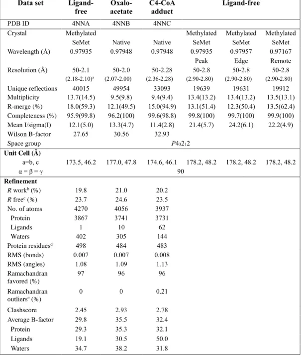
Table 2. ObcA data collection and refinement statistics.
Data set Ligand-
free acetate Oxalo- C4-CoA adduct Ligand-free
PDB ID 4NNA 4NNB 4NNC
Crystal Methylated Methylated Methylated Methylated
SeMet Native Native SeMet SeMet SeMet
Wavelength (Å) 0.97935 0.97948 0.97948 0.97935 0.97957 0.97167
Peak Edge Remote
Resolution (Å) 50-2.1 50-2.0 50-2.28 50-2.8 50-2.8 50-2.8 (2.18-2.10)a (2.07-2.00) (2.36-2.28) (2.90-2.80) (2.90-2.80) (2.90-2.80) Unique reflections 40015 49954 33093 19639 19631 19912 Multiplicity 13.7(14.5) 9.5(9.8) 9.4(9.4) 13.4(13.2) 13.4(13.2) 13.5(13.1) R-merge (%) 18.0(59.3) 12.1(49.5) 15.0(94.9) 13.1(51.4) 12.3(50.4) 13.5(62.4) Completeness (%) 95.9(99.8) 96.2(100) 99.6(98.8) 99.8(100) 99.7(100) 99.9(100) Mean I/sigma(I) 12.1(5.0) 13.3(4.7) 11.4(2.8) 21.4(5.7) 24.2(6.1) 22.2(4.9) Wilson B-factor 27.65 30.56 32.93 Space group P43212 Unit Cell (Å ) a=b, c 173.5, 46.2 177.0, 47.8 174.6, 46.1 178.2, 48.2 178.2, 48.2 178.2, 48.2 α = β = γ 90 Refinement R workb (%) 19.8 21.0 20.2 R freec (%) 23.7 24.6 23.5 No. of atoms 4270 4056 3937 Protein 3867 3741 3731 Ligands 1 10 62 Waters 402 305 144 Protein residuesd 498 484 483 RMS (bonds) 0.007 0.007 0.008 RMS (angles) 1.08 1.09 1.13 Ramachandran favored (%) 97 96 96 Ramachandran outlierse (%) 0 0 0.21 Clashscore 2.45 2.93 2.78 Average B-factor 29.8 35.5 32.4 Protein 29.3 35.3 32.1 Ligands 19.1 30.5 50.0 Waters 34.7 38.2 31.8
- 26 -
aNumbers in parentheses refer to data in the highest resolution shell. bR
work = Σ ||Fobs|-k|Fcal||/ Σ|Fobs|
cRfree is the same as Robs for a selected subset (10%) of the reflections that was not included in prior refinement calculations.
dOrdered residues: ligand-free structure (Thr2 to Ile70 and Arg92 to Arg95 and His101 to Ile525), oxaloacetate bound structure (Thr2 to Ile70 and Ala111 to Ile525), and a C4-CoA adduct bound structure (Thr2 to Ile70 and Ala110 to Ala523).
- 27 -
Purification of Obc1* and various ObcA mutants
For the assay, the WT and mutant ObcA enzymes were expressed as described above. I also used a mutant enzyme of a bifunctional Obc1 from B.
thailandensis (hereafter indicated as Obc1*) to replace the function of ObcB in
the reaction due to an issue with solubility associated with B. glumae ObcB. Genes for the mutant ObcA and Obc1* were constructed by site-directed mutagenesis using mutagenic primers (Table 1). The N-terminal His-tagged enzyme was purified by immobilized metal affinity chromatography followed by a desalting step using a column of HiPrep 26/10 (GE Healthcare) and a buffer solution containing 50 mM Tris (pH 8.0), 100 mM NaCl, and 5% (v/v) glycerol for various ObcA mutants or 50 mM HEPES (pH 7.0), 300 mM NaCl, and 5% (v/v) glycerol for Obc1*. The enzymes were used without removing the His-tag. For measurement of the metal-dependent activity, purified WT ObcA and Obc1* enzymes were dialyzed against Buffer A plus an additional 10 mM EDTA and subsequently against an EDTA-free buffer A. Absence of metal ions, including Co2+, Mn2+, Ni2+, or Mg2+, in ObcA was further validated
- 28 - Activity assay of ObcA
An enzyme activity assay was performed using two different methods, each measuring a different product. First, a steady-state kinetic analysis for ObcA was carried out by monitoring the time-dependent production of free CoA using DCPIP, a dye that reacts with the sulfhydryl group of CoA to causes a linear decrease in absorbance at 600 nm (Raychaudhuri et al., 2005). In a second assay, total oxalic acid produced was measured using an oxalate kit (Trinity Biotech).
To monitor the production of free CoA, enzyme assays were performed at 30C using a UV-visible spectrophotometer (Jasco). The reaction mixture includes 50 mM Tris buffer (pH 8.0), 100 mM NaCl, 100 μM DCPIP, 100 μM CoCl2, and 400–800 μM acetyl-CoA. The mixture was incubated for 20 min at
30C, followed by the addition of 50–500 nM WT ObcA or its mutant and 800 nM Obc1*, after which the mixture was incubated for an additional 4 min. Enzyme reaction was initiated by adding 1–10 mM oxaloacetateto the resulting reaction mixture, and the initial velocity was determined by measuring the linear decrease in absorbance at 600 nm from the time range of 60 to 105 s; nonlinear decreases in absorbance were observed in the first 60 s. The 800 nM Obc1* was confirmed as the saturating concentration for the coupled reaction of a steady-state kinetic assay for ObcA. Free CoA concentration produced per minute was calculated from the standard reaction curve. Specifically, the initial velocity as a function of free CoA concentration was obtained using my assay mixture but in the absence of enzymes and substrates. The KM and Vmax values
were obtained using SigmaPlot, and kcat values were computed by dividing Vmax
by the ObcA concentration used.
- 29 -
protocol. The enzyme reaction was also conducted at 30C, and the reaction mixture was identical with that in the first method, except for DCPIP, which contained 50 mM Tris buffer (pH 8.0), 100 mM NaCl, 100 μM CoCl2, 400 μM
acetyl-CoA, and 1 mM oxaloacetate. The assay solution containing both substrates was incubated for 1 min, and then 50 nM ObcA or its mutant enzyme and 800 nM Obc1* were added to the mixture, incubating for another 5 min. I found that 5 min is sufficient to complete the reaction when using WT ObcA. After the ObcA and Obc1* reactions, 30 μL of the resulting reaction mixture was added to 660 μL of the assay reagent from the manufacturer, and the absorbance at 590 nm was measured after 5 min. Total oxalic acid produced was calculated from the standard curve. Specifically, the standard curve as a function of oxalic acid concentration was obtained in the absence of two enzymes and two substrates.
- 30 -
Results
Structure of ObcA in the ligand-free form
ObcA (residues Thr2 to Ile525) consists of two structurally distinct domains: a small cap and a large barrel domain (Figs. 3 and 6A). The overall structure comprises a (β/α)8-barrel fold with the cap domain (Pro15 to Tyr198) inserted
into a loop between β1 and α1 of the barrel fold (Fig. 6B). This topology represents a unique structural architecture. Homologous structure searches using DALI (Holm and Rosenstrom, 2010) indicated that despite the abundance of (β/α)8-barrel folds in the Protein Data Bank, no other structures resemble the
overall features of ObcA. In particular, DALI search using the cap domain indicates that the most homologous structure found exhibits a Z-score of only 1.3. The N-terminal 14 residues form β1 in the barrel fold. Other insertions occur in the loop regions of the barrel domain, specifically in the loops following β2,β6, and β8 (Figs. 3 and 6B). First, two long antiparallel β-strands (β21 and β22) following β2 protrude vertically from the barrel domain and
belong structurally to the cap domain (hereafter, subscripts used in this manuscript represent additional structural elements present in the corresponding region of the canonical (β/α)8-barrel fold, and the cap domain
structural elements are indicated by “C”). The second insertion consists of two β-strands and one α-helix inserted into the loop connecting β6 and α6. The resulting antiparallel β-strands (β61 and β62) protrude horizontally from the
barrel, and the α61 is packed in an antiparallel orientation to the N-terminal
region of α6. Additional alterations are seen in a loop following β8, in which two antiparallel β-strands (β81 and β82) project horizontally from the barrel. In
- 31 -
the successive α8, a loop runs through the bottom of the barrel, and the C-terminal α9 turns nearly 90° relative to the preceding loop, thus sealing off any possible opening in the bottom of the barrel fold. In addition to these obvious modifications, an additional helix α51 exists between α5 and β6.
The cap domain is locatedalong the C-terminal ends of the central β-strands in the barrel fold. It does not share any structural similarity with the known folds and consists of two segments (Fig. 6A). In one segment, two antiparallel β-strands (β21 and β22) from the barrel domain and three twisted
β-strands (βC5, βC1, and βC4) are arranged perpendicularly along the long axis of
the strands, with two helices (αC1 and αC2) filling the open space between them.
In the second segment, a layer of helices and β-strands is packed on the other side of the three twisted β-strands from the first segment, forming a funnel-shaped fold. The potential small opening in the funnel-like structure is located toward the barrel domain but completely occluded by two β-strands (β81 and
β82) from the barrel fold, while the larger opening at the opposite end comprises
- 32 -
Figure 6. The overall structure and topology of ObcA.
(A) The overall structure of ObcA in a ligand-free form is displayed, indicating an cap domain (blue) and a C-terminal (β/α)8-barrel fold (magenta). Additional structural elements from the barrel domain (green), β1 (yellow), and a metal ion (green sphere) are also indicated. (B) The topology of ObcA is shown with color coding identical to that described in (A). The C-terminus of βC1 to the N-terminus of αC3 is highly disordered and not modeled. Those termini are marked with a black dot. Secondary structure elements are shown in Fig. 3.
- 34 - The active site in ligand-free ObcA
The putative active site of ObcA was indicated by the presence of a metal-binding site in a cavity of the C-terminal barrel fold, consistent with a previous observation that ObcA activity requires a metal ion (Li et al., 1999). My functional analysis also provided evidence for a metal-dependent activity of ObcA, in which the Co2+ ion is the strongest (see below). Given that purified
ObcA does not contain metal ions characterized by inductively coupled plasma atomic emission spectroscopy analysis and that the crystallization condition of a ligand-free ObcA requires 200 mM Mg2+, the metal-binding site identified in
a ligand-free ObcA is likely occupied by the Mg2+ ion. The active site is ~20 Å
from the surface of the enzyme (Fig. 7, A and B). Three residues, including His222 and His224 in β2, and Glu473 in the loop of β8, along with three water molecules, ligate the metal ion, representing an octahedral coordination (Fig. 7C). In particular, His224 and Glu473, as well as two water molecules spanning these residues, form a square coordination for the equatorial plane, while His222 in the interior floor of the cavity and a water molecule proximal to the surface of the enzyme represent the axial ligands. Arg279 on the tip of the loop following β3 covers the metal-binding site, but the electron density for the loop, particularly Arg279 to Gly284, is relatively disordered, suggesting dynamic features of the loop. Many residues are present in the vicinity of the metal-coordinating ligands. In particular, water molecules present in the shell are within a hydrogen bonding distance from the nearby residues, including Ser275 and Ser308, for one trans to Glu473, and Arg469 for a water molecule across from His224 (Fig. 7D).
- 35 -
the metal-binding site and extending from the active site cavity into the enzyme surface along the central β-strands of the barrel fold (Fig. 7, A and B). Most pocket-forming residues are more than 4.5 Å distant from the equatorial water molecule trans to His224, and the wall of the pocket is lined with three ladders of residues: Glu346 and Arg469 constituted the bottom floor, and the second ladder consisted of Asp474, Phe348, Val376, and Leu447, while the rim of the pocket on the surface involved Phe316, Val379, and Pro449. Among those residues, the innermost residue Arg469 is within a hydrogen bonding distance from the equatorial water molecule.
- 36 -
Figure 7. The active site of ObcA in a ligand-free form.
(A) The active site is shown for ObcA in a ligand-free form with the nearby residues. Acetyl-CoA-interacting residues are indicated in gray, while metal-ligating and oxaloacetate-interacting residues (defined later) are colored in cyan. An octahedral geometry of the metal-coordinating shell is presented. Metal-coordinating water molecules (red sphere) and the metal ion (green sphere) are also indicated. (B) The cavity of the active site of ObcA is shown as surface representation in a ligand-free form, with the cap domain (blue) and barrel domain (gray). Water molecules in the metal-coordinating shell are indicated with red spheres. (C) The metal-coordinating shell is presented, with water molecules and metal ions overlaid with an Fo–Fc map (4σ). (D) A schematic drawing is presented for the interactions in the active site of ObcA in the ligand-free form. Dashed lines indicate possible hydrogen bonds within an interatomic distance of 3.6 Å. Note that Glu346 is 3.8 Å apart from the water molecule.
- 38 -
Structure of ObcA in complex with oxaloacetate
The structure of ObcA in complex with oxaloacetate shows that oxaloacetate is identified in the metal-binding site, with its oxalo group pointing toward Arg279 (Fig. 8A). The binding of oxaloacetate caused no noticeable change in conformation, relative to a ligand-free ObcA, with an RMSD of 0.41 Å for 484
Cα atoms. Specifically, the C4 carboxylate of oxaloacetate replaced the metal-ligating water molecule trans to Glu473 in the ligand-free ObcA, and the O2 atom in the oxalo group occupied the sixth coordination. The water molecule across from His224 is still present in the shell, maintaining its interaction with active site residues apparent in a ligand-free ObcA. Thus, the resulting interactions represent a typical octahedral coordination to the bound metal. The two carboxylates of oxaloacetate are stabilized by the nearby residues, including Ser277, Arg279, and Tyr322 for the C1 carboxylate, and Ser275 and Ser308 for the C4 carboxylate.
In addition to these nearby active site residues, possibly facilitating the binding of oxaloacetate, Tyr322 was brought into the immediate vicinity of the substrate, and the side chain hydroxyl group was placed ~3.3 Å distant from the C3 carbon of oxaloacetate (Fig. 8, A and B). In particular, its configuration fulfills the stereochemical requirements of an in-line projection to abstract a proton from the C3 methylene group, with an angle of 109° connecting the OH group in Tyr322, and C3 and C4 atoms of oxaloacetate. The binding of oxaloacetate stabilized the loop covering the metal-binding site, presenting a well-defined interaction between Arg279 and the C1 carboxylate of oxaloacetate. The pocket adjacent to the metal-coordinating shell exhibited no major changes, with the exception that the side chain of Glu346 was closer to
- 39 -
- 40 -
Figure 8. The active site of ObcA in an oxaloacetate bound form.
(A) The stereoview of the active site of ObcA in complex with oxaloacetate (yellow) is shown. The dashed line between Try322 and the C3 group of oxaloacetate does not represent a hydrogen bond. (B) The respective interactions are displayed. Dashed lines with an interatomic distance indicate possible hydrogen bonds. Oxaloacetate in the right of (B) is overlaid with an Fo–Fc map contoured at 2.3 σ.
- 42 -
Structure of ObcA in complex with the bisubstrate adduct
I unexpectedly identified a C4-CoA adduct and oxalate in the active site of ObcA at 2.28 Å resolution (Fig. 9). The electron density map in the active site indicated that it does not represent an authentic C6-CoA adduct as I had anticipated. Rather, the density was separated into two segments, one for a C4-CoA adduct lacking the oxalo group in the oxaloacetate moiety and a second one for oxalate (Figs. 10-12). Given that all three products, including oxalic acid, acetoacetate, and CoA, are readily produced only in ObcB-dependent reactions (Nakata and He, 2010), it is highly likely that under my experimental conditions, the oxalo group was spontaneously released from a C6-CoA adduct, forming oxalate, which was then trapped within the active site. Consistent with this crystallographic observation, my activity assay in solution indicate that oxalate is very slowly and spontaneously produced in an ObcA-dependent manner (Fig. 13A). Previous study on ObcA also supports spontaneous production of oxalic acid (Nakata and He, 2010). Therefore, I conclude that the C4-CoA adduct and oxalate are degradation products from the C6-CoA adduct, a proposed ObcA product (Fig. 2A).
The structure of ObcA in complex with a C4-CoA adduct and oxalate is almost identical with a ligand-free ObcA, with an RMSD of 0.40 Å for 483 Cα
atoms. The resulting C4-CoA adduct and oxalate occupy the metal-coordinating shell and its nearby pocket (Fig. 10B). The phosphopantetheine arm of the adduct is positioned in the pocket along the central β-strands of the barrel domain. Specifically, this arm is bound to a concave region between the cap and barrel domain composed of three different loops following β1, β7, and β8 in the barrel fold. This region was filled with a string of water molecules in the
- 43 -
oxaloacetate-bound ObcA complex. Several positively charged residues, such as Arg127, Arg128, Lys193, and Lys489, are localized around the phosphate group of the arm (Fig. 10C). Noticeably, the side chain of Lys193 underwent large changes that prompted its interaction with the phosphate group, while the 3'-phosphoadenosine monophosphate moiety interacted with few residues (Fig. 11).In contrast, oxalate is located near the C4-CoA adduct (Figs. 9 and 12, A and B), and its binding site is almost identical to the oxalo group in the oxaloacetate-bound ObcA but with a different orientation (Fig. 12C). As a result, oxalate is at least 2.6 Å away from the C4-CoA adduct. One carboxylate present in oxalate is within hydrogen bonding distance to Ser277 and Tyr322, as I observed in the oxaloacetate-bound ObcA (Fig. 8A), while the other carboxylate is nearly in the same position with the O2 atom present in oxaloacetate, occupying a position for the axial water molecule in a metal-coordinating shell (Fig. 11).
Notable features exist in the C4-CoA adduct. The carbon atom adjoined to the sulfhydryl group of CoA is in the tetrahedral configuration (Fig. 12B). The resulting tetrahedral C4-CoA adduct therefore consists of acetoacetate and CoA moieties (Fig. 2A), consistent with the observation that acetoacetate and free CoA moieties comprise the two other products in ObcB-dependent reactions. However, the adduct differs in chemical structure from a cognate acetoacetyl-CoA containing a thioester linkage between the sulfhydryl group of CoA and the carboxylate of acetoacetate. The acetoacetate moiety of the C4-CoA adduct occupies two adjacent positions in the equatorial plane of the metal coordination shell (Figs. 9 and 11). The carboxylate in the adduct replaced the C4 carboxylate position of oxaloacetate, while the oxygen atom on the
- 44 -
tetrahedral carbon occupied a position for the water molecule trans to His224. The resulting geometry around the metal ion is essentially identical with those of the ligand-free and oxaloacetate-bound ObcA. The binding of the phosphopantetheine arm was further stabilized by interactions with the pocket residues, for example, the Arg469 interaction with the oxygen atom in the adduct’s tetrahedral carbon, the Asp474 and Arg279 interactions with the arm of the adduct, and many other hydrophobic interactions.
Taken altogether, it is concluded that ObcA catalyzes the formation of a C6-CoA adduct by joining the oxaloacetate C3 carbon to the thioester carbonyl carbon of acetyl-CoA, resulting in a tetrahedral C6-CoA adduct, which in turn serves as a substrate for ObcB in the production of oxalic acid, acetoacetate, and CoA. Identification of a tetrahedral C4-CoA adduct in this study is unusual, considering that the tetrahedral CoA is thought to be a reaction intermediate in many CoA-dependent reactions, subsequently collapsing into a free CoA by protonating the leaving CoA thiolate anion (Dyda et al., 2000). In contrast, the presence of the tetrahedral C4-CoA adduct in ObcA is made possible by the fact that no obvious candidates for general acid(s) are positioned around the sulfur atom of the adduct in ObcA. Further inspection of the adduct interacting residues suggests that the oxygen atom on the tetrahedral carbon is possibly in a hydroxyl form rather than a labile anionic form, providing a structural foundation for the stability of the tetrahedral adduct. Specifically, the nearby Arg469, which is located within a hydrogen bonding distance from hydroxyl group of the adduct, likely protonates the oxygen atom on the tetrahedral carbon (Figs. 9 and 11; see Discussion). Identification of a bisubstrate adduct in the active site also suggests that the reaction proceeds via a ternary complex
- 45 -
- 46 -
Figure 9. The binding mode of the C4-CoA adduct and oxalate.
The stereoview of the active site is shown for ObcA in complex with the bisubstrate C4-CoA adduct (green) and oxalate (yellow), noting a tetrahedral carbon (black asterisk) and Co2+ ion (black sphere). Dashed lines are potential hydrogen bonds within an interatomic distance of 3.5 Å, and the schematic drawing for these interactions is shown in Fig. 11.
- 48 -
Figure 10. The electron density map and binding pocket of the C4-CoA adduct and oxalate.
(A) The model for the C4-CoA adduct, oxalate, and Co2+ is overlaid with an Fo–Fc electron density map at 2.3 σ. (B) The binding cavity of the C4-CoA adduct and oxalate is displayed in a surface representation of ObcA, with the metal ion indicated in gray. (C) The surface charge distribution of the adduct-binding region is shown in an orientation similar to Fig. 7C. Positive and negative charge distribution is indicted in blue and red, respectively. Note that the adenosine 3’, 5’-diphosphate moiety is placed in the vicinity of the positive charge.
- 50 -
Figure 11. The schematic representation of a C4-CoA adduct and oxalate in the active site of ObcA.
A schematic representation is shown for the interactions in the active site of the ObcA complexed with the C4-CoA adduct and oxalate. Dashed lines indicate possible hydrogen bonds within an interatomic distance of 3.5 Å among the metal-coordinating shell, ligand, and the nearby residues. Interaction distances between Lys489 and the phosphate and between Arg279 and the adduct are 3.8 and 3.6 Å, respectively. Many residues are within close enough distance to allow for hydrophobic interactions. Note that only a few interactions are identified in the vicinity of the adenosine 3’, 5’-diphosphate moiety.
- 52 -
Figure 12. The binding of a C4-CoA adduct and oxalate in the active site of ObcA.
(A) A magnified view for oxalate (yellow), C4-CoA adduct (green), and Co2+ (black sphere) is shown; the asterisk indicates the carbon atom in a tetrahedral configuration. In the left panel, the final model is overlaid with an Fo–Fc map (2.3 σ), showing the possible connection between the oxalate region and the C4-CoA adduct. However, after refinement, the density between the adduct and oxalate is clearly distinguished, which is shown in the right panel with a 2Fo–Fc map (1.7 σ). (B) Two different orientations with an Fo–Fc map (2.3 σ) are shown to indicate that the carbon atom joined to the sulfhydryl group of the adduct is in a tetrahedral configuration. (C) A magnified view is displayed for the relative positioning of the ligands, including oxaloacetate (yellow), as well as oxalate and the C4-CoA adduct, both in green. The relative positions were determined by superposing the two complexes. The C3 of oxaloacetate is ~2.6 Å away from the tetrahedral carbon in the C4-CoA adduct, implying that the enolate form of oxaloacetate undergoes a direct nucleophilic attack on the thioester carbonyl carbon of acetyl-CoA.
- 54 - Figure 13. Activity assay of ObcA.
(A) Total oxalic acid production was measured using an oxalate kit (Trinity Biotech). The presence of either Obc1* or ObcA failed to generate oxalic acid, while production of oxalic acid occurred slowly and spontaneously in solution with ObcA, only after continuing the reaction for a long time period (hours to days). Note that diffraction data was collected from ObcA crystals which were incubated at least two weeks. (B) The reaction profile is displayed for CoA production as a function of time. The decrease in absorbance at 600 nm resulted from 2, 6-dichlorophenolindophenol, a dye reacting with the free sulfhydryl group of CoA. Differences in the color code represent reactions in the absence of each component indicated. Initial velocity was determined between 60 and 105 s. (C) The metal dependency of ObcA activity was assayed but in the presence of different metal ion.
- 56 - Functional analysis of ObcA
An enzymatic analysis was performed using the WT and mutant ObcA enzymes. In particular, production of a free CoA and oxalic acid was measured in a coupled reaction with Obc1*. Inthe presence of the WT ObcA, Obc1*, and the Co2+ ion, the reaction completed in my assay produced oxalic acid as well as
CoA (Fig. 13, A-C). The Obc1* enzyme essentially lacks in its own ObcA activity, but not ObcB function, due to mutations at three residues located within the ObcA-like N-terminal domain.Those mutations are H227A, Y326A, and E350A, which correspond to His224, Tyr322, and Glu346, respectively, of
B. glumae ObcA (Fig. 3) and are essential for ObcA function (see below).
Measurement of the total oxalic acid produced, as well as kinetic analysis of CoA production by Obc1*, indicated that Obc1* exhibits less than 0.5% of the WT ObcA activity (Figs. 14 and 15A, and Table 3), with 176- and 328-fold decreases in the kcat value for acetyl-CoA and oxaloacetate, respectively,
relative to that of the WT ObcA (Fig. 15B).
A steady-state kinetic analysis for the production of free CoA and measurement of total oxalic acid production indicated that residues interacting with the metal ion as well as oxaloacetate or a bisubstrate adduct play an essential role in enzyme activity (Fig. 14). Greater activity for total oxalic acid production than CoA production is possibly attributable to the experimental condition of oxalic acid production in which the measurement was performed after 5 min of reaction time for each mutant ObcA. Mutations on metal binding residues (Fig. 7A), H222A, H224A and E473A significantly reduced ObcA functions; H224A became essentially inactive, although H222A and E473A has some marginal activity, suggesting that His224 is most important in metal
- 57 -
binding. Similar results were also observed for oxaloacetate-binding residues (Fig. 8A); mutants, such as S275A, S277A, S308A, R279K and R279A also largely defect ObcA activity, except for R279K. Unlike R279A which exhibits less than 5% of the WT ObcA activity, the R279K mutant maintained a nearly identical kcat value with the WT ObcA, but with a 35-fold increase in its KM for
oxaloacetate and only a 3-fold increase for acetyl-CoA(Fig. 8B and Table 3), indicating that the positive charge on Arg279 is crucial for the binding of oxaloacetate. Other mutations also largely affected the KM value for
oxaloacetate, consistent with their proposed structural roles. For example, S277A and S308A exhibited 8- and 27-fold increases, respectively, in the KM
value for oxaloacetate, but with much smaller changes (1- to 2-fold) in the KM
values for acetyl-CoA.
In additions to residues in the metal-coordination shell and oxaloacetate-binding environment, several residues in the acetyl-CoA-oxaloacetate-binding pocket (Fig. 9) such as Glu346, Leu447, Arg469, and Asp474 were also characterized as essential elements for activity (Fig. 14). Mutations on these residues caused their activities less than 3% of the WT ObcA, consistent with their proposed roles in the binding of acetyl-CoA or adduct. For example, Leu447 is within distance of the CoA moiety to mediate hydrophobic interactions, and Arg469 was suggested in this study to protonate the oxygen atom on the tetrahedral carbon of the adduct, resulting in the hydroxyl group (Fig. 11). Asp474 interacting with Arg469 appears to play a role in positioning of the Arg469 side chain toward the adduct and regenerating Arg469 in a catalytically competent form (see legend of Fig. 16).
- 58 -
substrate or a possible adduct, I conclude that Tyr322 is a key catalytic residue in ObcA reaction, based on its regiospecific location to the C3 atom of oxaloacetate (Fig. 8A; see Discussion). Mutation of Tyr322 with either alanine or phenylalanine also greatly diminished the enzyme activity (4~10% of the WT ObcA activity), suggesting a possible catalytic role of the side chain hydroxyl group in the reaction. In particular, further kinetic analysis of Y322F indicated that catalytic efficiency (kcat/KM) of the mutant is only 0.3~2.0% of
- 59 - Figure 14. Relative activity assay of ObcA.
The CoA (blue) and total oxalic acid (red) production were measured using the WT and mutant ObcAs. For the relative activity of the CoA production, the initial velocity was determined as shown in Fig. 13B, and those values were compared to that of the WT ObcA. Total oxalic acid produced was measured after 5 min of reaction time. In both assays, measurements were performed with 400 μM acetyl-CoA, 1 mM oxaloacetate, 100 μM CoCl2, 50 nM WT or mutant ObcA, and 800 nM Obc1*, and carried out in triplicate for each sample; error bars correspond to the SEs.
- 61 -
Figure 15. Functional features of ObcA and Obc1*.
(A) A steady-state kinetic analysis of Obc1* for the production of CoA was performed. The KM and Vmax values were obtained using SigmaPlot, with SEs noted in parenthesis. (B) A steady-state kinetic analysis of the WT ObcA was performed. The initial velocity was corrected by subtracting the value of Obc1* from that of ObcA, and the KM and
- 63 -
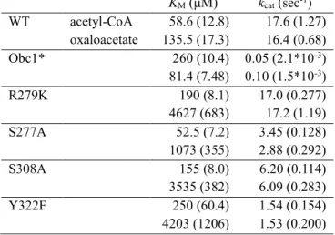
Table 3. Kinetic parameters of ObcA, ObcA mutants, and Obc1*.
The values for KM and kcat obtained by CoA production are listed for the WT ObcA and four mutant enzymes with marginal activity, with SEs in parenthesis.
KM (μM) kcat (sec-1) WT acetyl-CoA 58.6 (12.8) 17.6 (1.27) oxaloacetate 135.5 (17.3) 16.4 (0.68) Obc1* 260 (10.4) 0.05 (2.1*10-3) 81.4 (7.48) 0.10 (1.5*10-3) R279K 190 (8.1) 17.0 (0.277) 4627 (683) 17.2 (1.19) S277A 52.5 (7.2) 3.45 (0.128) 1073 (355) 2.88 (0.292) S308A 155 (8.0) 6.20 (0.114) 3535 (382) 6.09 (0.283) Y322F 250 (60.4) 1.54 (0.154) 4203 (1206) 1.53 (0.200)
- 64 - Figure 16. The proposed mechanism for ObcA.
Upon the binding of oxaloacetate, the side chain hydroxyl group of Tyr322 could be deprotonated by the C1 carboxylate of oxaloacetate, which is within 3.0 Å. The resulting anionic form of Tyr322 likely serves as a general base and abstracts a proton from the C3 carbon of oxaloacetate in a regiospecific manner, producing an enolate anion intermediate of oxaloacetate. The anion intermediate can be stabilized via interactions with a metal ion bound to the active site, followed by a direct nucleophilic attack on the thioester carbonyl carbon of acetyl-CoA to yield a tetrahedral C6-CoA adduct as a product. In an immediate vicinity of the oxygen atom on a tetrahedral carbon, Arg469 possibly protonates the oxygen atom. The hydrogen bond network from Arg469 to the water molecule via Asp474 could regenerate Arg469; in particular, Asp474 is a solvent-exposed residue, and the nearby water molecule is located in the binding pocket for acetyl-CoA. Note that mutation of either Tyr322, Arg469, or Asp474 greatly affects the enzyme activity (Fig.14).