저작자표시-비영리-변경금지 2.0 대한민국 이용자는 아래의 조건을 따르는 경우에 한하여 자유롭게 l 이 저작물을 복제, 배포, 전송, 전시, 공연 및 방송할 수 있습니다. 다음과 같은 조건을 따라야 합니다: l 귀하는, 이 저작물의 재이용이나 배포의 경우, 이 저작물에 적용된 이용허락조건 을 명확하게 나타내어야 합니다. l 저작권자로부터 별도의 허가를 받으면 이러한 조건들은 적용되지 않습니다. 저작권법에 따른 이용자의 권리는 위의 내용에 의하여 영향을 받지 않습니다. 이것은 이용허락규약(Legal Code)을 이해하기 쉽게 요약한 것입니다. Disclaimer 저작자표시. 귀하는 원저작자를 표시하여야 합니다. 비영리. 귀하는 이 저작물을 영리 목적으로 이용할 수 없습니다. 변경금지. 귀하는 이 저작물을 개작, 변형 또는 가공할 수 없습니다.
The prognostic value of volume status
assessment by bioelectrical impedance
analysis and lung ultrasound on
mortality in septic acute kidney injury
patients undergoing continuous renal
replacement therapy
Seung Gyu Han
Department of Medicine
The prognostic value of volume status
assessment by bioelectrical impedance
analysis and lung ultrasound on
mortality in septic acute kidney injury
patients undergoing continuous renal
replacement therapy
Directed by Professor Tae-Hyun Yoo
The Master's Thesis
submitted to the Department of Medicine,
the Graduate School of Yonsei University
in partial fulfillment of the requirements for the degree
of Master of Medical Science
Seung Gyu Han
December 2015
This certifies that the Master's Thesis of
Seung Gyu Han is approved.
---
Thesis Supervisor : Tae-Hyun Yoo
---
Thesis Committee Member#1 : Shin-Wook Kang
---
Thesis Committee Member#2 : Young-Jin Kim
The Graduate School
Yonsei University
ACKNOWLEDGEMENTS
This paper is dedicated to my wife. I love you.
I would like to appreciate professor Tae-Hyun Yoo for
supporting my back and guiding me to the goal. I learn a lot
from him.
I also appreciated professor Shin-Wook Kang. Now I began
to know why he is so much respected by other fellow doctors
and patients whom he takes care of. It was my honor to
apprentice under his teaching.
I thank my family who are always on my side, and my friends
that I can always lean on.
April 2015
Seung Gyu Han
<TABLE OF CONTENTS>
ABSTRACT ··· 1
I. INTRODUCTION ··· 3
II. MATERIALS AND METHODS ··· 4
1. Patients and ethics ··· 4
2. CRRT procedure ··· 7
3. Data collection ··· 7
4. Statistical analysis ··· 8
III. RESULTS ··· 10
1. Baseline characteristics ··· 10
2. Surrogates of volume status ··· 13
3. Correlations among three surrogates ··· 14
4. Outcomes ··· 15
IV. DISCUSSION ··· 22
V. CONCLUSION ··· 25
REFERENCES ··· 26
LIST OF FIGURES
Figure 1. Flow chart of the study population ··· 6
Figure 2. Correlations among used assessment tools ··· 14
Figure 3. Kaplan-Meier analysis of 28-day mortality according
to A) weight change, B) OH/ECW by BIA, and C) B-lines by
lung US ··· 19
LIST OF TABLES
Table 1. Baseline characteristics of patients with septic AKI on
CRRT ··· 11
Table 2. Surrogates of volume status at CRRT initiation ··· 13
Table 3. Patient characteristics according to weight change
tertiles ··· 16
Table 4. Patient characteristics according to OH/ECW tertiles by
BIA ··· 17
Table 5. Patient characteristics according to B-lines tertiles by
lung US ··· 18
Table 6. Univariate Cox proportional hazard model for 28-day
mortality ··· 20
Table 7. Multivariate Cox proportional hazard models for 28-day
mortality according to OH/ECW tertiles ··· 21
ABSTRACT
The prognostic value of volume status assessment by bioelectrical
impedance analysis and lung ultrasound on mortality in septic acute
kidney injury patients undergoing continuous renal replacement therapy
Seung Gyu Han
Department of Medicine
The Graduate School, Yonsei University
(Directed by Professor Tae-Hyun Yoo)
Background Septic acute kidney injury (AKI) is one of the most common causes in critically ill patients requiring continuous renal replacement therapy (CRRT). The fluid status of the patient is known as a significant risk factor for mortality in those patients. Therefore, it is necessary to find an objective assessment of volume status. The aim of present study is to elucidate the impact of fluid status assessed by bioelectrical impedance analysis (BIA) and lung ultrasound on clinical outcomes in septic AKI patients with CRRT.
Methods Septic AKI patients requiring CRRT between April 2014 and February 2015 at Yonsei University Health System were included. Surrogates of volume status were defined by 1) percent of body weight change between CRRT initiation and admission day, 2) over-hydration (OH)/extracellular water (ECW) measured by BIA, and 3) B-lines measured by lung ultrasound (US). Prognostic values of surrogates of volume status for 28-day mortality were evaluated.
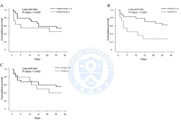
Results Among the 36 enrolled patients, 19 (52.8%) patients died during the follow-up duration. The mean percentage of weight change and OH/ECW measured by BIA was 5.3±20.7 % and 0.3±0.1 L/L. The median number of B-lines counted by lung US was 6 (interquartile range 4-10). Kaplan-Meier analysis showed that the risk for 28-day mortality was higher in patients with the highest OH/ECW tertile compared to patients with lower OH/ECW values (log-rank test, P=0.020). Percent of weight change and the number of B-lines were not significantly related with 28-day mortality risk (log-rank test, P=0.443
and P=0.450 respectively). Multivariate Cox proportional hazard regression analysis showed that higher OH/ECW measured by BIA was an independent risk factor for 28-day mortality after adjustment of confounding factors (HR=3.83, 95% CI=1.04-14.03, P=0.043).
Conclusion Higher OH/ECW measured by BIA was an independent risk factor for 28-day mortality in septic AKI patients undergoing CRRT. Determining fluid status by BIA could be a useful method to stratify mortality risk in this patient group.
---
Key words: continuous renal replacement therapy, bioelectrical impedance analysis, lung ultrasound
The prognostic value of volume status assessment by bioelectrical
impedance analysis and lung ultrasound on mortality in septic acute
kidney injury patients undergoing continuous renal replacement therapy
Seung Gyu Han
Department of Medicine
The Graduate School, Yonsei University
(Directed by Professor Tae-Hyun Yoo)
I. INTRODUCTION
Acute kidney injury (AKI) is one of the most common complications among critically ill patients, and occurs around up to 35% of this patient group. It was reported that one third of AKI is due to sepsis.1 Continuous renal replacement therapy (CRRT) is preferred as a treatment of septic AKI to eliminate toxic metabolite and overloaded fluid. According to previous study, CRRT was used in more than 80% of patients with severe kidney injury.2 In spite of many efforts to promote survival of this patient group, mortality rate of patients with septic AKI remains high.3
After Ronco et al. proposed that increased dose of ultrafiltration improved survival rate, several following studies were performed to evaluate the efficacy of CRRT dose modification on patient survival. But, it was recently reported by Bellomo et al. (RENAL trial) and Palevsky et al. (ATN trial) that increment of CRRT dose did not present any survival benefit.4 Therefore, novel approach to alleviate survival rate of these patient group is required. Meanwhile, fluid status of the patient with septic AKI was proved to be an independent factor for survival rate. Accordingly, it is predictable that control of patients’ fluid overload by way of CRRT be associated with improved survival rate.
bioelectrical impedance analysis (BIA), which can assess the fluid status of the patient as well as body composition by measuring the body’s electrical resistance varying with patient’s medical condition.5 BIA is already clinically used in patients undergoing intermittent hemodialysis to assess fluid status and modify dry weight, so that the patients’ long-term prognosis improves by optimizing blood pressure and cardiovascular parameters such as NT-pro BNP or pulse wave velocity. 6,7 Another novel method is lung ultrasound (US) which can estimate water content in the lung. The rationale of the technique is that under circumstance of pulmonary congestion, the US beam is reflected by thickened interlobular septa, a phenomenon generating hyper-echoic reverberation artifacts between edematous septa and the overlying pleura.8 These vertical US artifacts appear as the so-called lung comets on the screen, which can be considered as B-lines. A recent study also demonstrated that, in patients undergoing intermittent hemodialysis, B-lines on the lung US can estimate volume status before and after hemodialysis.9
Previous studies revealed that critically ill patients suffer fluid overload either in need of CRRT or not, and the extent of fluid overload is associated with the patient’s prognosis. And the same is relevant in patients with septic AKI who require CRRT.10 Therefore, this study is to elucidate the association of assessment of fluid status by BIA and lung US in patients with septic AKI in need of CRRT to the patient’s prognosis.
II. MATERIALS AND METHODS 1. Patients and ethics
A single center prospective observational study was conducted between April 2014 and February 2015. I included totally 36 patients in the study who underwent CRRT and were 20 years or older and satisfy following three criteria, 1) consensus criteria for sepsis, 2) AKI of Injury stage or severer stage in risk, injury, and failure; and loss and end-stage kidney disease (RIFLE) criteria. 3) AKI not explained other than sepsis. Sepsis is defined when patients have
evidence of infection and more than two of the following systemic inflammatory response syndrome (SIRS) criteria: body temperature >38 or <36 °C; heart rate >90/ min; hyperventilation evidenced by respiratory rate >20/ min or PaCO2 <32 mmHg; and white blood cell (WBC) count >12,000 cells/ ㎕, <4,000/㎕ or band forms > 10%.11
Infection is confirmed when patients satisfy one of the following: WBCs in normally sterile fluid; perforated viscus, source of infection detected on radiologic exams.12 AKI is diagnosed and staged according to RIFLE criteria: Risk: urine output (UO) < 0.5ml/kg/hr × 6 hour or serum creatinine (SCr) increased 1.5 to 2 times baseline; Injury: UO < 0.5ml/kg/hr × 12 hour or SCr increased 2 to 3 times baseline; Failure: UO < 0.3ml/kg/hr ×24 hour or SCr increased more than 3 times baseline; Loss of function: complete loss of kidney function > 4weeks (requiring dialysis).13 Patient who satisfies one of the following is excluded: 1) patients younger than 19 years or older than 80 years. 2) patients who have been undergoing hemodialysis due to end stage renal disease (ESRD) 3) patients who are in terminal stage malignancy. 4) patients with AKI from other causes than sepsis. 5) patients with intra-cardiac devices (pacemaker, implantable cardioverter- defibrillator etc.) 6) patients with CRRT for non-renal indications. 7) patients with negative OH value in BIA measurement. During the study, all patients were treated by a conventional CRRT regimen.14 I classified the patients into survivor and non-survivor according to 28-day mortality. The hospital ethics committee approved the study and waived the need for informed consent because of the anonymous and observational fashion of the study.
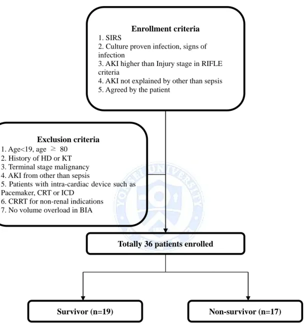
Figure 1. Flow chart of the study population Enrollment criteria
1. SIRS
2. Culture proven infection, signs of infection
3. AKI higher than Injury stage in RIFLE criteria
4. AKI not explained by other than sepsis 5. Agreed by the patient
Exclusion criteria
1. Age<19, age ≥ 80 2. History of HD or KT 3. Terminal stage malignancy 4. AKI from other than sepsis
5. Patients with intra-cardiac device such as Pacemaker, CRT or ICD
6. CRRT for non-renal indications 7. No volume overload in BIA
Totally 36 patients enrolled
Survivor (n=19) Non-survivor (n=17)
Abbreviations: SIRS, systemic inflammatory response syndrome; AKI, acute kidney
injury; HD, hemodialysis; KT, kidney transplantation; CRT, cardiac resynchronization therapy; ICD, implantable cardioverter-defibrillator, CRRT, continuous renal replacement therapy; BIA, bioelectrical impedance analysis
2. CRRT procedure
The treatment initiation was decided by a nephrologist. Vascular access for CRRT was placed via internal jugular vein or femoral vein. CRRT was performed with Prismaflex® (Gambro, Stockholm, Sweden) in the ICU.
Hemosol B0® (bicarbonate-buffered solution, Gambro) was used with a replacement fluid amounting 30-40 ml/kg/hr with a blood flow rate of 150-200 ml/min. ST100® (Gambro) is used as a polysulfone membrane filter. Changes in
maintenance of CRRT, blood flow rate, replacement fluid flow or ultrafiltration rate were made by the physician’s decision according to the patients’ clinical condition. CRRT is weaned when the patients satisfied on of the following: 1) systolic blood pressure is maintained >120 mmHg without use of inotropics and heart rate under 90 beats per minute. 2) UO > 1,000 ml/day with or without the use of diuretics. 3) conversion to conventional hemodialysis.
3. Data collection
Patient data were collected from the electronic medical record prospectively in Yonsei University Health System, Seoul, Korea. Demographic, clinical and biochemical data at the time of CRRT initiation were collected. In this study, I supposed three surrogates of volume status. Weight change (%) is defined as difference of body weight at CRRT initiation from body weight at the time of admission which is normalized by body weight at the time of admission. Body weight was measured by scale. Weight change (%) was obtained by following equation.
The other two surrogates were measured by devices at the time of CRRT initiation. OH was measured by BIA, and the value was normalized by ECW
(OH/ECW, L/L) to be kept relative. B-lines were also measured by lung US at the time of CRRT initiation. Lung US was performed with a portable US scanner (GE Logiq Book XP® General Electric Health care, New York, USA)
with a 2-3.6 MHz phased array cardiac assessment EM 3S-RS probe. The numbers of B-lines were measured from four points of the chest: the intercostal spaces between third and fourth ribs and sixth and seventh ribs on each mid-clavicular line. The numbers were recorded at each point and summed up as the number of B-lines. At the same time, BIA is performed with Body Composition Monitor-BCM® (Fresinius Medical Care, Bad Homburg,
Germany). Two each electrodes on ipsilateral arm and leg were attached for the measurement. Using body fluid model, BIA measures electric resistance of the body to assess the composition of the patient’s body. Body weight is composed of Lean tissue mass (LTM), Adipose tissue mass (ATM) and over-hydration (OH). In LTM and ATM, extracellular water (ECW) and intracellular water (ICW) exist at constant ratio, however, OH is assumed to be composed of solely ECW in the body fluid model, thus OH can be calculated by subtracting the average human’s ECW of the same height and body weight at one’s normal health condition from ECW of the patients. Acute Physiology and Chronic Health Evaluation (APACHE) II score was determined at the time of ICU admission. Estimated glomerular filtration rate (eGFR) was also calculated using the simplified Modification of Diet in Renal Disease equation.
4. Statistical analysis
Statistical analysis was performed by SPSS for Windows®, version 18.0 (SPSS, Inc, Chicago, IL, USA). Values are presented as mean ± SD for continuous variables, and as numbers and percentages for categorical variables. Baseline characteristics of the groups were compared using the Student t test or ANOVA for continuous and χ2 test for categorical variables. In the present study, I evaluated 28-day mortality as a primary end point.
Comparison of parameters of BIA and lung US between two groups was made using student T-test. The impact of variables on 28-day mortality in septic AKI patients was determined using Cox proportional hazard model analysis, and the results were presented as hazard ratios (HRs) and 95% confidence intervals (CIs).
III. RESULTS
1. Baseline characteristics
The baseline patient characteristics are shown in table 1. A total of 36 patients were included from April 2014 to February 2015. The mean age was 64.4±14.6 years, and 22 patients (61.1%) were male. The most common comorbidity is hypertension (18, 50.1%) followed by diabetes mellitus (14, 38.9%) and malignancy (12, 33.3%). At the time of CRRT initiation, the mean SCr level was 2.9±1.7 mg/dL. During the study period, 19 patients (52.8%) died. Heart rate, systolic blood pressure (SPB), diastolic blood pressure (DPB) and mean arterial pressure (MAP) did not differ significantly in both groups. Body weight on admission and CRRT initiation showed no difference between two groups.
However, weight change (%) did not show significant difference in survivor group and non-survivor group (5.7±21.7 vs. 4.9±20.0, P=0.913). The ratio of anuric patients whose UO was less than 100ml/day and UO at the time of CRRT initiation showed no difference, however, survivor group showed higher SCr (3.7±2.0 vs. 2.0±0.7, P=0.002) compared with non-survivor group. Other laboratory values did not differ statistically significant, but lactate (3.3±4.3 vs. 9.3±7.2, P=0.007) level was significantly lower in survivor group. APACHE Ⅱ score was significantly lower in survivor group (21.5±5.4 vs. 26.9±6.7, P=0.011).
Table 1. Baseline characteristics of patients with septic AKI on CRRT
Total Survivor Non-survivor p-value
No. of patients, n (%) 36 (100) 19 (52.8) 17 (47.2) Age at CRRT (yr) 64.6 ± 14.6 65.3 ± 13.1 63.4 ± 16.4 0.702 Male sex, n (%) 22 (61.1) 12 (63.2) 10 (58.8) 0.790 Vital sign SBP (mmHg) 131.4 ± 23.6 136.8 ± 24.6 125.4 ± 21.4 0.149 DBP (mmHg) 65.9 ± 13.5 67.6 ± 15.4 64.0 ± 11.2 0.790 MAP (mmHg) 87.7 ± 15.1 90.7 ± 16.3 84.5 ± 13.3 0.223 Heart rate (beats per minute) 101.4 ± 22.7 94.6 ± 19.1 109.0 ± 24.6 0.057 Body temperature (°C) 36.8 ± 0.9 36.8 ± 0.7 36.8 ± 1.0 0.991 Respiratory rate (per minute) 20.5 ± 4.3 19.3 ± 3.6 21.9 ± 4.7 0.073 Body weight
Body weight on admission (kg) 59.5 ± 8.7 57.8 ± 8.9 61.4 ± 8.2 0.213 Body weight on CRRT initiation (kg) 61.6 ± 9.3 59.9 ± 8.6 63.6 ± 9.8 0.239 Weight change (%) 5.3 ± 20.6 5.7 ± 21.7 4.9 ± 20.0 0.913 Urine output at CRRT initiation
Anuria, n (less than 100ml/day, %) 17 8 (42.1) 9 (52.9) 0.516* Average urine output (ml) 738.6 ± 1351.6 1017.9 ± 1545.2 426.5 ± 1055.5 0.194 Medical history CKD, n (%) 5 (13.9) 4 (21.1) 1 (5.9) 0.206* DM, n (%) 14 (38.9) 9 (47.4) 5 (29.4) 0.270** COPD, n (%) 4 (11.1) 3 (15.8) 1 (5.9) 0.345* Liver disease, n (%) 4 (11.1) 1 (5.3) 3 (17.6) 0.260* HTN, n (%) 18 (50.0) 10 (52.6) 8 (47.1) 0.500** CAOD, n (%) 6 (16.7) 3 (15.8) 3 (17.6) 0.614*
Initial Lab WBC (×103/㎕) 15.0 ± 10.0 15.7 ± 9.6 14.1 ± 10.7 0.644 Hemoglobin (mg/dL) 10.1 ± 1.7 10.4 ± 1.4 9.8 ± 2.0 0.294 Platelet count (×103/㎕) 136.0 ± 139.6 144.2± 142.8 126.8 ± 139.8 0.716 BUN (mg/dL) 49.3 ± 25.6 51.7 ± 25.6 46.6 ± 26.1 0.556 SCr (mg/dL) 2.9 ± 1.7 3.7 ± 2.0 2.0 ± 0.7 0.002 Sodium (mmol/L) 139.5 ± 4.9 138.9 ± 3.9 140.1 ± 5.9 0.505 Potassium (mmol/L) 4.3 ± 0.8 4.3 ± 0.6 4.3 ± 0.9 0.902 Total CO2 (mmol/L) 19.2 ± 5.9 20.6 ± 4.7 17.7 ± 6.9 0.149 Calcium (mg/dL) 8.4 ± 1.1 8.5 ± 0.9 8.4 ± 1.3 0.676 Phosphate (mg/dL) 4.5 ± 1.9 4.5 ± 1.7 4.4 ± 2.1 0.832 Albumin (g/dL) 2.7 ± 0.6 2.8 ± 0.4 2.6 ± 0.7 0.276 High sensitive-CRP (mg/dL, IQR) 101.5 (40.1-175.5) 81.4 (36.9-139.3) 136.0 (26.4-244.5) 0.149 Lactate (mmol/L) 6.0 ± 6.4 3.4 ± 4.3 9.3 ± 7.2 0.007 APACHE Ⅱ 24.1 ± 6.5 21.5 ± 5.4 26.9 ± 6.7 0.011
*
Fisher’s exact test **χ2 test
Abbreviations: CRRT, continuous renal replacement therapy; SBP, systolic blood pressure; DBP, diastolic blood pressure;
MAP, mean arterial pressure; CKD, chronic kidney disease; DM, diabetes mellitus; COPD, chronic obstructive pulmonary disease; HTN, hypertension; CAOD, coronary artery obstructive disease; WBC, white blood cell; BUN, blood urea nitrogen; SCr, serum creatinine concentration; CRP, C-reactive protein; IQR, interquartile range; APACHE Ⅱ, Acute Physiology and Chronic Health Evaluation Ⅱ
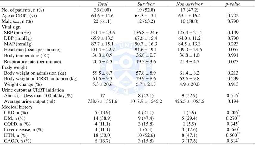
2. Surrogates of volume status
Values of three surrogates at the time of CRRT initiation are shown in last three rows from the bottom of Table 2 and other several values measured by BIA are also shown.
Table 2. Surrogates of volume status at CRRT initiation
Survivor (n=19) Non-survivor (n=17) p-value
OH (L) 5.4 ± 4.3 6.9 ± 3.8 0.275 VUrea (L) 38.5 ± 15.3 36.9 ± 17.4 0.816 ECW (L) 30.5 ± 29.3 24.7 ± 8.8 0.437 ICW (L) 26.7 ± 14.0 26.4 ± 15.3 0.938 Weight change (%) 5.7 ± 21.7 4.9 ± 20.0 0.913 OH/ECW (L/L) 0.21 ± 0.14 0.28 ± 0.13 0.110 B-lines on lung US 6.7 ± 3.7 7.0 ± 4.6 0.821
There was a tendency that survivors showed lower OH/ECW value (0.21±0.14 vs. 0.28±0.13, P=0.110) in BIA and the number of B-lines (6.7±3.7 vs. 7.0±4.6, P=0.821) in Lung US. However, there was no significant difference in volume status parameters of BIA and lung US between survivor group and non-survivor group.
Abbreviations: OH, over-hydration; VUrea, volume of distribution of urea; ECW, extra-cellular water; ICW intra-cellular water; E/I ratio, ECW/ICW ratio; lung US, lung ultrasound
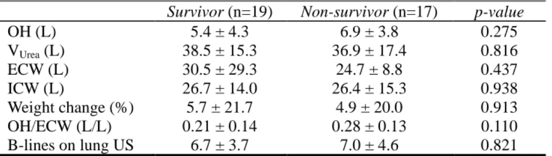
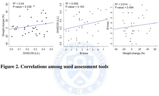
3. Correlations among three surrogates
As shown in Figure 2, three surrogates did not show significant correlations between each other, although each surrogate was supposed to be comparable to body status.
4. Outcomes
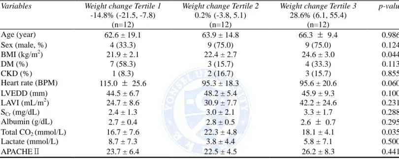
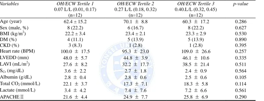
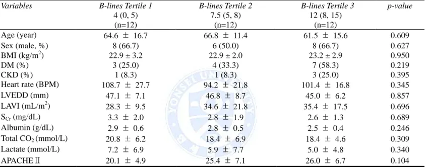
I classified the patients into three groups according to tertiles of each surrogates in order to evaluate primary outcome. Patient characteristics according to each surrogate are shown in Table 3, Table 4, and Table 5. Patient characteristics include demographic variables, echocardiographic variables that represent the patient’s cardiac function, and laboratory variables that are associated with severity of the patient’s status. Patient characteristics according to weight change tertiles did not show any significant difference among three tertiles except total CO2 (P=0.035), which was highest in tertile 2. However, there was no significant difference among tertiles when patients were divided according to OH/ECW by BIA and B-lines by lung US.
I analyzed 28-day mortality according to each surrogate by comparing mortality rate of highest tertile versus lower two third tertiles. Kaplan-Meier plots revealed that 28-day mortality according to weight change and B-lines by lung US showed trends that survival rate of highest tertile was lower than that of lower two thirds (log-rank test, P=0.443 and P=0.450 respectively, Figure 4 A and C). However, 28-day survival rate of highest OH/ECW tertile showed significantly lower than that of lower two thirds as shown in figure 4 (log-rank test, P=0.020, Figure 4 B).
Table 3. Patient characteristics according to weight change tertiles
Variables Weight change Tertile 1
-14.8% (-21.5, -7.8) (n=12)
Weight change Tertile 2
0.2% (-3.8, 5.1) (n=12)
Weight change Tertile 3
28.6% (6.1, 55.4) (n=12) p-value Age (year) 62.6 ± 19.1 63.9 ± 14.8 66.3 ± 9.4 0.986 Sex (male, %) 4 (33.3) 9 (75.0) 9 (75.0) 0.124 BMI (kg/m2) 21.9 ± 2.1 22.4 ± 2.7 24.6 ± 3.0 0.044 DM (%) 7 (58.3) 3 (15.7) 4 (33.3) 0.113 CKD (%) 1 (8.3) 2 (16.7) 3 (15.7) 0.855 Heart rate (BPM) 115.0 ± 25.6 95.3 ± 18.3 95.6 ± 20.6 0.060 LVEDD (mm) 44.5 ± 6.7 48.2 ± 5.4 45.9 ± 9.3 0.100 LAVI (mL/m2) 24.7 ± 8.6 30.9 ± 7.7 42.2 ± 24.6 0.231 SCr (mg/dL) 2.4 ± 1.3 3.0 ± 2.1 3.3 ± 1.7 0.288 Albumin (g/dL) 2.7 ± 0.4 2.8 ± 0.5 2.6 ± 0.7 0.295 Total CO2 (mmol/L) 16.7 ± 7.6 22.3 ± 4.8 18.1 ± 4.1 0.035 Lactate (mmol/L) 8.7 ± 7.3 3.8 ± 4.4 5.8 ± 7.1 0.500 APACHEⅡ 23.7 ± 6.4 22.5 ± 4.5 26.2 ± 8.3 0.441
Abbreviations: BMI, body mass index; DM, diabetes mellitus; CKD, chronic kidney disease; BPM, beat per minute;
LVEDD, left ventricular end diastolic diameter; LAVI, left atrial volume index; SCr, serum creatinine concentration; APACHE Ⅱ, Acute Physiology and Chronic Health Evaluation Ⅱ
Table 4. Patient characteristics according to OH/ECW tertiles by BIA
Variables OH/ECW Tertile 1
0.07 L/L (0.01, 0.17) (n=12) OH/ECW Tertile 2 0.27 L/L (0.18, 0.32) (n=12) OH/ECW Tertile 3 0.40.L/L (0.32, 0.45) (n=12) p-value Age (year) 62.4 ± 15.2 70.1 ± 8.8 60.3 ± 17.2 0.286 Sex (male, %) 8 (22.2) 6 (16.7) 8 (22.2) 0.627 BMI (kg/m2) 22.2 ± 3.4 23.4 ± 2.1 23.3 ± 2.9 0.530 DM (%) 4 (11.1) 5 (13.9) 5 (13.9) 0.890 CKD (%) 3 (8.3) 1 (2.8) 1 (2.8) 0.395 Heart rate (BPM) 100.0 ± 17.5 95.3 ± 23.0 109.0 ± 26.6 0.257 LVEDD (mm) 48.0 ± 5.7 44.8 ± 3.9 46.1 ± 10.6 0.335 LAVI (mL/m2) 27.6 ± 8.2 32.2 ± 17.7 38.5 ± 21.4 0.511 SCr (mg/dL) 3.6 ± 2.2 2.7 ± 1.8 2.4 ± 0.9 0.564 Albumin (g/dL) 2.8 ± 0.4 2.8 ± 0.6 2.5 ± 0.6 0.105 Total CO2 (mmol/L) 22.1 ± 3.7 17.3 ± 7.1 18.3 ± 5.8 0.114 Lactate (mmol/L) 3.4 ± 4.2 7.4 ± 7.6 7.2 ± 6.6 0.561 APACHEⅡ 21.6 ± 4.4 24.9 ± 7.7 25.8 ± 6.9 0.290
Abbreviations: BMI, body mass index; DM, diabetes mellitus; CKD, chronic kidney disease; BPM, beat per minute;
LVEDD, left ventricular end diastolic diameter; LAVI, left atrial volume index; SCr, serum creatinine concentration; APACHE Ⅱ, Acute Physiology and Chronic Health Evaluation Ⅱ
Table 5. Patient characteristics according to B-lines tertiles by lung US
Variables B-lines Tertile 1
4 (0, 5) (n=12) B-lines Tertile 2 7.5 (5, 8) (n=12) B-lines Tertile 3 12 (8, 15) (n=12) p-value Age (year) 64.6 ± 16.7 66.8 ± 11.4 61.5 ± 15.6 0.609 Sex (male, %) 8 (66.7) 6 (50.0) 8 (66.7) 0.627 BMI (kg/m2) 22.9 ± 3.2 22.9 ± 2.0 23.2 ± 2.9 0.950 DM (%) 3 (25.0) 4 (33.3) 7 (58.3) 0.219 CKD (%) 1 (8.3) 1 (8.3) 3 (25.0) 0.395 Heart rate (BPM) 108.7 ± 27.7 94.2 ± 21.8 101.4 ± 16.8 0.345 LVEDD (mm) 47.1 ± 7.1 46.8 ± 8.7 45.0 ± 6.2 0.857 LAVI (mL/m2) 28.3 ± 9.5 34.6 ± 21.8 35.4 ± 17.5 0.696 SCr (mg/dL) 3.3 ± 2.0 2.8 ± 1.9 2.6 ± 1.3 0.689 Albumin (g/dL) 2.9 ± 0.6 2.8 ± 0.5 2.5 ± 0.4 0.246 Total CO2 (mmol/L) 20.8 ± 6.2 18.4 ± 6.9 18.4 ± 4.6 0.309 Lactate (mmol/L) 7.2 ± 6.9 5.9 ± 7.7 5.0 ± 4.8 0.340 APACHEⅡ 20.1 ± 4.9 25.4 ± 7.1 26.0 ± 6.7 0.104
Abbreviations: BMI, body mass index; DM, diabetes mellitus; CKD, chronic kidney disease; BPM, beat per minute;
LVEDD, left ventricular end diastolic diameter; LAVI, left atrial volume index; SCr, serum creatinine concentration; APACHE Ⅱ, Acute Physiology and Chronic Health Evaluation Ⅱ
Figure 3. Kaplan-Meier analysis of 28-day mortality according to A) weight change, B) OH/ECW by BIA, and C) B-lines by lung US
A B
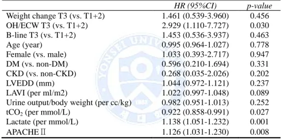
Univariate cox proportional hazard model was performed. Variables which affected 28-day mortality other than OH/ECW (HR 2.929, CI 1.110-7.727, P=0.030) were total CO2 level (HR 0.922, CI 0.858-0.991, P=0.027), lactate level (HR 1.138, CI 1.051-1.232, P=0.001), and APACHE Ⅱ score (HR 1.126, CI 1.031-1.230, P=0.008). Traditional risk factors such as Lactate level and APACHE Ⅱ score that indicates severity of critically ill patients also predicted 28-mortality in this patient group.
Table 6. Univariate Cox proportional hazard model for 28-day mortality
HR (95%CI) p-value Weight change T3 (vs. T1+2) 1.461 (0.539-3.960) 0.456 OH/ECW T3 (vs. T1+2) 2.929 (1.110-7.727) 0.030 B-line T3 (vs. T1+2) 1.453 (0.536-3.937) 0.463 Age (year) 0.995 (0.964-1.027) 0.778 Female (vs. male) 1.033 (0.393-2.717) 0.947 DM (vs. non-DM) 0.596 (0.210-1.694) 0.331 CKD (vs. non-CKD) 0.268 (0.035-2.026) 0.202 LVEDD (mm) 1.044 (0.972-1.121) 0.237 LAVI (per ml/m2) 1.022 (0.997-1.048) 0.089 Urine output/body weight (per cc/kg) 0.982 (0.951-1.013) 0.252 tCO2 (per mmol/L) 0.922 (0.858-0.991) 0.027 Lactate (per mmol/L) 1.138 (1.051-1.232) 0.001 APACHEⅡ 1.126 (1.031-1.230) 0.008
Abbreviations: T3, highest tertile; T1+2, lower two-thrids tertiles; HR, hazard
ratio; CI, confidence interval; OH, over-hydration; ECW, extra-cellular water; DM, diabetes mellitus; CKD, chronic kidney disease; LVEDD, left ventricular end diastolic diameter; LAVI, left atrial volume index; APACHE Ⅱ, Acute Physiology and Chronic Health Evaluation Ⅱ
Table 7. Multivariate Cox proportional hazard models for 28-day mortality according to OH/ECW tertiles.
Variables Model 1 Model 2 Model 3
HR (95% CI) P-value HR (95% CI) P-value HR (95% CI) P-value OH/ECW T3 (vs. T1+2) 2.929 (1.110-7.727) 0.030 4.924 (1.408-17.22) 0.013 3.826 (1.043-14.03) 0.043
Age (per year) 1.019
(0.981-1.058) 0.331 1.023 (0.981-1.066) 0.289 Female (vs. male) 0.877 (0.326-2.355) 0.794 1.094 (0.379-3.156) 0.868 DM (vs. non-DM) 0.323 (0.082-1.271) 0.106 0.314 (0.088-1.120) 0.074 CKD (vs. non-CKD) 0.319 (0.041-2.477) 0.274 0.219 (0.025-1.905) 0.169 APACHE Ⅱ 0.079 (0.989-1.213) 0.079
tCO2 (per mmol/L) 0.946
(0.861-1.039) 0.247
Abbreviations: HR, hazard ratio; CI, confidence interval; OH, over-hydration; ECW, extra-cellular water; DM, diabetes
Multivariate Cox proportional hazard model analyses were conducted. Analyses were performed according to each surrogate. Model 1 was crude model. Model 2 included demographic factors such as age, sex, DM and CKD history. Finally, model 3 included total CO2 level and APACHE Ⅱ score as shown in Table 7. OH/ECW predicted significant increase of 28-day mortality after adjustment of confounding factors (HR 3.826, CI 1.043-14.03, P=0.043).
IV. DISCUSSION
This study was performed to investigate the prognostic value of changes in weight as a volume parameter and volume status assessment by BIA and lung US on mortality in septic AKI patients undergoing CRRT. In present study, three surrogates were used to measure volume status of the septic AKI patients undergoing CRRT. These surrogates were under investigation to verify their effectiveness in various circumstances. Previous studies revealed that fluid overload is associated with mortality of patients with septic AKI as well as treated with chronic dialysis.15 Several studies were performed on predictive use of BIA on chronic kidney disease on hemodialysis. Some of those studies revealed that values measured by BIA are associated with biochemical and parameters of hemodialysis such as dry weight and ultrafiltration volume.16 Other recent study showed that fluid assessment and adjustment of dry weight by BIA improves clinical outcomes and all-cause mortality in patients undergoing conventional hemodialysis and peritoneal dialysis.17 However, usage of BIA in critically ill patients such as patients with septic shock is yet to be explored. Although numerous previous studies on critically ill patients emphasized of volume control, assessment tool for volume status is still elusive. An observational study performed on 37 patients with septic shock treated with CRRT showed that net balanced ultrafiltration reduced intra-abdominal pressure, total body water, ECW, and ICW in this patient group.18 In present study, a positive trend that increasing weight change and B-lines measured by lung US were associated with increasing risk of 28-day mortality, despite of lack of
statistical power. However, OH/ECW measured by BIA showed significant prognostic power on 28-day mortality in this patient group (HR=2.929, CI=1.110-7.727, P=0.030). In the future, large scale randomized control study on volume control using BIA is required for verification of effectiveness of BIA in this patient group for prediction of overall mortality.
As for the number of B-lines in lung US, it did not predict 28-day mortality with statistical significance in present study group. Since lung US was introduced, there have been many efforts to assess volume status in various patient groups. Though a few studies were performed on lung US for volume assessment, they dealt mainly with patients with chronic kidney disease undergoing intermittent hemodialysis. However, those studies verified usefulness of lung US only for prediction of pulmonary congestion and fail to verify predictive power of hemodynamic congestions such as pulmonary artery occlusion pressure.19 Potential use of lung US in the management of acute circulatory failure such as septic AKI seems yet to be examined.20 This study did not verify that the number of B-lines affect 28-day mortality. Meanwhile, other recent study also concluded the impact of integrated cardiopulmonary sonography including lung US on prognoses requires further study.21 That is, the role of lung US in critically ill patients such as patients with septic AKI is not clearly identified.
Previous studies on the similar patient groups who were chronic hemodialysis patients showed conflicting results. One study of Basso et al. on reproducibility of BIA and lung US concluded correlation between BIA and lung US was statistically significant,22 while other study of Donadio et al. showed correlation between these two techniques was not significantly associated.23 In this study, lack of significant correlation of these two techniques may be because of relatively small sample size and non-homogeneous characteristics of the patients. In addition, B-lines in lung US can be detected when pulmonary edema or effusion arise from other etiologies than volume overload. Lung congestion due to different etiologies such as cardiac failure or liver failure, pneumonia, post-operative status could commonly result in increased number of
B-lines in lung US.
The device that we used in this study calculates and displays OH in comparison with the healthy individuals with same height and body weight by using equations which was suggested by Moissl et al.24 This OH value required to be normalized by ECW to enhance accuracy of OH because OH values are dependent on physiological hydration properties of body tissues.25 Especially, previous study reported discrepancy among OH/ECW, OH/BMI and OH/body weight and accuracy of OH/ECW over OH/BMI or OH/body weight.26 So, I also chose OH/ECW as a surrogate of volume status in this study.
In this study, there are several potential limitations that should be noted. First the study was performed small number of patients with heterogeneous characteristics. I did not take into consideration of various etiologies that generated B-lines in lung US, that is, B-lines could have been observed without obvious volume overload. In addition, it should be determined whether one performs measurement right before or long enough after administration of CRRT because administration of CRRT could be urgent to some patients with unstable vital sign.
Finally, BIA values are known to be affected by numerous variables including body position, hydration status, consumption of food and beverages, ambient air and skin temperature, recent physical activity, and conductance of examining table.27 However, because this study involved critically ill patients and so many life-supporting devices were attached to the patients, the standardization of measurement condition was difficult. Minimization of interruption in BIA and Lung US measurement should be taken regarded in future studies.
V. CONCLUSION
Higher OH/ECW measured by BIA was an independent risk factor for 28-day mortality in septic AKI patients undergoing CRRT. Determining fluid status by BIA could be a useful method to stratify mortality risk in this patient group.
The large number of patients and prospective randomized control study
should be needed to figure out the usefulness of a novel assessment for
fluid status objectively.
REFERENCES
1. Wan L, Bagshaw SM, Langenberg C, Saotome T, May C, Bellomo R. Pathophysiology of septic acute kidney injury: what do we really know? Crit Care Med 2008; 36(4 Suppl):S198-203.
2. Uchino S, Kellum JA, Bellomo R, Doig GS, Morimatsu H, Morgera S et al. Beginning and Ending Supportive Therapy for the Kidney (BEST Kidney) Investigators Acute renal failure in critically ill patients: A multinational, multicenter study. JAMA 2005; 294:813–8.
3. Uchino S, Bellomo R, Morimatsu H, Morgera S, Schetz M, Tan I et al. Continuous renal replacement therapy: a worldwide practice survey. The beginning and ending supportive therapy for the kidney (B.E.S.T. kidney) investigators. Intensive Care Med 2007; 33(9):1563–70.
4. Kellum JA, Ronco C. Dialysis: results of RENAL--what is the optimal CRRT target dose? Nat Rev Nephrol 2010; 6(4):191-2.
5. Piccoli A. Bioelectric impedance measurement for fluid status assessment. Contrib Nephrol 2010; 164:143-52.
6. Basso F, Milan Manani S, Cruz DN, Teixeira C, Brendolan A, Nalesso F et al. Comparison and Reproducibility of Techniques for Fluid Status Assessment in Chronic Hemodialysis Patients. Cardiorenal Med 2013; 3(2):104-12
7. Onofriescu M, Mardare NG, Segall L, Voroneanu L, Cuşai C, Hogaş S et al. Randomized trial of bioelectrical impedance analysis versus clinical criteria for guiding ultrafiltration in hemodialysis patients: effects on blood pressure, hydration status, and arterial stiffness. Int Urol Nephrol 2012; 44(2):583-91.
8. Zoccali C, Torino C, Tripepi R, Tripepi G, D'Arrigo G, Postorino M et al. Lung US in CKD Working Group. Pulmonary Congestion Predicts Cardiac Events and Mortality in ESRD J Am Soc Nephrol 2013; 24(4):639-46.
9. Trezzi M, Torzillo D, Ceriani E, Costantino G, Caruso S, Damavandi PT et al. Lung ultrasonography for the assessment of rapid extravascular water variation: evidence from hemodialysis patients.
Intern Emerg Med 2013; 8(5):409-15.
10. Kellum JA, Ronco C. Dialysis: results of RENAL--what is the optimal CRRT target dose? Nat Rev Nephrol 2010; 6(4):191-2.
11. Bone RC, Balk RA, Cerra FB, Dellinger RP, Fein AM, Knaus WA et al. Definitions for sepsis and organ failure and guidelines for the use of innovative therapies in sepsis. The ACCP/SCCM Consensus Conference Committee. American College of Chest Physicians/Society of Critical Care Medicine. 1992. Chest 2009; 136(5 Suppl):e28
12. Cohen J, Cristofaro P, Carlet J, Opal S. New method of classifying infections in critically ill patients Crit Care Med 2004; 32(7):1510-26 13. Bellomo R, Ronco C, Kellum JA, Mehta RL, Palevsky P; Acute
Dialysis Quality Initiative workgroup. Acute renal failure - definition, outcome measures, animal models, fluid therapy and information technology needs: the Second International Consensus Conference of the Acute Dialysis Quality Initiative (ADQI) Group. Crit Care 2004; 8(4):R204-12
14. Oh HJ, Lee MJ, Kim CH, Kim DY, Lee HS, Park JT et al. The benefit of specialized team approaches in patients with acute kidney injury undergoing continuous renal replacement therapy: propensity score matched analysis. Crit Care 2014; 18(4): 454
15. Çelik G, Kara I, Yilmaz M, Apiliogullari S. The relationship between bioimpedance analysis, haemodynamic parameters of haemodialysis, biochemical parameters and dry weight. J Int Med Res 2011; 39(6):2421-8.
16. Onofriescu M, Hogas S, Voroneanu L, Apetrii M, Nistor I, Kanbay M et al. Bioimpedance-Guided Fluid Management in Maintenance Hemodialysis: A Pilot Randomized Controlled Trial Am J Kidney dis 2014; 64(1):111-8.
17. Haapio M, Lentini P, House AA, de Cal M, Cruz DN, Gong D et al. Bioelectrical impedance analysis in the assessment of hydration status in peritoneal dialysis patients. Contrib Nephrol 2012; 178:238-45. 18. Wojciech D, Edyta KH, Daniel S, Wojciech Z, Ziemowit R, Bart DK et
al. Continuous veno-venous hemofiltration to adjust fluid volume excess in septic shock patients reduces intra-abdominal pressure Clinical Nephrology 2014; 82(1):41-50
19. Donadio C, Bozzoli L, Colombini E, Pisanu G, Ricchiuti G, Picano E et al. Effective and Timely Evaluation of Pulmonary Congestion: Qualitative Comparison Between Lung Ultrasound and Thoracic Bioelectrical Impedance in Maintenance Hemodialysis Patients. Medicine(Baltimore) 2015; 94(6):e473
20. Lichtenstein D Fluid administration limited by lung sonography: the place of lung ultrasound in assessment of acute circulatory failure (the FALLS-protocol) Expert Rev Respir Med 2012; 6(2):155-62
21. Wang XT, Liu DW, Zhang HM, Chai WZ. Integrated cardiopulmonary sonography: a useful tool for assessment of acute pulmonary edema in the intensive care unit. J Ultrasound Me. 2014; 33(7):1231-9.
22. Basso F, Milan Manani S, Cruz DN, Teixeira C, Brendolan A, Nalesso F et al. Comparison and Reproducibility of Techniques for Fluid Status Assessment in Chronic Hemodialysis Patients. Cardiorenal Med 2013; 3(2):104-12.
23. Donadio C, Bozzoli L, Colombini E, Pisanu G, Ricchiuti G, Picano E et al. Effective and Timely Evaluation of Pulmonary Congestion: Qualitative Comparison Between Lung Ultrasound and Thoracic Bioelectrical Impedance in Maintenance Hemodialysis Patients. Medicine(Baltimore) 2015; 94(6):e473
24. Moissl UM, Wabel P, Chamney PW, Bosaeus I, Levin NW, Bosy-Westphal A et al. Body fluid volume determination via body composition spectroscopy in health and disease. Physiol Meas 2006; 27(9):921-33
25. Cridlig J, Nadi M, Kessler M. Bioimpedance Measurement in the Kidney Disease Patient. In: Technical Problems in Patients on Hemodialysis. Online edition. Rijeka, Croatia: InTech Europe: 2011. pp.174-5.
multi-frequency bioimpedance spectroscopy in peritoneal dialysis patients: independent predictor of patient survival. Nephrol Dial Transplant 2014; 29(7):1430-7.
27. Rombeau JL, Bandini L, Barr R, Bier DM, Bistrian BR, Blair SN et al. National Institutes of Health Technology Assessment Conference Statement, Bioelectrical Impedance Analysis in Body Composition Measurement December; 1994. Am J Clin Nutr 1996; 64(3Suppl): 387S-532S
ABSTRACT(IN KOREAN)
CRRT를 필요로 하는 패혈성 급성 신손상 환자에서
생체전기임피던스와 폐초음파를 이용한 체액상태의 평가가
가지는 예후평가적 가치
<지도교수 유태현>
연세대학교 대학원 의학과
한 승 규
배경 패혈성 급성 신손상은 중환자실 치료를 필요로 하는 환자에서 일어나는 가장 흔한 합병증 중 하나이다. CRRT 치료를 요하는 환자에서 전신의 수분 상태는 환자의 예후를 평가하는데 있어 유의한 위험요소로 밝혀져 있다. 이 연구에서는 CRRT를 필요로 하는 패혈성 급성 신손상 환자에서 생체전기임피던스와 폐초음파를 이용한 체액상태의 평가가 가지는 예후평가적 가치를 분석하였다. 방법 2014년 4월부터 2015년 2월 사이에 본원에서 CRRT를 필요로 하는 패혈성 급성 신손상 환자 36명을 대상으로 하여 전향적 관찰연구를 시행하였다. 생체전기임피던스와 폐초음파는 CRRT의 시작 시점에 시행하였다. 수분 상태를 평가하는 표지자로는 1) 입원일과 CRRT 시작일의 체중변화, 2) 생체전기임피던스로 측정한 over-hydration(OH)/extracellular water(ECW), 3) 폐 초음파로 측정한 B-line의 수를 사용하였다. 1차 종점은 28일 생존률로 설정하였다. 결과 36명 대상 환자 중, 연구 기간 동안 19(48.7%) 명의 환자가 사망했다. 평균 체중변화와 생체전기임피던스로 측정된 OH/ECW 는 각각 5.3±20.7 % 와 0.3±0.1 L/L 였다. 폐초음파로 측정한 B-line 개수의 중앙값은 6 (사분위수범위 4-10)이었다. Kaplan-Meier 분석을 시행하였을 때, 상위 삼분위의 OH/ECW 를 보이는 환자들이 나머지 환자들에비해 28일 생존률이 유의하게 더 낮은 것으로 나타났다(log-rank test, P=0.020). 체중변화와 B-line의 수는 28일 생존률과 유의한 상관관계를 보이지는 않았다 (각각 log-rank test, P=0.443 and P=0.450). 다변량 Cox proportional hazard model analysis 를시행한 결과 상위 삼분위수의 OH/ECW 는 혼란변수를 보정하고 난 뒤에도 독립적인 위험요소인 것으로 밝혀졌다 (HR=3.83, 95% CI=1.04-14.03, P=0.043). 결론 생체전기임피던스로 측정한 OH/ECW 값이 높은 것은 CRRT를 필요로 하는 패혈성 급성 신손상 환자에서 독립적인 위험요소인 것으로 밝혀졌다. 본 연구에서는 생체전기임피던스로 측정한 체내 수분상태를 평가하는 것이 이 환자군의 생존률을 결정하는데 있어 유용한 방법이 될 수 있다는 결과를 얻을 수 있었다. 이 연구는 이들 새로운 측정방법을 활용하여 CRRT로 환자의 수분상태를 조절하는 대규모의 무작위 제어 연구를 시행하는 데 대한 근거를 마련해 주고 있다. --- 핵심되는 말: 지속적신대체 요법, 생체전기임피던스, 폐초음파