Review Article
Functional Roles of p38 Mitogen-Activated Protein Kinase in
Macrophage-Mediated Inflammatory Responses
Yanyan Yang,
1Seung Cheol Kim,
2Tao Yu,
1Young-Su Yi,
1Man Hee Rhee,
3Gi-Ho Sung,
4Byong Chul Yoo,
5and Jae Youl Cho
11Department of Genetic Engineering, Sungkyunkwan University, Suwon 440-746, Republic of Korea
2Division of Gynecologic Oncology Department of Obstetrics and Gynecology, Ewha Womans University
Mokdong Hospital College of Medicine, Ewha Womans University, Seoul 158-710, Republic of Korea
3College of Veterinary Medicine, Kyungpook National University, Daegu 702-701, Republic of Korea
4Department of Herbal Crop Research, National Institutes of Horticultural & Herbal Science,
Rural Development Administration, Suwon 441-707, Republic of Korea
5Colorectal Cancer Branch, Research Institute, National Cancer Center, Goyang, Gyeonggi 410-769, Republic of Korea
Correspondence should be addressed to Byong Chul Yoo; yoo [email protected] and Jae Youl Cho; [email protected] Received 29 September 2013; Revised 27 November 2013; Accepted 11 February 2014; Published 20 March 2014 Academic Editor: Pham My-Chan Dang
Copyright © 2014 Yanyan Yang et al. This is an open access article distributed under the Creative Commons Attribution License, which permits unrestricted use, distribution, and reproduction in any medium, provided the original work is properly cited. Inflammation is a natural host defensive process that is largely regulated by macrophages during the innate immune response. Mitogen-activated protein kinases (MAPKs) are proline-directed serine and threonine protein kinases that regulate many physiological and pathophysiological cell responses. p38 MAPKs are key MAPKs involved in the production of inflammatory mediators, including tumor necrosis factor-𝛼 (TNF-𝛼) and cyclooxygenase-2 (COX-2). p38 MAPK signaling plays an essential role in regulating cellular processes, especially inflammation. In this paper, we summarize the characteristics of p38 signaling in macrophage-mediated inflammation. In addition, we discuss the potential of using inhibitors targeting p38 expression in macrophages to treat inflammatory diseases.
1. Introduction
Inflammatory response is a basic protective immune process of the organism and is accompanied by symptoms such as redness, heat, swelling, and pain associated with damage to tissues or organs [1]. This is one of the mechanisms by which our body defends us from pathogens such as parasites, bacteria, viruses, and other harmful microorgan-isms. Diseases induced by chronic inflammation, including gastritis, colitis, dermatitis, rheumatoid arthritis, pulmonary diseases, and type II diabetes, damage millions of people’s health every year. Of concern is the increase in prevalence of these chronic inflammatory diseases. Furthermore, there is growing evidence that inflammation is a critical initiation factor inducing a variety of other major diseases such as
cancer, atherosclerosis, Alzheimer’s disease, cardiovascular disease, neurological disorders, and pulmonary diseases [2–
7]. Therefore, a better understanding of inflammation is clinically significant and could improve treatment strategies.
Macrophages within tissues play an essential role in the initiation, development, and resolution of inflammation [8–
11]. Macrophages are white blood cells that are differentiated from monocytes. Their roles are to clean up damaged cells and pathogens by phagocytosis and to activate immune cells, such as neutrophils, dendritic cells, macrophages, and monocytes, in response to pathogens and diseases. They can be activated or deactivated during inflammatory processes depending on the signaling molecules produced. Stimu-lation signals include lipopolysaccharide (LPS), cytokines (interleukin-1 (IL-1) and tumor necrosis factor-𝛼 (TNF-𝛼)),
Volume 2014, Article ID 352371, 13 pages http://dx.doi.org/10.1155/2014/352371
Table 1: p38 family members and their functions in inflammatory responses. p38 isoform
(molecular weight, kDa)
Distribution in tissue Expressing cells Inflammatory responses Reference
p38𝛼 (38) Ubiquitous Macrophages,
neutrophils
Cytokine production (IL-1𝛽, TNF-𝛼, and IL-6); regulation of enzymes (iNOS, COX2); involvement of cell proliferation
and differentiation; induction of cardiomyocyte apoptosis.
[21,27,73]
p38𝛽 (39) Ubiquitous Endothelial
cells, T cells
Regulation of cell differentiation;
induction of cardiomyocyte hypertrophy. [21,27,73]
p38𝛾 (43) Skeletal muscle Not detected Muscle differentiation. [25,27,73]
p38𝛿 (40) Lung, kidney, testis,pancreas, and small intestine
T cells, endothelial cells,
and macrophages
Developmentally regulated; involvement
of cell differentiation. [26,27,73]
other chemical mediators, and extracellular matrix proteins. A variety of membrane receptors are expressed on the sur-faces of macrophages, including pattern recognition recep-tors (PRRs) such as dectin-1 and Toll-like receprecep-tors (TLRs) [12, 13]. These receptors recognize activation signals and subsequently activate downstream protein kinases, eventually resulting in the stimulation of transcription factors including activator protein-1 (AP-1), nuclear factor-kappa B (NF-𝜅B), and cAMP response element-binding protein (CREB).
Various intracellular proteins can initiate inflamma-tion. p38 proteins are a class of mitogen-activated protein kinases (MAPKs) that are major players during inflammatory responses, especially in macrophages. p38, also called RK or cytokinin-specific binding protein (CSBP), was identified in 1994 and is the mammalian ortholog of the yeast Hog1p MAP kinase [14]. p38 was isolated as a 38 kDa protein that is rapidly phosphorylated at a tyrosine residue in response to LPS stimulation, and the p38 gene was cloned through binding of the p38 protein with pyridinyl imidazole derivatives [15]. p38 expression is upregulated in response to inflammatory and stress stimuli, such as cytokines, ultraviolet irradiation, osmotic shock, and heat shock, and is involved in autophagy, apoptosis, and cell differentiation [16–20]. Accumulating evidence suggests that p38 plays an important role in arthritis and inflammation of the liver, kidney, brain, and lung and that it acts as a critical player in inflammatory diseases mediated by macrophages [21–23].
In this paper, we summarize the characteristics of p38 and highlight the physiological significance of p38 activation in macrophage-mediated inflammatory responses. Moreover, we discuss the possibility of using plant extracts, natural products, and chemicals that target p38 as therapeutic drug candidates for the treatment of inflammatory diseases.
2. Structure and Function of p38 Kinases
2.1. The p38 Family. p38 family members are classified into four subtypes:𝛼 (MAPK14), 𝛽 (MAPK11), 𝛾 (MAPK12/ER-K6), and 𝛿 (MAPK13/SAPK4) (Table 1). Genes encoding p38𝛼 and p38𝛽 show 74% sequence homology, whereas 𝛾and 𝛿 are more distant relatives, with approximately 62% sequence identity [24–26]. Genes encoding p38𝛼 and p38𝛽
are ubiquitously expressed within tissues, and especially highly expressed in heart and brain. However, p38𝛾 and p38𝛿 show tissue-specific expression patterns; p38𝛾 is highly expressed in skeletal muscle, whereas p38𝛿 expression is concentrated in the kidneys, lungs, pancreas, testis, and small intestine [27]. In addition, p38𝛾 expression can be induced
during muscle differentiation, and its expression can also be developmentally regulated. Moreover, we demonstrated very high expression of the active form of p38 in inflammatory dis-eases, such as gastritis, colitis, arthritis, and hepatitis [28,29] (unpublished data). p38𝛼 and p38𝛿 are abundantly expressed in macrophages, whereas p38𝛽 is undetectable. p38𝛼 and p38𝛿 are also expressed in endothelial cells, neutrophils, and CD4+ T cells, whereas p38𝛽 is abundant in endothelial cells. These findings indicate that, even though the four p38 family members share sequence homology, their expression is cell-and tissue dependent cell-and their functions may therefore be different.
2.2. p38 Structure and Domains. p38 kinases have two do-mains: a 135 amino acid N-terminal domain and a 225 amino acid C-terminal domain. The main secondary structure of the N-terminal domain is 𝛽-sheets, while the C-terminal domain has a𝛼-helical structure. The catalytic site is located in the region linking the two domains. The phosphorylation lip of p38 consists of 13 residues, Leu-171–Val-183, and the protein is activated by phosphorylation of a single threonine (Thr-180) and a single tyrosine residue (Tyr-182) in the lip [30]. Moreover, in Drosophila p38 MAPK, phosphorylation of tyrosine-186 was detected exclusively in the nucleus fol-lowing osmotic stress [31]. p38 isoforms show various three-dimensional structures with differences in the orientation of the N- and C-terminal domains, resulting in different sized ATP-binding pockets [32].
2.3. Activation of the p38 Response. p38 kinases are activated by environmental and cellular stresses including pathogens, heat shock, growth factors, osmotic shock, ultraviolet irradi-ation, and cytokines. Moreover, various signaling events are
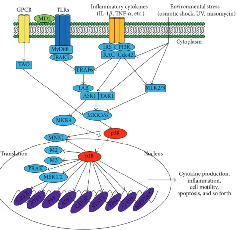
p38 Inflammatory cytokines MSK1/2 p38 MKK3/6 MKK4 Nucleus Cytoplasm Environmental stress (osmotic shock, UV, anisomycin) GPCR TAO RAC Cdc42 IRS PI3K MLK2/3 Cytokine production, inflammation, cell motility, apoptosis, and so forth ASK1 TAK1 TAB TRAF6 TLRs MD2 MyD88 IRAK1 M2 M3 Translation PRAK MNK1 (IL-1𝛽, TNF-𝛼, etc.) CREB ATF1 P53 ATF2 CHOP STAT 1
Eik1 C-Fos MEF2A/C
Figure 1: p38-regulated signaling pathways in inflammatory responses. Inflammation-derived cytokines such as TNF-𝛼 and IL-1, TLR ligands such as LPS, poly(I:C), and peptidoglycan, as a environmental stresses, stimulate the phosphorylation of p38, leading to the activation of transcription factors such as AP-1 family. Subsequent expression of inflammatory genes by these transcription factors mediates various inflammatory responses including cytokine production, migration, and apoptosis of macrophages, monocytes, and neutrophils.
able to stimulate p38 kinases, for example, insulin signaling. Interestingly, with respect to inflammatory responses, a num-ber of studies have reported p38 regulation in macrophages treated with LPS, endothelial cells stimulated with TNF-𝛼, U1 monocytic cells treated with IL-18, and human neutrophils activated with phorbol 12-myristate 13-acetate (PMA), LPS, TNF-𝛼, and fMLP [33,34]. It should also be noted that p38 activation in different cell types is dependent on the type of stimulus.
In addition, a number of studies have reported that distinct upstream kinases selectively activate p38 isoforms. p38 family kinases are all activated by MAP kinase kinases (MKKs). MKK6 activates all four p38 isoforms, while MKK3 can activate p38𝛼, 𝛽, and 𝛿, but not p38𝛾 [35], and MKK4 activates p38𝛼 and 𝛿 [36]. This implies that p38 isoforms can be coactivated by the same upstream regulators and regulated specifically through different regulators.
2.4. p38 Deficiency. p38𝛼 deficiency affects placental devel-opment and erythropoietin expression and can result in embryonic lethality [37–40]. Tetraploid rescue of placental defects in p38𝛼−/−embryos indicated that p38𝛼 was required for extraembryonic development, while it was not neces-sary for embryo development or adult mice survival. In accordance with the phenotype of p38𝛼 knockouts, knock-out of two p38 activators, namely, MKK3 and MKK6, led
to placental and vascular defects and induced embryonic lethality [41]. In contrast, p38𝛽−/− mice were viable and exhibited no obvious health defects. Neither transcription of p38-dependent immediate-early genes, such as TNF-𝛼 and IL-1𝛽, nor T cell development was influenced by the loss of p38𝛽 [42, 43]. Furthermore, mice harboring a T106M mutation in p38𝛼 resisted the drug inhibitory effect of collagen antibody-induced arthritis and LPS-induced TNF production, whereas the same mutation in p38𝛽 had the opposite effect [44], and p38𝛽 knockout mice responded
normally to inflammatory stimuli. Single knockouts of either p38𝛾 or p38𝛿, and even a double knockout, were viable [45]. However, reduced production of TNF-𝛼, IL-1𝛽, and IL-10 in stimulated macrophages isolated from p38𝛾/𝛿 null mice has been observed, which indicates that p38𝛾/𝛿 are important regulatory components of the innate immune response [46]. Taken together, these findings suggest that p38𝛼 is the critical isoform in inflammatory responses but that other subtypes also play important roles.
2.5. Regulation of p38-Activated Signaling. Because p38 sig-naling can be activated by a variety of stimuli, the receptors and downstream pathways are diverse (Figure 1). MTK1, mixed lineage kinase (MLK) 2/3, apoptosis signal-regulating kinase (ASK) 1, and transforming growth factor𝛽-activated kinase (TAK) 1 are all MKK kinases (MAP3Ks) that have
been demonstrated to activate p38 signaling [47–54]. Fur-thermore, different kinases can mediate different signals. Among upstream proteins, Cdc42 and Rac are recognized as critical intermediates of p38 activity [55–57]. Many studies have also reported that p21-activated kinases (PAKs) can be stimulated by binding to Cdc42 and Rac in vitro and subsequently activate a p38 response [58–61]. In addition, Mst1, a mammalian homologue of Ste20, was reported to stimulate MKK6, p38, MKK7, and JNK [62]. However, there are no reports of the involvement of MTK1 and Mst1 in p38 responses in macrophages.
There are numbers of substrates downstream of p38 signaling pathways. MAP kinase-activated protein kinase 2 (M2) and M3 were the first p38 substrates identified [63,64]. Phosphorylated M2 or M3 can activate a variety of substrates, such as small heat shock protein 27 (HSP27), CREB, and activating transcription factor (ATF) 1 [65,66]. To date, sev-eral other proteins have also been identified as downstream substrates of p38, such as mitogen- and stress-activated kinase (MSK), p38-regulated/activated kinase (PRAK), and MAP kinase interaction protein kinase (MNK1) [67–70]. Various novel proteins have also been shown to be direct substrates of p38𝛼, including Ahnak, Iws1, Grp78, Pgrmc, Prdx6, and Ranbp2 [71]. Additionally, TPL2/ERK1/2 has been shown to be downstream kinases controlled by p38𝛾 and 𝛿 isoforms [46].
Phosphatases that downregulate p38 activity have also been identified. Mitogen-activated protein kinase phos-phatases (MKPs) can recognize MAPKs by recognizing the TXY amino acid motif and consequently dephosphorylate and deactivate them. MKP-1, MKP-4, MKP-5, and MKP7 can effectively dephosphorylate p38𝛼 and p38𝛽 [72–75]. However, right now, MKPs cannot dephosphorylate p38𝛾 or p38𝛿 as shown by other researchers [73,76].
Several transcription factors in the nucleus can be phos-phorylated and activated by p38 MAPKs, such as activat-ing transcription factor 1 and 2 (ATF-1, ATF-2), myocyte enhancer factor 2 (MEF2), CCAAT/enhancer-binding pro-teins (C/EBPs), SRF accessory protein-1 (Sap1), p53, and E26 transformation-specific sequence-1 (ETS-1) [77–82]
(Figure 1).
3. p38 Functions in Macrophage-Mediated
Inflammatory Responses and Diseases
Macrophages are the first line of defense of organisms against pathogens. They represent a major cell population distributed in most tissues, and their numbers increase massively in inflammatory diseases. In particular, macrophages are crit-ically involved in the pathogenesis of rheumatoid arthritis (RA) and produce a variety of proinflammatory cytokines and chemokines that contribute to cartilage and bone degra-dation. They are also the predominant cells in the synovial lining and sublining of patients with RA [83]. Macrophages also play a central role in the development of type 2 diabetic nephropathy. Macrophage accumulation in kidney, coronary arteries, nerves, and epiretinal membrane is regarded as one of major causing factors in terms of type 2 diabetic compli-cations, including nephropathy, atherosclerosis, neuropathy,and retinopathy [84–88]. Components of the diabetic milieu, including high glucose, advanced glycation end products, and oxidized low-density lipoprotein, promote macrophage accumulation and activation within diabetic tissues [89]. Macrophage depletion studies have also demonstrated the crucial role of macrophages in the development of diabetic complications [89]. Moreover, macrophages play a pivotal role in the clearance of pulmonary pathogens. Alveolar macrophages (AM) constitute more than 90% of the cells present in bronchoalveolar lavage of na¨ıve tissues [90]. AM can rapidly clear bacteria from airways and cellular debris, help to depress the immune characteristics of the airways, and aid in lung parenchyma modeling [90]. Furthermore, macrophages have significant roles in metabolic diseases, atherosclerosis, bowel disease, and liver fibrosis [91–94]. The fundamental roles of macrophages in inflammation highlight the need for macrophage-targeted studies and therapeutics.
Accumulating evidence suggests that p38 plays an essen-tial role in macrophage-mediated inflammation. p38𝛼 is involved in the expression of proinflammatory mediators in macrophages such as IL-1𝛽, TNF-𝛼, PGE2, and IL-12 [95–
97] as well as COX-2, IL-8, IL-6, IL-3, IL-2, and IL-1, all of which contain AU-rich elements (AREs) in their 3 untrans-lated regions to which p38 binds [98]. Moreover, p38 can regulate the production of endothelial vascular cell adhesion molecule-1 (VCAM-1), which participates in cell proliferation and differentiation of the immune response [99]. Further-more, p38 is associated with various inflammatory dis-eases, including endotoxin-induced shock, collagen-induced arthritis, granuloma, diabetes, and acute lung inflammation [100–103], as well as joint diseases, including synovial inflam-mation, cartilage damage, and bone loss [104]. In contrast, p38𝛽 and 𝛿 also play important roles in regulation of TPA-induced skin inflammation and tumor development [105,
106]. In addition, a large number of reports have suggested a close correlation between p38 and cell apoptosis, cell cycle progression, and differentiation [107–110].
4. Development of p38-Targeted Drugs as
New Anti-Inflammatory Therapeutics
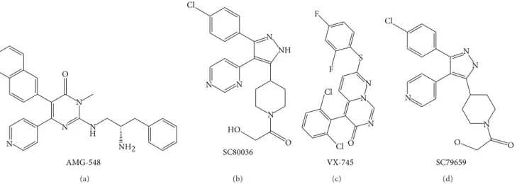
p38 MAPK signaling plays a significant role in the inflam-matory response and other physiological processes. A better understanding of the functional and biological significance of p38 in inflammation has led to the development of p38 inhibitors. Currently, a number of p38 inhibitors have been developed such as AMG-548, SC-80036, SC-79659, and VXs
(Figure 2) [111]; however, few studies have reported their
effects on macrophages.
4.1. Discovery. p38 signaling and specific p38 inhibitors were identified simultaneously. A series of pyridinyl imidazole anti-inflammatory agents, such as bicyclic pyridinyl imida-zoles SKF-86002, SB203580, and SB202190 [15,112–116], were first found to inhibit p38 activity [104,117]. SB inhibitors can antagonize p38 by competing for the ATP-binding pocket, and it has been suggested that Thr-106 could be important for this interaction [115].
AMG-548 NH2 O N N N N H (a) NH N N N N O HO Cl SC80036 (b) S N N N O F F Cl Cl VX-745 (c) O O N N N N Cl SC79659 (d)
Figure 2: Chemical structures of representative novel p38 inhibitors. Promising therapeutic activities of these inhibitors against inflammatory diseases such as RA encourage the continuous progression for clinical trials.
4.2. Crude Plant Extracts. Natural plant extracts that target p38 are promising therapeutics for the treatment of inflam-matory diseases (Table 2). For example, Scutellaria baicalensis extract attenuates MAPK phosphorylation, especially p38 activity, resulting in inhibition of inflammatory mediators such as COX-2, iNOS, L-1𝛽, IL-12, IL-6, IL-2, PGE2, and TNF-𝛼 in RAW 264.7 cells treated with LPS [118]. Phaseolus angularis ethanol extract suppressed the release of PGE2 and NO in macrophages activated by LPS-, Poly(I:C)-, or pam3CSK through regulation of TAK1/p38 pathways and, moreover, it ameliorated gastritis induced by EtOH/HCl in mice, which implies a close relationship between p38 and gastritis [119]. Archidendron clypearia extract suppressed the production of PGE2 in activated RAW264.7 and peritoneal macrophages, as well as gastritis lesions in mouse stomachs exposed to EtOH/HCl [28]. Unfortunately, p38 is not the only target of these extracts; they contain several other active ingredients and therefore are not good candidates for the development of p38-specific inhibitors. However, they are effective at treating inflammatory diseases because of their multiple targets and their ability to improve body’s homeo-static defense responses [120–123]. Meanwhile, as reported previously [124], during covering years 1981–2006, of the 974 small molecule new chemical entities, 63% were naturally derived or semisynthetic derivatives of naturally occurring products, which indicate the importance of plant extract in the drug development [124]. In addition, we and other groups have found that various traditional plant extracts that target p38 kinase can reduce the symptoms of inflammatory diseases (unpublished data), such as gastritis, colitis, arthritis, and hepatitis [28,29]. Plant extract data are summarized in
Table 2.
4.3. Plant-Derived Compounds. Several compounds from natural products inhibit p38 activity and inflammatory responses (Table 3). Sugiol, an aditerpene that was isolated and purified from alcohol extracts of the bark of Calocedrus formosana, effectively decreased the production of intracellu-lar reactive oxygen species (ROS), IL-1𝛽, and TNF-𝛼 in LPS-stimulated macrophages through regulation of MAPKs [125].
Quercetin, a plant-derived flavonoid that is widely distributed in fruits and vegetables, strongly decreased the expression of the inflammatory cytokines iNOS and TNF-𝛼 by targeting both MAPK (ERK and p38) and I𝜅B𝛼 signaling pathways [126,127]. Sulfur-containing compounds from garlic inhib-ited the production of NO, PGE2, and proinflammatory cytokines such as TNF-𝛼, IL-1𝛽, and IL-6 in macrophages by suppressing p38 transduction pathways [128]. A summary of natural products targeting p38 is provided in Table 3. These studies indicate that natural products inhibiting p38 activity exhibit strong anti-inflammatory properties, and are therefore potential therapeutic drug candidates for inflam-matory diseases. Moreover, studies of natural compounds, in addition to elucidating why these extracts have strong anti-inflammatory effects, can also aid the design of novel p38 inhibitors to treat inflammatory diseases.
4.4. Novel Inhibitors. Pharmaceutical companies and re-searchers have worked hard to develop novel, safe, and specific p38 inhibitors. Based on the importance of p38𝛼 in inflammation, people have focused on inhibitors for this isoform rather than the other isoforms. ML3403, a SB203580 analogue, represses the expression of TNF-𝛼, IL-6, and IL-8. It can bind to both active and inactive forms of p38𝛼 kinase, which may reduce asthma-induced airway inflammation and remodeling [129]. AS1940477 has been shown to inhibit the production of proinflammatory cytokines such as TNF-𝛼, IL-1𝛽, IL-6, PEG2, and MMP3 at very low concentrations. Moreover, it can reduce the enzyme activity of both p38𝛼 and𝛽 but has no effect on 100 other kinases, including p38𝛾 and𝛿. It has been shown in rats experiment that low doses of this compound can also reduce the expression of LPS-and Con A-stimulated proinflammatory cytokines, including TNF-𝛼 and IL-6 [130]. Pamapimod strongly suppresses p38 𝛼 and 𝛽 activity and therefore the expression of TNF-𝛼, IL-1𝛽, and IL-6. It also shows high specific activity; when tested for binding to 350 kinases, it only bound to four other kinases in addition to p38. Furthermore, it can reduce clinical signs of inflammatory diseases, such as arthritis, bone loss, and renal diseases. Consistent with this, it inhibited
Table 2: Plant extracts that inhibit the p38 signaling in macrophages.
Plant Action target of p38 Reference
Archidendron clypearia Suppression of PGE2production; amelioration of EtOH/HCl-induced
gastritis [28]
Scutellaria baicalensis Inhibition of iNOS, COX-2, PGE2, IL-1𝛽, IL-2, IL-6, IL-12, and TNF-𝛼
expression [118]
Phaseolus angularis Suppression of the release of PGE2and NO; amelioration of
EtOH/HCl-induced gastritis [119]
Artemisia vestita Inhibition of TNF-𝛼 release; beneficial for the treatment of endotoxin
shock or sepsis [141]
Boswellia serrata Inhibition of TNF-𝛼, IL-1𝛽, and IL-6 [142]
Hibiscus sabdariffa Suppression of nitrite, PGE2release, and hepatic inflammation [143]
Clinopodium vulgare Suppression of NO production; MMP-9 activation [144]
Eriobotryae folium Suppression of LPS-induced NO and PGE2production [145]
Elaeocarpus petiolatus Inhibition of the production of PGE2, TNF-𝛼, and IL-1𝛽 [146]
Polygonum cuspidatum Inhibition of IL-6, TNF-𝛼, NO, and PGE2 [147]
Ginkgo biloba Inhibition of LPS-induced iNOS and COX-2 expression [148]
Lycium chinense Inhibition of LPS-induced NO, PGE2, TNF-𝛼, and IL-6 production [149]
Hopea odorata Inhibition of NO, PGE2, and TNF-𝛼 release; amelioration of gastritis
and ear edema [150]
Table 3: Naturally occurring compounds that inhibit p38 signaling in macrophages.
Compound Action target of p38 Reference
Sugiol Inhibition of IL-1𝛽, TNF-𝛼, and ROS production [151]
Quercetin Inhibition of NO and TNF-𝛼 [114]
Ajoenes Inhibition of NO, PGE2, TNF-𝛼, IL-1𝛽, and IL-6 production [116]
Ginsan Enhanced phagocytic activity; downregulation of TNF-𝛼, IL-1𝛽, IL-6,
IFN-𝛾, and IL-18 [152]
4-Methoxyhonokiol Inhibition of iNOS and COX-2 expression; inhibition of dye leakage
and paw swelling [153]
Schisandrin Suppression of NO production and PGE2release [154]
Rengyolone Inhibition of iNOS and COX-2 expression [155]
Pseudocoptisine Inhibition of proinflammatory mediators such as iNOS, COX-2,
TNF-𝛼, and IL-6 [156]
Mycoepoxydiene Inhibition of LPS-induced proinflammatory mediators including
TNF-𝛼, IL-1𝛽, IL-6, and NO [157]
Britanin Suppression of NO, PGE2, TNF-𝛼, IL-1𝛽, and IL-6 [158]
Hyperin Inhibition of NO production through suppression of iNOS expression [159] Carnosol Inhibition of LPS-stimulated NO production; antioxidative activity [160]
Table 4: p38 inhibitors under human clinical trials.
Compound Clinical trials Reference
PH797804 Chronic obstructive pulmonary disease (COPD) [132]
Losmapimod (GW856553) COPD; atherosclerosis [133,134]
Adalimumab Antipsoriatic [135]
Pamapimod Rheumatoid arthritis [136]
VX-745 Werner syndrome [137]
BMS-582949 Rheumatoid arthritis [138]
TNF-𝛼 production in RA synovial explants and reduced bone loss in murine collagen-induced arthritis. Meanwhile, it increased pain tolerance in a rat model of hyperalgesia [131]. Examples of other newly synthesized compounds are GSK-681323 to treat rheumatoid arthritis, SCIO-469 to treat multiple myeloma and dental pain, and RWJ67657 that was developed as an anti-inflammatory drug, all of which inhibit p38 activity [98]. In summary, most of these inhibitors were designed based on the structure of SB203580 but show more specific and stronger activity. They are therefore promising therapeutic agents for inflammatory diseases.
4.5. Inhibitors in Human Clinical Trials. Based on the impor-tance of p38 MAPK in disease development, inhibition of p38 was regarded as a promising therapeutic strategy to control inflammatory diseases. Right now, effectiveness of some p38 inhibitors is currently under evaluation in clinical trials to treat human diseases. For example, it has been reported that PH797804 and losmapimod were able to improve lung function parameters and to attenuate dyspnoea in patients with chronic obstructive pulmonary disease symptoms [132,
133]. Also, losmapimod was reported to reduce vascular inflammation in the most inflamed regions in patients with atherosclerosis [134]. Clinical and histological improvements linked to the inhibition of TNF-𝛼 level were clearly seen by p38 MAPK inhibitor adalimumab in lesional psoriatic skin [135]. Moreover, it was found that pamapimod can clearly alleviate various rheumatoid arthritis symptoms when coadministered with methotrexate [136]. Besides, there are still many other inhibitors which are ongoing clinical trials as summarized inTable 4[137–140].
5. Summary and Perspectives
Inflammation has attracted great interest because of its significant role in several major diseases and the need to develop better ways to treat these diseases. Importantly, because inflammatory responses are largely mediated by macrophages, functional studies of macrophages in inflam-mation are crucial. Investigation of the roles of p38 MAPKs is particularly relevant as these are essential protein kinases in macrophage-mediated inflammatory responses. A number of studies have indicated that p38 plays a significant role in inflammatory diseases mediated by macrophages, and, as a consequence, several p38 inhibitors have been developed to treat inflammatory diseases. However, most of these inhibitors have shortcomings, such as low specificity, low efficacy, and high toxicity. As a result, new p38 inhibitors are urgently required. We are optimistic that novel and safe p38 inhibitors that possess strong anti-inflammatory properties will be developed in the near future to treat inflammatory diseases.
Abbreviations
MAPKs: Mitogen-activated protein kinases COX2: Cyclooxygenase-2
TNF-𝛼: Tumor necrosis factor alpha
PGE2: Prostaglandin E2 NF-𝜅B: Nuclear factor
kappa-light-chain-enhancer of activated B cells
AP-1: Activator protein-1 CREB: cAMP response
element-binding protein CSBP: Cytokinin-specific binding
protein
PMA: Phorbol myristate acetate LPS: Lipopolysaccharide MKPs: MAP kinase phosphatases MKKs: MAPK kinases
PAK: p21-activated kinases HSP27: Heat shock protein 27 TLRs: Toll-like receptors
PRRs: Pattern recognition receptors MNK1: MAP kinase interaction protein
kinase
ATF-2: Activating transcription factor-2 MEF2C: Myocyte enhancer factor 2C PRAK: p38-regulated/activated kinase MSK: Mitogen- and stress-activated
kinase
ARE: AU-rich elements ICAM: Cell adhesion molecule.
Conflict of Interests
The authors report no conflict of interests. The authors alone are responsible for the content and writing of the paper.
Authors’ Contribution
Yanyan Yang, Seung Cheol Kim, and Tao Yu contributed equally to this work.
Acknowledgment
This study was supported by a Grant (HI12C0050) of the Korean Health Technology R&D Project, Ministry of Health and Welfare, Republic of Korea.
References
[1] L. Ferrero-Miliani, O. H. Nielsen, P. S. Andersen, and S. E. Girardin, “Chronic inflammation: importance of NOD2 and NALP3 in interleukin-1𝛽 generation,” Clinical and Experimental
Immunology, vol. 147, no. 2, pp. 227–235, 2007.
[2] N. Eiro and F. J. Vizoso, “Inflammation and cancer,” World
Journal of Gastrointestinal Surgery, vol. 4, no. 3, pp. 62–72, 2012.
[3] Y. W. Lee, P. H. Kim, H. L. Won, and A. A. Hirani, “Interleukin-4, oxidative stress, vascular inflammation and atherosclerosis,”
Biomolecules and Therapeutics, vol. 18, no. 2, pp. 135–144, 2010.
[4] T. Wyss-Coray and J. Rogers, “Inflammation in Alzheimer disease-a brief review of the basic science and clinical literature,”
Cold Spring Harbor Perspectives in Medicine, vol. 2, no. 1, Article
[5] P. Urrutia, P. Aguirre, A. Esparza et al., “Inflammation alters the expression of DMT1, FPN1 and hepcidin, and it causes iron accumulation in central nervous system cells,” Journal of
Neurochemistry, vol. 126, no. 4, pp. 541–549, 2013.
[6] V. Wee Yong, “Inflammation in neurological disorders: a help or a hindrance?” Neuroscientist, vol. 16, no. 4, pp. 408–420, 2010. [7] M. Provinciali, M. Cardelli, and F. Marchegiani, “Inflammation,
chronic obstructive pulmonary disease and aging,” Current
Opinion in Pulmonary Medicine, vol. 17, supplement 1, pp. S3–
S10, 2011.
[8] T. Yu, Y. S. Yi, Y. Yang, J. Oh, D. Jeong, and J. Y. Cho, “The pivotal role of TBK1 in inflammatory responses mediated by macrophages,” Mediators of Inflammation, vol. 2012, Article ID 979105, 8 pages, 2012.
[9] N. Fujiwara and K. Kobayashi, “Macrophages in inflammation,”
Current Drug Targets, vol. 4, no. 3, pp. 281–286, 2005.
[10] J. S. Duffield, “The inflammatory macrophage: a story of Jekyll and Hyde,” Clinical Science, vol. 104, no. 1, pp. 27–38, 2003. [11] J. J. Jeong, S. E. Jang, E. H. Joh, M. J. Han, and D. H. Kim,
“Kalo-panaxsaponin B ameliorates TNBS-induced colitis in mice,”
Biomolecules & Therapeutics, vol. 20, no. 5, pp. 457–462, 2012.
[12] P. R. Taylor, L. Martinez-Pomares, M. Stacey, H.-H. Lin, G. D. Brown, and S. Gordon, “Macrophage receptors and immune recognition,” Annual Review of Immunology, vol. 23, pp. 901– 944, 2005.
[13] S. Batbayar, D. H. Lee, and H. W. Kim, “Immunomodulation of fungal beta-glucan in host defense signaling by dectin-1,”
Biomolecules & Therapeutics, vol. 20, no. 5, pp. 433–445, 2012.
[14] S. M. O’Rourke and I. Herskowitz, “The Hog1 MAPK prevents cross talk between the HOG and pheromone response MAPK pathways in Saccharomyces cerevisiae,” Genes and Development, vol. 12, no. 18, pp. 2874–2886, 1998.
[15] J. C. Lee, J. T. Laydon, P. C. McDonnell et al., “A protein kinase involved in the regulation of inflammatory cytokine biosynthesis,” Nature, vol. 372, no. 6508, pp. 739–746, 1994. [16] J. E. Huh, I. T. Jung, J. Choi et al., “The natural flavonoid
galangin inhibits osteoclastic bone destruction and osteoclasto-genesis by suppressing NF-kappaB in collagen-induced arthritis and bone marrow-derived macrophages,” European Journal of
Pharmacology, vol. 698, no. 1–3, pp. 57–66, 2013.
[17] M. Kim, Y. X. Li, P. Dewapriya, B. Ryu, and S. K. Kim, “Flori-doside suppresses pro-inflammatory responses by blocking MAPK signaling in activated microglia,” BMB Reports, vol. 46, no. 8, pp. 398–403, 2013.
[18] G. L. Schieven, “The biology of p38 kinase: a central role in inflammation,” Current Topics in Medicinal Chemistry, vol. 5, no. 10, pp. 921–928, 2005.
[19] G. Rajashekhar, M. Kamocka, A. Marin et al., “Pro-inflam-matory angiogenesis is mediated by p38 MAP kinase,” Journal
of Cellular Physiology, vol. 226, no. 3, pp. 800–808, 2011.
[20] A. R. Clark and J. L. Dean, “The p38 MAPK pathway in rheumatoid arthritis: a sideways look,” The Open Rheumatology
Journal, vol. 6, pp. 209–219, 2012.
[21] F. Ren, H. Y. Zhang, Z. F. Piao et al., “Inhibition of glycogen syn-thase kinase 3b activity regulates Toll-like receptor 4-mediated liver inflammation,” Zhonghua Gan Zang Bing Za Zhi, vol. 20, no. 9, pp. 693–697, 2012.
[22] A. K. Lim and G. H. Tesch, “Inflammation in diabetic nephropa-thy,” Mediators of Inflammation, vol. 2012, Article ID 146154, 12 pages, 2012.
[23] H. M. Ko, S. H. Joo, P. Kim et al., “Effects of Korean red ginseng extract on tissue plasminogen activator and plasminogen acti-vator inhibitor-1 expression in cultured rat primary astrocytes,”
Journal of Ginseng Research, vol. 37, no. 4, pp. 401–412, 2013.
[24] Y. Jiang, C. Chen, Z. Li et al., “Characterization of the structure and function of a new mitogen- activated protein kinase (p38𝛽),” The Journal of Biological Chemistry, vol. 271, no. 30, pp. 17920–17926, 1996.
[25] Z. Li, Y. Jiang, R. J. Ulevitch, and J. Han, “The primary structure of p38𝛾: a new member of p38 group of MAP kinases,”
Bio-chemical and Biophysical Research Communications, vol. 228,
no. 2, pp. 334–340, 1996.
[26] S. Kumar, P. C. McDonnell, R. J. Gum, A. T. Hand, J. C. Lee, and P. R. Young, “Novel homologues of CSBP/p38 MAP kinase: activation, substrate specificity and sensitivity to inhibition by pyridinyl imidazoles,” Biochemical and Biophysical Research
Communications, vol. 235, no. 3, pp. 533–538, 1997.
[27] K. K. Hale, D. Trollinger, M. Rihanek, and C. L. Manthey, “Dif-ferential expression and activation of p38 mitogen-activated protein kinase𝛼, 𝛽, 𝛾, and 𝛿 in inflammatory cell lineages,”
Journal of Immunology, vol. 162, no. 7, pp. 4246–4252, 1999.
[28] W. S. Yang, D. Jeong, G. Nam et al., “AP-1 pathway-targeted inhi-bition of inflammatory responses in LPS-treated macrophages and EtOH/HCl-treated stomach by Archidendron clypearia methanol extract,” Journal of Ethnopharmacology, vol. 146, no. 2, pp. 637–644, 2013.
[29] T. Yu, J. Shim, Y. Yang et al., “3-(4-(tert-Octyl)phenoxy)pro-pane-1,2-diol suppresses inflammatory responses via inhibition of multiple kinases,” Biochemical Pharmacology, vol. 83, no. 11, pp. 1540–1551, 2012.
[30] Z. Wang, P. C. Harkins, R. J. Ulevitch, J. Han, M. H. Cobb, and E. J. Goldsmith, “The structure of mitogen-activated protein kinase p38 at 2.1- ˚A resolution,” Proceedings of the National
Academy of Sciences of the United States of America, vol. 94, no.
6, pp. 2327–2332, 1997.
[31] S.-J. Han, K.-Y. Choi, P. T. Brey, and W.-J. Lee, “Molecular cloning and characterization of a Drosophila p38 mitogen-activated protein kinase,” The Journal of Biological Chemistry, vol. 273, no. 1, pp. 369–374, 1998.
[32] S. B. Patel, P. M. Cameron, S. J. O’keefe et al., “The three-dimensional structure of MAP kinase p38Β: different features of the ATP-binding site in p38Β compared with p38𝛼,” Acta
Crystallographica D, vol. 65, no. 8, pp. 777–785, 2009.
[33] J. A. Nick, N. J. Avdi, P. Gerwins, G. L. Johnson, and G. S. Worthen, “Activation of a p38 mitogen-activated protein kinase in human neutrophils by lipopolysaccharide,” Journal of
Immunology, vol. 156, no. 12, pp. 4867–4875, 1996.
[34] L. Shapiro, A. J. Puren, H. A. Barton et al., “Interleukin 18 stimulates HIV type 1 in monocytic cells,” Proceedings of the
National Academy of Sciences of the United States of America,
vol. 95, no. 21, pp. 12550–12555, 1998.
[35] G. A. Keesler, J. Bray, J. Hunt et al., “Purification and activation of recombinant p38 isoforms𝛼, 𝛽, 𝛾 and 𝛿,” Protein Expression
and Purification, vol. 14, no. 2, pp. 221–228, 1998.
[36] Y. Jiang, H. Gram, M. Zhao et al., “Characterization of the structure and function of the fourth member of p38 group mitogen-activated protein kinases, p38𝛿,” The Journal of
Biolog-ical Chemistry, vol. 272, no. 48, pp. 30122–30128, 1997.
[37] R. H. Adams, A. Porras, G. Alonso et al., “Essential role of p38𝛼 MAP kinase in placental but not embryonic cardiovascular development,” Molecular Cell, vol. 6, no. 1, pp. 109–116, 2000.
[38] M. Allen, L. Svensson, M. Roach, J. Hambor, J. McNeish, and C. A. Gabel, “Deficiency of the stress kinase p38𝛼 results in embryonic lethality: characterization of the kinase dependence of stress responses of enzyme-deficient embryonic stem cells,”
The Journal of Experimental Medicine, vol. 191, no. 5, pp. 859–
869, 2000.
[39] J. S. Mudgett, J. Ding, L. Guh-Siesel et al., “Essential role for p38𝛼 mitogen-activated protein kinase in placental angiogene-sis,” Proceedings of the National Academy of Sciences of the United
States of America, vol. 97, no. 19, pp. 10454–10459, 2000.
[40] K. Tamura, T. Sudo, U. Senftleben, A. M. Dadak, R. Johnson, and M. Karin, “Requirement for p38𝛼 in erythropoietin expres-sion: a role for stress kinases in erythropoiesis,” Cell, vol. 102, no. 2, pp. 221–231, 2000.
[41] D. Brancho, N. Tanaka, A. Jaeschke et al., “Mechanism of p38 MAP kinase activation in vivo,” Genes and Development, vol. 17, no. 16, pp. 1969–1978, 2003.
[42] V. A. Beardmore, H. J. Hinton, C. Eftychi et al., “Generation and characterization of p38𝛽 (MAPK11) gene-targeted mice,”
Molecular and Cellular Biology, vol. 25, no. 23, pp. 10454–10464,
2005.
[43] B. Xing, A. D. Bachstetter, and L. J. van Eldik, “Deficiency in p38beta MAPK fails to inhibit cytokine production or protect neurons against inflammatory insult in in vitro and in vivo mouse models,” PLOS ONE, vol. 8, no. 2, Article ID e56852, 2013. [44] S. J. O’Keefe, J. S. Mudgett, S. Cupo et al., “Chemical genetics define the roles of p38𝛼 and p38𝛽 in acute and chronic inflammation,” The Journal of Biological Chemistry, vol. 282, no. 48, pp. 34663–34671, 2007.
[45] G. Sabio, J. S. C. Arthur, Y. Kuma et al., “p38𝛾 regulates the localisation of SAP97 in the cytoskeleton by modulating its interaction with GKAP,” The EMBO Journal, vol. 24, no. 6, pp. 1134–1145, 2005.
[46] A. Risco, C. del Fresno, A. Mambol et al., “p38gamma and p38delta kinases regulate the Toll-like receptor 4 (TLR4)-induced cytokine production by controlling ERK1/2 protein kinase pathway activation,” Proceedings of the National Academy
of Sciences of the United States of America, vol. 109, no. 28, pp.
11200–11205, 2012.
[47] A. Cuenda and D. S. Dorow, “Differential activation of stress-activated protein kinase kinases SKK4/MKK7 and SKK1/MKK4 by the mixed-lineage kinase-2 and mitogen-activated protein kinase kinase (MKK) kinase-1,” The Biochemical Journal, vol. 333, part 1, pp. 11–15, 1998.
[48] M. Takekawa, F. Posas, and H. Saito, “A human homolog of the yeast Ssk2/Ssk22 MAP kinase kinase kinases, MTK1, mediates stress-induced activation of the p38 and JNK pathways,” The
EMBO Journal, vol. 16, no. 16, pp. 4973–4982, 1997.
[49] S.-I. Hirai, M. Katoh, M. Terada et al., “MST/MLK2, a member of the mixed lineage kinase family, directly phosphorylates and activates SEK1, an activator of c-Jun N-terminal kinase/stress-activated protein kinase,” The Journal of Biological Chemistry, vol. 272, no. 24, pp. 15167–15173, 1997.
[50] L. A. Tibbles, Y. L. Ing, F. Kiefer et al., “MLK-3 activates the SAPK/JNK and p38/RK pathways via SEK1 and MKK3/6,” The
EMBO Journal, vol. 15, no. 24, pp. 7026–7035, 1996.
[51] H. Ichijo, E. Nishida, K. Irie et al., “Induction of apoptosis by ASK1, a mammalian MAPKKK that activates SAPK/JNK and p38 signaling pathways,” Science, vol. 275, no. 5296, pp. 90–94, 1997.
[52] S. Lund, P. Porzgen, A. L. Mortensen et al., “Inhibition of microglial inflammation by the MLK inhibitor CEP-1347,”
Journal of Neurochemistry, vol. 92, no. 6, pp. 1439–1451, 2005.
[53] H. L. Liu, H. Q. Zhang, K. E. Iles et al., “The Adp-stimulated NADPH oxidase activates the ASK-1/MMK4/JNK pathway in alveolar macrophages,” Free Radical Research, vol. 40, no. 8, pp. 865–874, 2006.
[54] Y. Yang, S. H. Moh, T. Yu et al., “Methanol extract of Osbeckia stellata suppresses lipopolysaccharide- and HCl/ethanol-induced inflammatory responses by inhibiting Src/Syk and IRAK1,” Journal of Ethnopharmacology, vol. 143, no. 3, pp. 876–883, 2012.
[55] S. Zhang, J. Han, M. A. Sells et al., “Rho family GTPases regulate p38 mitogen-activated protein kinase through the downstream mediator Pak1,” The Journal of Biological Chemistry, vol. 270, no. 41, pp. 23934–23936, 1995.
[56] S. Bagrodia, B. Derijard, R. J. Davis, and R. A. Cerione, “Cdc42 and PAK-mediated signaling leads to Jun kinase and p38 mitogen- activated protein kinase activation,” The Journal of
Biological Chemistry, vol. 270, no. 47, pp. 27995–27998, 1995.
[57] M. Yano, T. Matsumura, T. Senokuchi et al., “Statins acti-vate peroxisome proliferator-actiacti-vated receptor 𝛾 through extracellular signal-regulated kinase 1/2 and p38 mitogen-activated protein kinase-dependent cyclooxygenase-2 expres-sion in macrophages,” Circulation Research, vol. 100, no. 10, pp. 1442–1451, 2007.
[58] G. A. Martin, G. Bollag, F. McCormick, and A. Abo, “A novel serine kinase activated by rac1/CDC42Hs-dependent autophos-phorylation is related to PAK65 and STE20,” The EMBO Journal, vol. 14, no. 17, p. 4385, 1995.
[59] E. Manser, T. Leung, H. Salihuddin, Z.-S. Zhao, and L. Lim, “A brain serine/threonine protein kinase activated by Cdc42 and Rac1,” Nature, vol. 367, no. 6458, pp. 40–46, 1994.
[60] U. G. Knaus, S. Morris, H.-J. Dong, J. Chernoff, and G. M. Bokoch, “Regulation of human leukocyte p21-activated kinases through G protein-coupled receptors,” Science, vol. 269, no. 5221, pp. 221–223, 1995.
[61] S. Rousseau, I. Dolado, V. Beardmore et al., “CXCL12 and C5a trigger cell migration via a PAK1/2-p38𝛼 MAPK-MAPKAP-K2-HSP27 pathway,” Cellular Signalling, vol. 18, no. 11, pp. 1897– 1905, 2006.
[62] J. D. Graves, Y. Gotoh, K. E. Draves et al., “Caspase-mediated activation and induction of apoptosis by the mammalian Ste20-like kinase Mst1,” The EMBO Journal, vol. 17, no. 8, pp. 2224– 2234, 1998.
[63] Y.-L. Zu, F. Wu, A. Gilchrist, Y. Ai, M. E. Labadia, and C.-K. Huang, “The primary structure of a human MAP kinase acti-vated protein kinase 2,” Biochemical and Biophysical Research
Communications, vol. 200, no. 2, pp. 1118–1124, 1994.
[64] M. M. McLaughlin, S. Kumar, P. C. McDonnell et al., “Identi-fication of mitogen-activated protein (MAP) kinase-activated protein kinase-3, a novel substrate of CSBP p38 MAP kinase,”
The Journal of Biological Chemistry, vol. 271, no. 14, pp. 8488–
8492, 1996.
[65] J. Guay, H. Lambert, G. Gingras-Breton, J. N. Lavoie, J. Huot, and J. Landry, “Regulation of actin filament dynamics by p38 map kinase-mediated phosphorylation of heat shock protein 27,” Journal of Cell Science, vol. 110, part 3, pp. 357–368, 1997. [66] Y. Tan, J. Rouse, A. Zhang, S. Cariati, P. Cohen, and M. J.
Comb, “FGF and stress regulate CREB and ATF-1 via a pathway involving p38 MAP kinase and MAPKAP kinase-2,” The EMBO
[67] R. Fukunaga and T. Hunter, “MNK1, a new MAP kinase-activated protein kinase, isolated by a novel expression screen-ing method for identifyscreen-ing protein kinase substrates,” The
EMBO Journal, vol. 16, no. 8, pp. 1921–1933, 1997.
[68] A. J. Waskiewicz, A. Flynn, C. G. Proud, and J. A. Cooper, “Mitogen-activated protein kinases activate the serine/threo-nine kinases Mnk1 and Mnk2,” The EMBO Journal, vol. 16, no. 8, pp. 1909–1920, 1997.
[69] L. New, Y. Jiang, M. Zhao et al., “PRAK, a novel protein kinase regulated by the p38 MAP kinase,” The EMBO Journal, vol. 17, no. 12, pp. 3372–3384, 1998.
[70] M. Deak, A. D. Clifton, J. M. Lucocq, and D. R. Alessi, “Mitogen-and stress-activated protein kinase-1 (MSK1) is directly acti-vated by MAPK and SAPK2/p38, and may mediate activation of CREB,” The EMBO Journal, vol. 17, no. 15, pp. 4426–4441, 1998. [71] J. D. Knight, R. Tian, R. E. Lee et al., “A novel whole-cell lysate kinase assay identifies substrates of the p38 MAPK in differentiating myoblasts,” Skeletal Muscle, vol. 2, p. 5, 2012. [72] S. M. Keyse, “An emerging family of dual specificity MAP kinase
phosphatases,” Biochimica et Biophysica Acta, vol. 1265, no. 2-3, pp. 152–160, 1995.
[73] K. Ono and J. Han, “The p38 signal transduction pathway Activation and function,” Cellular Signalling, vol. 12, no. 1, pp. 1–13, 2000.
[74] M. Camps, A. Nichols, C. Gillieron et al., “Catalytic activation of the phosphatase MKP-3 by ERK2 mitogen-activated protein kinase,” Science, vol. 280, no. 5367, pp. 1262–1265, 1998. [75] A. Theodosiou, A. Smith, C. Gillieron, S. Arkinstall, and A.
Ashworth, “MKP5, a new member of the MAP kinase pho-sphatase family, which selectively dephosphorylates stress-activated kinases,” Oncogene, vol. 18, no. 50, pp. 6981–6988, 1999. [76] T. Tanoue, T. Yamamoto, R. Maeda, and E. Nishida, “A Novel MAPK Phosphatase MKP-7 Acts Preferentially on JNK/SAPK and p38𝛼 and 𝛽 MAPKs,” The Journal of Biological Chemistry, vol. 276, no. 28, pp. 26629–26639, 2001.
[77] R. Janknecht and T. Hunter, “Convergence of MAP kinase pathways on the ternary complex factor Sap-1a,” The EMBO
Journal, vol. 16, no. 7, pp. 1620–1627, 1997.
[78] X. Wang and D. Ron, “Stress-induced phosphorylation and activation of the transcription factor CHOP (GADD153) by p38 MAP kinase,” Science, vol. 272, no. 5266, pp. 1347–1349, 1996. [79] J. Han, Y. Jiang, Z. Li, V. V. Kravchenko, and R. J. Ulevitch,
“Activation of the transcription factor MEF2C by the MAP kinase p38 in inflammation,” Nature, vol. 386, no. 6622, pp. 296– 299, 1997.
[80] M. Zhao, L. New, V. V. Kravchenko et al., “Regulation of the MEF2 family of transcription factors by p38,” Molecular and
Cellular Biology, vol. 19, no. 1, pp. 21–30, 1999.
[81] C. Huang, W.-Y. Ma, A. Maxiner, Y. Sun, and Z. Dong, “p38 kinase mediates UV-induced phosphorylation of p53 protein at serine 389,” The Journal of Biological Chemistry, vol. 274, no. 18, pp. 12229–12235, 1999.
[82] R. P. Rehfuss, K. M. Walton, M. M. Loriaux, and R. H. Goodman, “The cAMP-regulated enhancer-binding protein ATF-1 activates transcription in response to cAMP-dependent protein kinase A,” The Journal of Biological Chemistry, vol. 266, no. 28, pp. 18431–18434, 1991.
[83] Y. Ma and R. M. Pope, “The role of macrophages in rheumatoid arthritis,” Current Pharmaceutical Design, vol. 11, no. 5, pp. 569– 580, 2005.
[84] G. H. Tesch, “Role of macrophages in complications of Type 2 diabetes,” Clinical and Experimental Pharmacology and
Physiol-ogy, vol. 34, no. 10, pp. 1016–1019, 2007.
[85] D. Nguyen, F. Ping, W. Mu, P. Hill, R. C. Atkins, and S. J. Chadban, “Macrophage accumulation in human progressive diabetic nephropathy,” Nephrology, vol. 11, no. 3, pp. 226–231, 2006.
[86] H. Fukumoto, Z. Naito, G. Asano, and T. Aramaki, “Immuno-histochemical and morphometric evaluations of coronary atherosclerotic plaques associated with myocardial infarction and diabetes mellitus,” Journal of Atherosclerosis and
Thrombo-sis, vol. 5, no. 1, pp. 29–35, 1998.
[87] G. Said, C. Lacroix, P. Lozeron, A. Ropert, V. Plant´e, and D. Adams, “Inflammatory vasculopathy in multifocal diabetic neuropathy,” Brain, vol. 126, part 2, pp. 376–385, 2003. [88] S. Tang, O. F. Scheiffarth, S. R. Thurau, and G. Wildner,
“Cells of the immune system and their cytokines in epiretinal membranes and in the vitreous of patients with proliferative diabetic retinopathy,” Ophthalmic Research, vol. 25, no. 3, pp. 177–185, 1993.
[89] B. Qin, B. Xiao, D. Liang, Y. Li, T. Jiang, and H. Yang, “MicroRNA let-7c inhibits Bcl-xl expression and regulates ox-LDL-induced endothelial apoptosis,” BMB Reports, vol. 45, no. 8, pp. 464–469, 2012.
[90] E. G. Findlay and T. Hussell, “Macrophage-mediated inflamma-tion and disease: a focus on the lung,” Mediators of
Inflamma-tion, vol. 2012, Article ID 140937, 6 pages, 2012.
[91] A. Chawla, K. D. Nguyen, and Y. P. S. Goh, “Macrophage-mediated inflammation in metabolic disease,” Nature Reviews
Immunology, vol. 11, no. 11, pp. 738–749, 2011.
[92] T. Gui, A. Shimokado, Y. J. Sun, T. Akasaka, and Y. Mura-gaki, “Diverse roles of macrophages in atherosclerosis: from inflammatory biology to biomarker discovery,” Mediators of
Inflammation, vol. 2012, Article ID 693083, 14 pages, 2012.
[93] O. Grip, S. Janciauskiene, and S. Lindgren, “Macrophages in inflammatory bowel disease,” Current Drug Targets, vol. 2, no. 2, pp. 155–160, 2003.
[94] T. A. Wynn and L. Barron, “Macrophages: master regulators of inflammation and fibrosis,” Seminars in Liver Disease, vol. 30, no. 3, pp. 245–257, 2010.
[95] W. S. Yang, Y. C. Park, J. H. Kim et al., “Nanostructured, self-assembling peptide K5 blocks TNF- and PGE 2 production by suppression of the AP-1/p38 pathway,” Mediators of
Inflamma-tion, vol. 2012, Article ID 489810, 8 pages, 2012.
[96] S. E. Byeon, J. Lee, B. C. Yoo et al., “P38-Targeted inhibition of interleukin-12 expression by ethanol extract from Cordy-ceps bassiana in lipopolysaccharide-activated macrophages,”
Immunopharmacology and Immunotoxicology, vol. 33, no. 1, pp.
90–96, 2011.
[97] J. Garcia, B. Lemercier, S. Roman-Roman, and G. Rawadi, “A Mycoplasma fermentans-derived synthetic lipopeptide induces AP-1 and NF-𝜅B activity and cytokine secretion in macrophages via the activation of mitogen-activated protein kinase path-ways,” The Journal of Biological Chemistry, vol. 273, no. 51, pp. 34391–34398, 1998.
[98] A. Amirouche, H. Tadesse, J. A. Lunde, G. B´elanger, J. Cˆot´e, and B. J. Jasmin, “Activation of p38 signaling increases utrophin A expression in skeletal muscle via the RNA-binding protein KSRP and inhibition of AU-rich element-mediated mRNA decay: implications for novel DMD therapeutics,” Human
[99] A. Pietersma, B. C. Tilly, M. Gaestel et al., “P38 mitogen acti-vated protein kinase regulates endothelial VCAM-1 expression at the post-transcriptional level,” Biochemical and Biophysical
Research Communications, vol. 230, no. 1, pp. 44–48, 1997.
[100] M. Guma, D. Hammaker, K. Topolewski et al., “Antiinflamma-tory functions of p38 in mouse models of rheumatoid arthritis advantages of targeting upstream kinases MKK-3 or MKK-6,”
Arthritis and Rheumatism, vol. 64, no. 9, pp. 2887–2895, 2012.
[101] J. R. Jackson, B. Bolognese, L. Hillegass et al., “Pharmacological effects of SB 220025, a selective inhibitor of p38 mitogen-activated protein kinase, in angiogenesis and chronic inflamma-tory disease models,” Journal of Pharmacology and Experimental
Therapeutics, vol. 284, no. 2, pp. 687–692, 1998.
[102] A. C. Brando Lima, A. L. Machado, P. Simon et al., “Anti-inflammatory effects of LASSBio-998, a new drug candidate designed to be a p38 MAPK inhibitor, in an experimental model of acute lung inflammation,” Pharmacological Reports, vol. 63, no. 4, pp. 1029–1039, 2011.
[103] R. Menghini, V. Casagrande, S. Menini et al., “TIMP3 over-expression in macrophages protects from insulin resistance, adipose inflammation, and nonalcoholic fatty liver disease in mice,” Diabetes, vol. 61, no. 2, pp. 454–462, 2012.
[104] G. Schett, J. Zwerina, and G. Firestein, “The p38 mitogen-activated protein kinase (MAPK) pathway in rheumatoid arthritis,” Annals of the Rheumatic Diseases, vol. 67, no. 7, pp. 909–916, 2008.
[105] L. Langer Lilleholt, C. Johansen, J. S. C. Arthur et al., “Role of p38 mitogen-activated protein kinase isoforms in murine skin inflammation induced by 12-O-tetradecanoylphorbol 13-acetate,” Acta Dermato-Venereologica, vol. 91, no. 3, pp. 271–278, 2011.
[106] E. M. Schindler, A. Hindes, E. L. Gribben et al., “p38𝛿 mitogen-activated protein kinase is essential for skin tumor development in mice,” Cancer Research, vol. 69, no. 11, pp. 4648–4655, 2009. [107] J. L. Kummer, P. K. Rao, and K. A. Heidenreich, “Apoptosis
induced by withdrawal of trophic factors is mediated by p38 mitogen-activated protein kinase,” The Journal of Biological
Chemistry, vol. 272, no. 33, pp. 20490–20494, 1997.
[108] K. Takenaka, T. Moriguchi, and E. Nishida, “Activation of the protein kinase p38 in the spindle assembly checkpoint and mitotic arrest,” Science, vol. 280, no. 5363, pp. 599–661, 1998. [109] T. Morooka and E. Nishida, “Requirement of p38
mitogen-activated protein kinase for neuronal differentiation in PC12 cells,” The Journal of Biological Chemistry, vol. 273, no. 38, pp. 24285–24288, 1998.
[110] J. A. Engelman, M. P. Lisanti, and P. E. Scherer, “Specific inhibitors of p38 mitogen-activated protein kinase block 3T3-L1 adipogenesis,” The Journal of Biological Chemistry, vol. 273, no. 48, pp. 32111–32120, 1999.
[111] J. F. Schindler, J. B. Monahan, and W. G. Smith, “P38 pathway kinases as anti-inflammatory drug targets,” Journal of Dental
Research, vol. 86, no. 9, pp. 800–811, 2007.
[112] J. C. Lee, D. E. Griswold, B. Votta, and N. Hanna, “Inhibition of monocyte IL-1 production by the anti-inflammatory com-pound, SK&F 86002,” International Journal of
Immunopharma-cology, vol. 10, no. 7, pp. 835–843, 1988.
[113] J. C. Lee, S. Kassis, S. Kumar, A. Badger, and J. L. Adams, “p38 Mitogen-activated protein kinase inhibitors-mechanisms and therapeutic potentials,” Pharmacology and Therapeutics, vol. 82, no. 2-3, pp. 389–397, 1999.
[114] T. F. Gallagher, S. M. Fier-Thompson, R. S. Garigipati et al., “2,4,5-triarylimidazole inhibitors of IL-1 biosynthesis,”
Bioor-ganic and Medicinal Chemistry Letters, vol. 5, no. 11, pp. 1171–
1176, 1995.
[115] J. Saklatvala, “The p38 MAP kinase pathway as a therapeutictar-get in inflammatory disease,” Current Opinion in Pharmacology, vol. 4, no. 4, pp. 372–377, 2004.
[116] S. Nemoto, J. Xiang, S. Huang, and A. Lin, “Induction of apoptosis by SB202190 through inhibition of p38𝛽 mitogen-activated protein kinase,” The Journal of Biological Chemistry, vol. 273, no. 26, pp. 16415–16420, 1998.
[117] I. Lantos, P. E. Bender, K. A. Razgaitis et al., “Antiinflammatory activity of 5,6-diaryl-2,3-dihydroimidazo[2,1-b]thiazoles. Iso-meric 4-pyridyl and 4-substituted phenyl derivatives,” Journal
of Medicinal Chemistry, vol. 27, no. 1, pp. 72–75, 1984.
[118] E. H. Kim, B. Shim, S. Kang et al., “Anti-inflammatory effects of Scutellaria baicalensis extract via suppression of immune modulators and MAP kinase signaling molecules,” Journal of
Ethnopharmacology, vol. 126, no. 2, pp. 320–331, 2009.
[119] T. Yu, H. M. Ahn, T. Shen et al., “Anti-inflammatory activity of ethanol extract derived from Phaseolus angularis beans,”
Journal of Ethnopharmacology, vol. 137, no. 3, pp. 1197–1206, 2011.
[120] H. D. Ma, Y. R. Deng, Z. Tian, and Z. X. Lian, “Traditional Chinese medicine and immune regulation,” Clinical Reviews in
Allergy & Immunology, vol. 44, no. 3, pp. 229–241, 2013.
[121] Z. Amirghofran, “Medicinal plants as immunosuppressive agents in traditional Iranian medicine,” Iranian Journal of
Immunology, vol. 7, no. 2, pp. 65–73, 2010.
[122] Z. Y. Shen, “Reviewing and evaluating of the traditional Chinese medicine affecting on immunological function,” Zhongguo
Zhong Xi Yi Jie He Za Zhi, vol. 12, no. 7, pp. 443–446, 1992.
[123] J.-X. Yang and X.-M. Wang, “Progress in studies on anti-hepatoma effect of traditional Chinese medicine by adjusting immune function,” Zhongguo Zhongyao Zazhi, vol. 32, no. 4, pp. 281–284, 2007.
[124] D. J. Newman and G. M. Cragg, “Natural products as sources of new drugs over the last 25 years,” Journal of Natural Products, vol. 70, no. 3, pp. 461–477, 2007.
[125] K.-P. Chao, K.-F. Hua, H.-Y. Hsu, Y.-C. Su, and S.-T. Chang, “Anti-inflammatory activity of sugiol, a diterpene isolated from Calocedrus formosana bark,” Planta Medica, vol. 71, no. 4, pp. 300–305, 2005.
[126] S.-Y. Cho, S.-J. Park, M.-J. Kwon et al., “Quercetin sup-presses proinflammatory cytokines production through MAP kinases and NF-𝜅B pathway in lipopolysaccharide-stimulated macrophage,” Molecular and Cellular Biochemistry, vol. 243, no. 1-2, pp. 153–160, 2003.
[127] T. L. Wadsworth, T. L. McDonald, and D. R. Koop, “Effects of Ginkgo biloba extract (EGb 761) and quercetin on lipopolysaccharide-induced signaling pathways involved in the release of tumor necrosis factor-𝛼,” Biochemical Pharmacology, vol. 62, no. 7, pp. 963–974, 2001.
[128] D. Y. Lee, H. Li, H. J. Lim, H. J. Lee, R. Jeon, and J. H. Ryu, “Anti-inflammatory activity of sulfur-containing compounds from garlic,” Journal of Medicinal Food, vol. 15, no. 11, pp. 992–999, 2012.
[129] L. Munoz, E. E. Ramsay, M. Manetsch et al., “Novel p38 MAPK inhibitor ML3403 has potent anti-inflammatory activity in airway smooth muscle,” European Journal of Pharmacology, vol. 635, no. 1–3, pp. 212–218, 2010.
[130] M. Terajima, T. Inoue, K. Magari, H. Yamazaki, Y. Higashi, and H. Mizuhara, “Anti-inflammatory effect and selectivity profile of AS1940477, a novel and potent p38 mitogen-activated protein kinase inhibitor,” European Journal of Pharmacology, vol. 698, no. 1–3, pp. 455–462, 2013.
[131] R. J. Hill, K. Dabbagh, D. Phippard et al., “Pamapimod, a novel p38 mitogen-activated protein kinase inhibitor: preclinical analysis of efficacy and selectivity,” Journal of Pharmacology and
Experimental Therapeutics, vol. 327, no. 3, pp. 610–619, 2008.
[132] W. MacNee, R. J. Allan, I. Jones, M. C. de Salvo, and L. F. Tan, “Efficacy and safety of the oral p38 inhibitor PH-797804 in chronic obstructive pulmonary disease: a randomised clinical trial,” Thorax, vol. 68, no. 8, pp. 738–745, 2013.
[133] D. A. Lomas, D. A. Lipson, B. E. Miller et al., “An oral inhibitor of p38 MAP kinase reduces plasma fibrinogen in patients with chronic obstructive pulmonary disease,” Journal of Clinical
Pharmacology, vol. 52, no. 3, pp. 416–424, 2012.
[134] M. Elkhawad, J. H. Rudd, L. Sarov-Blat et al., “Effects of p38 mitogen-activated protein kinase inhibition on vascular and systemic inflammation in patients with atherosclerosis,” JACC:
Cardiovascular Imaging, vol. 5, no. 9, pp. 911–922, 2012.
[135] L. Soegaard-Madsen, C. Johansen, L. Iversen, and K. Kragballe, “Adalimumab therapy rapidly inhibits p38 mitogen-activated protein kinase activity in lesional psoriatic skin preceding clinical improvement,” British Journal of Dermatology, vol. 162, no. 6, pp. 1216–1223, 2010.
[136] X. Zhang, Y. Huang, M. T. Navarro, G. Hisoire, and J. P. Caulfield, “A proof-of-concept and drug-drug interaction study of pamapimod, a novel p38 MAP kinase inhibitor, with methotrexate in patients with rheumatoid arthritis,” Journal of
Clinical Pharmacology, vol. 50, no. 9, pp. 1031–1038, 2010.
[137] M. C. Bagley, T. Davis, M. C. Dix et al., “Gram-scale synthesis of the p38𝛼 MAPK-inhibitor VX-745 for preclinical studies into Werner syndrome,” Future Medicinal Chemistry, vol. 2, no. 9, pp. 1417–1427, 2010.
[138] C. Liu, J. Lin, S. T. Wrobleski et al., “Discovery of 4-(5-(Cyclo- propylcarbamoyl)-2-methylphenylamino)-5-methyl-N-propy-lpyrrolo[1,2-f][1,2,4]triazine-6-carboxamide (BMS-582949), a clinical p38𝛼 MAP kinase inhibitor for the treatment of infla-mmatory diseases,” Journal of Medicinal Chemistry, vol. 53, no. 18, pp. 6629–6639, 2010.
[139] L. Sarov-Blat, J. M. Morgan, P. Fernandez et al., “Inhibition of p38 mitogen-activated protein kinase reduces inflammation after coronary vascular injury in humans,” Arteriosclerosis,
Thrombosis, and Vascular Biology, vol. 30, no. 11, pp. 2256–2263,
2010.
[140] D. M. Goldstein, A. Kuglstatter, Y. Lou, and M. J. Soth, “Selective p38𝛼 inhibitors clinically evaluated for the treatment of chronic inflammatory disorders,” Journal of Medicinal Chemistry, vol. 53, no. 6, pp. 2345–2353, 2010.
[141] Y. Sun, Y.-H. Li, X.-X. Wu et al., “Ethanol extract from Artemisia vestita, a traditional Tibetan medicine, exerts anti-sepsis action through down-regulating the MAPK and NF-𝜅B pathways,”
International Journal of Molecular Medicine, vol. 17, no. 5, pp.
957–962, 2006.
[142] B. Gayathri, N. Manjula, K. S. Vinaykumar, B. S. Lakshmi, and A. Balakrishnan, “Pure compound from Boswellia serrata extract exhibits anti-inflammatory property in human PBMCs and mouse macrophages through inhibition of TNF𝛼, IL-1𝛽, NO and MAP kinases,” International Immunopharmacology, vol. 7, no. 4, pp. 473–482, 2007.
[143] E.-S. Kao, J.-D. Hsu, C.-J. Wang, S.-H. Yang, S.-Y. Cheng, and H.-J. Lee, “Polyphenols extracted from Hibiscus sabdariffa L. inhibited lipopolysaccharide-induced inflammation by improv-ing antioxidative conditions and regulatimprov-ing cyclooxygenase-2 expression,” Bioscience, Biotechnology and Biochemistry, vol. 73, no. 2, pp. 385–390, 2009.
[144] D. R. Burk, P. Senechal-Willis, L. C. Lopez, B. G. Hogue, and S. M. Daskalova, “Suppression of lipopolysaccharide-induced inflammatory responses in RAW 264.7 murine macrophages by aqueous extract of Clinopodium vulgare L. (Lamiaceae),” Journal
of Ethnopharmacology, vol. 126, no. 3, pp. 397–405, 2009.
[145] T. Uto, N. Suangkaew, O. Morinaga, H. Kariyazono, S. Oiso, and Y. Shoyama, “Eriobotryae folium extract suppresses LPS-induced iNOS and COX-2 expression by inhibition of NF-𝜅B and MAPK activation in murine macrophages,” American
Journal of Chinese Medicine, vol. 38, no. 5, pp. 985–994, 2010.
[146] O.-K. Kwon, K.-S. Ahn, J.-W. Park et al., “Ethanol extract of Elaeocarpus petiolatus inhibits lipopolysaccharide-induced inflammation in macrophage cells,” Inflammation, vol. 35, no. 2, pp. 535–544, 2012.
[147] H.-J. Seo, J.-E. Huh, J.-H. Han et al., “Polygoni rhizoma inhibits inflammatory response through inactivation of nuclear factor-kappaB and mitogen activated protein kinase signaling pathways in RAW264.7 mouse macrophage cells,” Phytotherapy
Research, vol. 26, no. 2, pp. 239–245, 2012.
[148] S.-H. Jang, E. K. Lee, M. J. Lim et al., “Suppression of lipopolysaccharide-induced expression of inflammatory indi-cators in RAW 264.7 macrophage cells by extract prepared from Ginkgo biloba cambial meristematic cells,” Pharmaceutical
Biology, vol. 50, no. 4, pp. 420–428, 2012.
[149] Y.-C. Oh, W.-K. Cho, G. Y. Im et al., “Anti-inflammatory effect of Lycium Fruit water extract in lipopolysaccharide-stimulated RAW 264.7 macrophage cells,” International
Immunopharma-cology, vol. 13, no. 2, pp. 181–189, 2012.
[150] Y. Yang, T. Yu, Y. G. Lee et al., “Methanol extract of Hopea
odor-ata suppresses inflammatory responses via the direct inhibition
of multiple kinases,” Journal of Ethnopharmacology, vol. 145, no. 2, pp. 598–607, 2013.
[151] L. K. Chao, K. F. Hua, H. Y. Hsu, Y. C. Su, and S. T. Chang, “Bioactivity assay of extracts from Calocedrus macrolepis var. formosana bark,” Bioresource Technology, vol. 97, no. 18, pp. 2462–2465, 2006.
[152] J.-Y. Ahn, I.-S. Choi, J.-Y. Shim et al., “The immunomodulator ginsan induces resistance to experimental sepsis by inhibiting Toll-like receptor-mediated inflammatory signals,” European
Journal of Immunology, vol. 36, no. 1, pp. 37–45, 2006.
[153] H. Y. Zhou, E. M. Shin, L. Y. Guo et al., “Anti-inflammatory activity of 4-methoxyhonokiol is a function of the inhibition of iNOS and COX-2 expression in RAW 264.7 macrophages via NF-𝜅B, JNK and p38 MAPK inactivation,” European Journal of
Pharmacology, vol. 586, no. 1–3, pp. 340–349, 2008.
[154] L. Y. Guo, T. M. Hung, K. H. Bae et al., “Anti-inflammatory effects of schisandrin isolated from the fruit of Schisandra
chinensis Baill,” European Journal of Pharmacology, vol. 591, no.
1–3, pp. 293–299, 2008.
[155] J. H. Kim, D. H. Kim, S. H. Baek et al., “Rengyolone inhibits inducible nitric oxide synthase expression and nitric oxide production by down-regulation of NF-𝜅B and p38 MAP kinase activity in LPS-stimulated RAW 264.7 cells,” Biochemical
Phar-macology, vol. 71, no. 8, pp. 1198–1205, 2006.
[156] K.-J. Yun, J.-S. Shin, J.-H. Choi, N.-I. Back, H.-G. Chung, and K.-T. Lee, “Quaternary alkaloid, pseudocoptisine isolated
from tubers of Corydalis turtschaninovi inhibits LPS-induced nitric oxide, PGE2, and pro-inflammatory cytokines produc-tion via the down-regulaproduc-tion of NF-𝜅B in RAW 264.7 murine macrophage cells,” International Immunopharmacology, vol. 9, no. 11, pp. 1323–1331, 2009.
[157] Q. Chen, T. H. Chen, W. J. Li et al., “Mycoepoxydiene inhibits lipopolysaccharide-induced inflammatory responses through the of TRAF6 polyubiquitination,” PLOS ONE, vol. 7, no. 9, Article ID e44890, 2012.
[158] H. H. Park, M. J. Kim, Y. Li et al., “Britanin suppresses LPS-induced nitric oxide, PGE(2) and cytokine production via NF-kappa B and MAPK inactivation in RAW 264.7 cells,”
International Immunopharmacology, vol. 15, no. 2, pp. 296–302,
2013.
[159] S. Lee, H.-S. Park, Y. Notsu et al., “Effects of hyperin, isoquercitrin and quercetin on lipopolysaccharide- induced nitrite production in rat peritoneal macrophages,” Phytotherapy
Research, vol. 22, no. 11, pp. 1552–1556, 2008.
[160] S.-C. Huang, C.-T. Ho, S.-Y. Lin-Shiau, and J.-K. Lin, “Carnosol inhibits the invasion of B16/F10 mouse melanoma cells by sup-pressing metalloproteinase-9 through down-regulating nuclear factor-kappaB and c-Jun,” Biochemical Pharmacology, vol. 69, no. 2, pp. 221–232, 2005.
Submit your manuscripts at
http://www.hindawi.com
Stem Cells
International
Hindawi Publishing Corporationhttp://www.hindawi.com Volume 2014
Hindawi Publishing Corporation
http://www.hindawi.com Volume 2014
INFLAMMATION
Hindawi Publishing Corporation
http://www.hindawi.com Volume 2014
Behavioural
Neurology
Endocrinology
International Journal ofHindawi Publishing Corporation
http://www.hindawi.com Volume 2014
Hindawi Publishing Corporation
http://www.hindawi.com Volume 2014
Disease Markers
Hindawi Publishing Corporation
http://www.hindawi.com Volume 2014 BioMed
Research International
Oncology
Journal of Hindawi Publishing Corporationhttp://www.hindawi.com Volume 2014
Hindawi Publishing Corporation
http://www.hindawi.com Volume 2014 Oxidative Medicine and Cellular Longevity Hindawi Publishing Corporation
http://www.hindawi.com Volume 2014
PPAR Research
The Scientific
World Journal
Hindawi Publishing Corporationhttp://www.hindawi.com Volume 2014
Immunology Research Hindawi Publishing Corporation
http://www.hindawi.com Volume 2014 Journal of
Obesity
Journal ofHindawi Publishing Corporation
http://www.hindawi.com Volume 2014
Hindawi Publishing Corporation
http://www.hindawi.com Volume 2014 Computational and Mathematical Methods in Medicine
Ophthalmology
Journal of Hindawi Publishing Corporationhttp://www.hindawi.com Volume 2014
Diabetes Research
Journal ofHindawi Publishing Corporation
http://www.hindawi.com Volume 2014
Hindawi Publishing Corporation
http://www.hindawi.com Volume 2014
Research and Treatment
AIDS
Hindawi Publishing Corporation
http://www.hindawi.com Volume 2014
Gastroenterology Research and Practice
Hindawi Publishing Corporation
http://www.hindawi.com Volume 2014