INTRODUCTION
Clostridium difficileis a major cause of nosocomial antibiotic-associated diarrhea [1]. Toxins A (enterotoxin; TcdA) and B (cytotoxin; TcdB) are well-known primary virulence factors of C. difficile[2]. These toxins are encod-ed by 2 separate genes, tcdAand tcdB, which are located
491 491
Investigation of Toxin Gene Diversity, Molecular Epidemiology,
and Antimicrobial Resistance of Clostridium difficile Isolated from 12
Hospitals in South Korea
Heejung Kim, M.D.
1, Seok Hoon Jeong, M.D.
1, Kyoung Ho Roh, M.D.
2, Seong Geun Hong, M.D.
3, Jong Wan Kim, M.D.
4,
Myung-Geun Shin, M.D.
5, Mi-Na Kim, M.D.
6, Hee Bong Shin, M.D.
7, Young Uh, M.D.
8, Hyukmin Lee, M.D.
9, Kyungwon Lee, M.D.
1Department of Laboratory Medicine and Research Institute of Bacterial Resistance1, Yonsei University College of Medicine, Seoul; Department of Laboratory Medicine2, Korea University College of Medicine, Seoul; Department of Laboratory Medicine3, CHA University, Seongnam; Department of Laboratory Medicine4, Dankook University Hospital, Cheonan; Department of Laboratory Medicine5, Chonnam National University Medical School, Gwangju; Department of Laboratory Medicine6, University of Ulsan College of Medicine and Asan Medical
Center, Seoul; Department of Laboratory Medicine7, Soonchunhyang University College of Medicine, Bucheon; Department of Laboratory Medicine8, Yonsei University Wonju College of Medicine, Wonju; Department of Laboratory Medicine9, Kwandong University College of
Medicine, Goyang, Korea
491 491
Background: Clostridium difficile is a major cause of antibiotic-associated diarrhea. The objective of this study was to characterize clinical isolates of C. difficile obtained from various regions in Korea with regard to their toxin status, molecular type, and antimicrobial susceptibility.
Methods: We analyzed a total of 408 C. difficile isolates obtained between 2006 and 2008 from 408 patients with diarrhea in 12 South Korean teaching hospitals. C. difficile toxin genes tcdA, tcdB,
cdtA, and cdtB were detected by PCR. Molecular genotyping was performed by PCR ribotyping.
Antimicrobial susceptibilities of the 120 C. difficile isolates were assessed by agar dilution methods. Results: Among 337 toxigenic isolates, 105 were toxin A-negative and toxin B-positive (A
-B+ ) and 29 were binary toxin-producing strains. PCR ribotyping showed 50 different ribotype patterns. The 5 most frequently occurring ribotypes comprised 62.0% of all identified ribotypes. No isolate was susceptible to cefoxitin, and all except 1 were susceptible to piperacillin and piperacillin-tazobac-tam. The resistance rates of isolates to imipenem, cefotetan, moxifloxacin, ampicillin, and clindamycin were 25%, 34%, 42%, 51%, and 60%, respectively. The isolates showed no resistance to metron-idazole or vancomycin.
Conclusions: This is the first nationwide study on the toxin status, including PCR ribotyping and antimicrobial resistance, of C. difficile isolates in Korea. The prevalence of A
-B+
strains was 25.7%, much higher than that reported from other countries. Binary toxin-producing strains accounted for 7.1% of all strains, which was not rare in Korea. The most prevalent ribotype was ribotype 017, and all A
-B+
strains showed this pattern. We did not isolate strains with decreased susceptibility to metron-idazole or vancomycin. (Korean J Lab Med 2010;30:491-7)
Key Words : Clostridium difficile, toxin A, toxin B, Ribotyping, Drug Resistance, Epidemiology
Received :June 17, 2010 Manuscript No :KJLM10-109 Revision received :August 9, 2010
Accepted :August 12, 2010
Corresponding author :Kyungwon Lee, M.D.
Department of Laboratory Medicine, Research Institute of Bacterial Resistance, Yonsei University College of Medicine, 134 Sinchon-dong, Seodaemun-gu, Seoul 120-752, Korea Tel : +82-2-2228-2446 , Fax : +82-2-313-0908 E-mail : [email protected]
*This work was supported by the Korea Research Foundation Grant funded by the Korean Government (MOEHRD, Basic Research Promotion Fund) (KRF-2007-313-E00440).
in the pathogenicity locus of the chromosome called PaLoc [3, 4].
Toxigenic isolates of C. difficileusually produce both toxins A and B. Toxin A-negative and toxin B-positive (A-B+) strains ofC. difficilewere first described in the early 1990s [5,6]. A
-B+
strains fail to produce detectable amounts of toxin A due to a deletion in the repeating sequence of the tcdAgene. However, A-B+strains have been associ-ated with clinical conditions ranging from asymptomatic carriage to fatal pseudomembranous colitis. Alfa et al. [7] reported convincing evidence that indicates that these strains have been responsible for outbreaks in hospitals. Some isolates of C. difficileproduce an additional binary toxin (actin-specific ADP-ribosyltransferase toxin, CDT), whose role in C. difficile-associated disease (CDAD) is unclear [8]. The 2 genes cdtA and cdtBencode the enzy-matic (CDTa) and binding (CDTb) components of the binary toxin. These genes are located on the CDT locus of the chromosome but are not part of the PaLoc [8, 9]. The prevalence of A-B+, and binary toxin-producing C. difficilestrains varies geographically [10].
Recently, outbreaks of CDAD due to an emerging strain of C. difficile(PCR ribotype 027) associated with high mor-bidity and mortality have been reported in Canada, the United States, and Europe [11]. This strain produces a binary toxin and has deletions in tcdC, a putative nega-tive regulator for toxins A and B [11, 12]. The epidemic strain is resistant to gatifloxacin and moxifloxacin, and increasing use of fluoroquinolone has been considered a risk factor in these outbreaks [11]. The most commonly used drugs for the treatment of CDAD are metronidazole (MTZ) and vancomycin (VAN). C. difficileis considered to be susceptible to both agents, and therefore, the in vitro activity of these agents against C. difficileisolates is rarely performed in most centers. However, a few reports have been published regarding elevated minimum inhibitory concentrations (MIC) of MTZ and VAN against C. difficile [13]. Moreover, increased resistance to antimicrobial agents has played a role in their selection in hospital environ-ments [14].
The objective of this study was to characterize clinical
isolates of C. difficileassociated with diarrhea through-out Sthrough-outh Korea with regard to their toxin status, molec-ular typing, and antimicrobial susceptibility.
MATERALS AND METHODS
1. Bacterial strains
We obtained and analyzed 408 unduplicated isolates of C. difficilerecovered between 2006 and 2008 from 408 patients with diarrhea in 12 tertiary teaching hospitals in 7 regions of Korea. We received C. difficileisolates or frozen stool samples from all 12 hospitals. Stool samples were cultured anerobically on C. difficileselective agar (CDSA, Becton Dickinson and Company, Sparks, MD, USA) for 48 hr at 37℃. Species identification was performed on the basis of typical morphology on agar plates as well as characteristic odor and ATB 32A system results (BioMerieux SA, Marcy I’Etoile, France). The reference strains VPI 10463, 3608/03, SE844, 48489, 1470, and UK078 were supplied by Dr. Maja Rupnik, Michel Delmee, and Thomas V. Riley.
2. Toxin analysis by PCR
C. difficile toxin genes were detected by PCR as described previously [15, 16]. The primer pairs used were NK9-NK11 for the repetitive domain of tcdA, NK104-NK105 for tcdB, cdtA pos-cdtA rev for cdtA, and cdtB pos-cdtB rev for cdtB.
3. PCR ribotyping
PCR ribotyping was performed as previously described with the primers CTGGGGTGAAGTCGTAACAAGG-3′(position 1445 to 1466 of the 16S rRNA gene) and 5′-GCGCCCTTTGTAGCTTGACC-3′(position 20 to 1 of the 23S rRNA gene) [17]. Comparison of the PCR ribotyping patterns was performed visually. Ribotype patterns that differed by at least 1 band were assigned to different types. Ribotype groups were designated by upper- and
lower-case letters combined with a number.
4.
tcdC sequencing
The tcdCgene was PCR-amplified with the primers PaL15 and PaL16 on the ribotype 027 strain as previous-ly described [18]. Amplicons were sequenced commer-cially (Macrogen, Seoul, Korea). The analyzed amino acid sequences were compared to the published tcdC sequence for strain VPI10463 [18].
5. Antimicrobial susceptibility testing
Antimicrobial susceptibility tests were performed with 120 C. difficileisolates using 10 randomly selected iso-lates per hospital and the agar dilution method on Bru-cella blood agar according to the recommendations of the CLSI [19]. Quality control strains used for susceptibility testing included Bacteroides thetaiotaomicron(ATCC 29741) and B. fragilis (ATCC 25285). Antimicrobial agents used were ampicillin (Sigma-Aldrich Co., St. Louis, MO, USA), piperacillin and tazobactam (Yuhan, Seoul, Korea), cefoxitin (Merck Sharp & Dohme, West Point, PA, USA), cefotetan (Daiichi Pharmaceutical, Tokyo, Japan), clin-damycin (Korea Upjohn, Seoul, Korea), imipenem and
metronidazole (Choong Wae, Seoul, Korea), moxifloxacin (Bayer Korea, Seoul, Korea), and vancomycin (Chong Kun Dang, Seoul, Korea). For the combination of piperacillin and tazobactam, a constant amount of tazobactam (final concentration, 4 mg/mL) was added to piperacillin. The CLSI breakpoints were used for the analysis. However, the CLSI guidelines do not recommend a breakpoint for VAN, and therefore the breakpoint suggested by the European Committee on Antimicrobial Susceptibility Testing (EUCAST; www.escmid.org/research_projects/ eucast) was used.
RESULTS
1. Toxin analysis by PCR
Of the total 408 isolates, 337 (82.6%) were toxigenic C. difficile(A+ B+ and A -B+ ). We identified 232 (56.9%) A+ B+ strains and 105 (25.7%) A -B+
strains. The recovery rates of the toxigenic strains were 70-100% according to the hospitals studied. The proportion of A-B+strains differed between the hospitals during the study period (from 0% to 37.9%).
Twenty-nine (7.1%) strains were CDT+. The proportion of CDT+strains varied between the hospitals (from 0% to
*data from the Korean Hospital Association 2007.
Abbreviations: A+B+, toxin A-positive, toxin B-positive; A-B+, toxin A-negative, toxin B-positive; A-B-, toxin A-negative, toxin B-negative, CDT+, binary
toxin-positive; CDT-, binary toxin-negative.
Hospitals N of isolates tested Study period Number of beds*
N (%) A+B+CDT- A+B+CDT+ A-B+CDT- A-B-CDT -Seoul A 145 Jan.2007-Dec. 2007 2,064 52 (35.9) 8 (5.5) 55 (37.9) 30 (20.7) Seoul B 37 Jan.2007-Jun.2008 758 19 (51.4) 4 (10.8) 9 (24.3) 5 (13.5) Seoul C 30 Jan.2007-Feb.2008 938 13 (43.3) 6 (20.0) 6 (20.0) 5 (16.7) Seoul D 20 Jan.2008-Feb.2008 2,200 8 (40.0) 3 (15.0) 3 (15.0) 6 (30.0) Gyeonggi A 41 Jun.2007-Mar.2008 589 23 (56.1) 2 (4.9) 9 (21.9) 7 (17.1)
Gyeonggi B 17 Jan. 2008-May. 2008 920 14 (82.3) 1 (5.9) 2 (11.8) 0 (0)
Gyeonggi C 15 Jan.2006-Dec. 2006 550 8 (53.3) 0 (0) 3 (20.0) 4 (26.7) Chungnam 22 Oct.2007-May.2008 803 12 (54.5) 2 (9.1) 6 (27.3) 2 (9.1) Daejeon 25 Mar.2008-Jun.2008 813 17 (68.0) 2 (8.0) 3 (12.0) 3 (12.0) Busan 20 Feb.2007-Dec. 2007 912 12 (60.0) 0 (0) 6 (30.0) 2 (10.0) Gwangju 20 Mar.2008-Jun.-2008 555 15 (75.0) 0 (0) 0 (0) 5 (25.0) Gangwon 16 Nov.2007-May.2008 816 10 (62.5) 1 (6.3) 3 (18.8) 2 (12.5) Total 408 203 (56.9) 29 (7.1) 105 (25.7) 71 (17.4)
20.0%). All CDT+strains were A+B+(Table 1).
2. PCR ribotyping
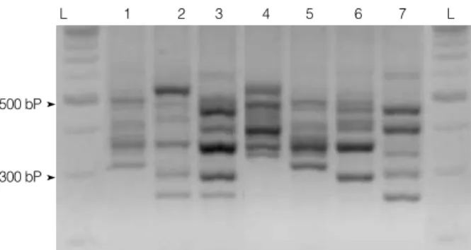
A total of 50 different ribotype patterns were found. We identified 24 patterns of A+ B+ CDT -strains (ribotype AB1-AB24), 12 A+ B+ CDT+ (ribotype C1-C12), and 13 A -B -(ribotype ab1-ab13). The PCR ribotypes aB, C5, and C2 are equivalent to the PCR ribotypes 017, 027, and 078 by O’Neill’s method, respectively [17] (Fig. 1).
All A -B+
strains showed the same banding pattern (ribo-type aB) in ribotyping, which was identical to the pat-tern of the C. difficile1470 strain (ribotype 017). Ribo-type aB was the predominant Ribo-type (105 isolates, 25.7%). The most frequently observed ribotypes of C. difficilein decreasing order were as follows: ribotype AB2 (71, 17.4%), ribotype AB3 (30, 7.4%), ribotype AB1 (27, 6.6%), and ribo-type AB17 (20, 4.9%). These 5 riboribo-types comprised 62.0% of the total. Ribotypes of C. difficileisolates were differ-ent from their toxin statuses.
Only 1 C. difficileisolate showed a pattern (ribotype C5) identical to PCR ribotype 027. Sequence analysis of tcdC in this isolate showed a single-base pair deletion at posi-tion 117 as well as a well-documented 18-bp deleposi-tion, which was identical to the sequence results of the epidemic strain of C. difficile 027. When examined by the E-test, the isolate was susceptible to moxifloxacin (MIC=0.5 mg/mL). Thirteen strains of PCR ribotype 078 (ribotype C2) were identified in 6 hospitals, making ribotype 078 the most
prevalent ribotype among CDT+
strains (13/29 CDT+ strains, 44.8%; 13/408 isolates, 3.1%).
3. Antimicrobial susceptibility testing
The in vitroactivities of antimicrobial agents against C. difficileisolates are summarized in Table 2. No iso-lates were susceptible to cefoxitin and all except 1 were susceptible to piperacillin and piperacillin-tazobactam. The resistance rates to imipenem, cefotetan, moxifloxacin, ampicillin, and clindamycin were 25%, 34%, 42%, 51%, and 60%, respectively. All strains were susceptible to metronidazole and vancomycin.
DISCUSSION
We conducted this study to enhance the knowledge on the nationwide epidemiology of C. difficile. This study included data from 12 hospitals in 7 different areas of South Korea.
The prevalence of A -B+
strains differs according to the country studied. In Europe, 6.2% of toxigenic C. difficile isolates recovered in 2005 were A-B+ [10]. In a recent study, A
-B+
strains comprised 33.3% of 75 toxigenic iso-lates from Shanghai and 0% of 80 isoiso-lates from Stock-holm [20]. The prevalence of A-B+strains was 25.7% (0-37.9%, according to the data obtained from hospitals) in Fig. 1. PCR ribotype patterns of the Clostridium difficile isolates
representing PCR ribotypes AB24, C11, C5, C2, AB14, AB23, and aB (Lane 1 to 7, respectively). Lane L refers to 100 bp lad-der. Banding patterns of the C5, C2, and aB ribotypes were iden-tical to the pattern of C. difficile ribotype 027, 078, and 017 strains.
L 1 2 3 4 5 6 7 L
500 bP
300 bP
Abbreviations: MIC, minimum inhibitory concentration; S, Susceptible; I, Intermediate; R, Resistant.
Antimicrobials MIC (mg/mL) S I R Range MIC50 MIC90
Ampicillin 1-8 2 2 0 49 51 Piperacillin 2-64 8 16 99 1 0 Piperacillin- 4-64 8 16 99 1 0 tazobactam Cefoxitin 64->128 128 >128 0 0 100 Cefotetan 8->128 32 128 26 40 34 Clindamycin 0.5->128 128 >128 11 29 60 Imipenem 2-64 8 16 22 53 25 Metronidazole 0.12-8 1 4 100 0 0 Moxifloxacin 1->128 2 16 53 5 42 Vancomycin 0.25-2 0.5 1 100 0 0
Table 2. The MICs of 10 antimicrobial agents for 120 Korean
this study. In our previous study, the prevalence of A-B+ strains increased steadily (4.2% in 1995, 39.6% in 2004) [21], and in another multicenter study conducted in Korea, 17.6-54.8% of the isolated strains were A-B+in 2005 [22]. The prevalence of A-B+strains in Korea and Shanghai was much higher than in European countries.
The prevalence of CDT+
strains was 7.1% (0-20.0%) in this study. Before the epidemics caused by ribotype 027, a binary toxin was identified in about 6% of clinical C. difficileisolates obtained in the United States and Europe [16, 23]. The prevalence of CDT+
C. difficilestrains increased to 34.6% due to the ribotype 027 epidemics in Canada [24]. In our previous study, the prevalence of CDT+ strains increased from 0% in 2003 to 3.9% in 2006 [21]. There-fore, we thought the prevalence of CDT+
strains had steadily increased without evidence of a C. difficile epi-demic. All CDT+strains were A+B+. Therefore, no addi-tional binary toxin test was required for the diagnosis of CDAD.
A total of 408 C. difficile isolates were successfully typed with our PCR ribotyping method. Predominant ribotypes among the participating hospitals were not significantly different.
All 105 A-B+strains showed the same ribotyping pattern (aB), which was the most common ribotype (105/408, 25.7%) and indistinguishable from the pattern of C. difficile1470 (ribotype 017). It was previously reported that most A
-B+ strains yield this distinct ribotype pattern in many stud-ies, suggesting a worldwide clonal spread [7, 10, 21].
Only 1 PCR ribotype 027 strain was identified in hos-pital Seoul A. In contrast to epidemic 027 strains resis-tant to fluoroquinolone, this isolate was susceptible, which is in accordance with a report on 027 isolates obtained before 2001 in North America [11].
PCR ribotype 078 is the predominant ribotype in calves and pigs, and is an emerging new hypervirulent strain [25]. The prevalence of CDAD caused by a PCR ribotype 078 strain increased from 3% to 13% during 2005-2008 in The Netherlands. CDAD caused by type 078 strains has a similar severity of CDAD caused by type 027 strains [26]. Thirteen strains of PCR ribotype 078 were
identi-fied in our study, which was the most prevalent ribotype among CDT+
strains (44.8% of CDT+
strains, 3.1% of all isolates).
Antimicrobial therapy plays a central role in the devel-opment of CDAD. The increasing use of fluoroquinolones in US health care facilities may have provided a selec-tive advantage for the fluoroquinolone-resistant 027 strain and promoted its widespread emergence [11]. MTZ and VAN remain the most active agents in this study. No resistance to piperacillin-tazobactam was found in isolates from Shanghai and Stockholm [20] and only 1 non-toxigenic isolate showed intermediate resistance in this study. Resistance to other antimicrobials varies widely between countries [29]. The resistance rate to moxifloxacin was 42% in our study, which was lower than that in Scot-land (87.5%, 2007) and higher than that in Sweden (15.0%, 2009). The resistance rate to clindamycin was 60% in our study, which was lower than in Canada (90.9%, 2009) and higher than that in Hungary (27.5%, 2009) [27].
The MICs of ampicillin, piperacillin, piperacillin-tazobactam, cefoxitin, cefotetan, imipenem, metronida-zole, and vancomycin were not significantly different according to the toxin status. However, the MIC50values of clindamycin and moxifloxacin in A-B+strains were sig-nificantly higher than those of A+B+strains: 128 and 16 in A
-B+
versus 4 and 1 in A+ B+
, respectively (data not shown). It was reported that higher MICs of antimicro-bial agents for predominant C. difficilestrains may have played a role in their persistence and dissemination in hospitals [28, 29]. Therefore, the increased prevalence of A
-B+
strains in this study may reflect their higher MICs and the selective advantage it allows. This is the first nationwide study on the toxigenic status, including molecular genotyping and antimicrobial susceptibility pattern, of C. difficile isolates in South Korea. The prevalence of A-B+and CDT+strains was 25.7% and 7.1%, respectively. Surveys of all A-B+strains showed that the most common ribotype was ribotype 017. We isolated 1 ribotype 027 strain, which is regarded as a historic iso-late, with susceptibility to moxifloxacin. The prevalence of ribotype 078 was 3.1%, which was higher than that of
ribotype 027. We did not isolate strains with decreased susceptibility to MTZ or VAN, since these 2 antimicro-bial agents can be used without an antimicroantimicro-bial sus-ceptibility test.
ACKNOWLEDGEMENTS
We thank Gwanghee Byun (Kyunghee University, Yon-gin, Korea) for laboratory assistance.
REFERENCES
1. Bartlett JG. Clostridium difficile: history of its role as an enteric pathogen and the current state of knowledge about the organism. Clin Infect Dis 1994;18(S4):S265-72.
2. Borriello SP, Davies HA, Kamiya S, Reed PJ, Seddon S. Virulence factors of Clostridium difficile. Rev Infect Dis 1990;12(S2):S185-91. 3. Braun V, Hundsberger T, Leukel P, Sauerborn M, von
Eichel-Streiber C. Definition of the single integration site of the pathogenic-ity locus in Clostridium difficile. Gene 1996;181:29-38.
4. Hammond GA and Johnson JL. The toxigenic element of
Clostridi-um difficile strain VPI 10463. Microb Pathog 1995;19:203-13.
5. Borriello SP, Wren BW, Hyde S, Seddon SV, Sibbons P, Krishna MM, et al. Molecular, immunological, and biological characterization of a toxin A-negative, toxin B-positive strain of Clostridium difficile. Infect Immun 1992;60:4192-9.
6. Lyerly DM, Barroso LA, Wilkins TD, Depitre C, Corthier G. Char-acterization of a toxin A-negative, toxin B-positive strain of
Clostridi-um difficile. Infect Immun 1992;60:4633-9.
7. Alfa MJ, Kabani A, Lyerly D, Moncrief S, Neville LM, Al-Barrak A, et al. Characterization of a toxin A-negative, toxin B-positive strain of
Clostridium difficile responsible for a nosocomial outbreak of Clostrid-ium difficile-associated diarrhea. J Clin Microbiol 2000;38:2706-14.
8. Popoff MR, Rubin EJ, Gill DM, Boquet P. Actin-specific ADP-ribo-syltransferase produced by a Clostridium difficile strain. Infect Immun 1988;56:2299-306.
9. Perelle S, Gibert M, Bourlioux P, Corthier G, Popoff MR. Produc-tion of a complete binary toxin (actin-specific ADP-ribosyltrans-ferase) by Clostridium difficile CD196. Infect Immun 1997;65:1402-7. 10. Barbut F, Mastrantonio P, Delmée M, Brazier J, Kuijper E, Poxton
I.. Prospective study of Clostridium difficile infections in Europe with
phenotypic and genotypic characterisation of the isolates. Clin Micro-biol Infect 2007;13:1048-57.
11. McDonald LC, Killgore GE, Thompson A, Owens RC Jr, Kazakova SV, Sambol SP, et al. An epidemic, toxin gene-variant strain of
Clostridium difficile. N Engl J Med 2005;353:2433-41.
12. Warny M, Pepin J, Fang A, Killgore G, Thompson A, Brazier J, et al. Toxin production by an emerging strain of Clostridium difficile associated with outbreaks of severe disease in North America and Europe. Lancet 2005;366:1079-84.
13. Baines SD, O’Connor R, Freeman J, Fawley WN, Harmanus C, Mastrantonio P, et al. Emergence of reduced susceptibility to metron-idazole in Clostridium difficile. J Antimicrob Chemother 2008;62: 1046-52.
14. Muto CA, Pokrywka M, Shutt K, Mendelsohn AB, Nouri K, Posey K, et al. A large outbreak of Clostridium difficile-associated disease with an unexpected proportion of deaths and colectomies at a teaching hospital following increased fluoroquinolone use. Infect Control Hosp Epidemiol 2005;26:273-80.
15. Kato H, Kato N, Watanabe K, Iwai N, Nakamura H, Yamamoto T, et al. Identification of toxin A-negative, toxin B-positive Clostridium
difficile by PCR. J Clin Microbiol 1998;36:2178-82.
16. Stubbs S, Rupnik M, Gibert M, Brazier J, Duerden B, Popoff M. Pro-duction of actin-specific ADP-ribosyltransferase (binary toxin) by strains of Clostridium difficile. FEMS Microbiol Lett 2000;186:307-12. 17. O’Neill GL, Ogunsola FT, Brazier JS, Duerden BI. Modification of a
PCR ribotyping method for application as a routine typing scheme for Clostridium difficile. Anaerobe 1996;2:205-9.
18. Spigaglia P and Mastrantonio P. Comparative analysis of
Clostridi-um difficile clinical isolates belonging to different genetic lineages
and time periods. J Med Microbiol 2004;53:1129-36.
19. Clinical and Laboratory Standards Institute. Methods for antimi-crobial susceptibility testing of Anaerobic bacteria; Approved stan-dard. 7th ed. CLSI document M11-A7. Wayne, PA: Clinical and Laboratory Standards Institute, 2007.
20. Huang H, Fang H, Weintraub A, Nord CE. Distinct ribotypes and rates of antimicrobial drug resistance in Clostridium difficile from Shanghai and Stockholm. Clin Microbiol Infect 2009;15:1170-3. 21. Kim H, Riley TV, Kim M, Kim CK, Yong D, Lee K, et al. Increasing
prevalence of toxin A-negative, toxin B-positive isolates of
Clostridi-um difficile in Korea: impact on laboratory diagnosis. J Clin
22. Shin BM, Kuak EY, Yoo HM, Kim EC, Lee K, Kang JO, et al. Multi-centre study of the prevalence of toxigenic Clostridium difficile in Korea: results of a retrospective study 2000-2005. J Med Microbiol 2008;57:697-701.
23. Geric B, Rupnik M, Gerding DN, Grabnar M, Johnson S. Distribu-tion of Clostridium difficile variant toxinotypes and strains with binary toxin genes among clinical isolates in an American hos-pital. J Med Microbiol 2004;53:887-94.
24. Martin H, Willey B, Low DE, Staempfli HR, McGeer A, Boerlin P, et al. Characterization of Clostridium difficile strains isolated from patients in Ontario, Canada, from 2004 to 2006. J Clin Microbiol 2008;46:2999-3004.
25. Keel K, Brazier JS, Post KW, Weese S, Songer JG. Prevalence of PCR ribotypes among Clostridium difficile isolates from pigs, calves, and other species. J Clin Microbiol 2007;45:1963-4.
26. Goorhuis A, Bakker D, Corver J, Debast SB, Harmanus C, Noter-mans DW, et al. Emergence of Clostridium difficile infection due to a new hypervirulent strain, polymerase chain reaction ribotype 078. Clin Infect Dis 2008;47:1162-70.
27. Huang H, Weintraub A, Fang H, Nord CE. Antimicrobial resistance in Clostridium difficile. Int J Antimicrob Agents 2009;34:516-22. 28. Drudy D, Quinn T, O’Mahony R, Kyne L, O’Gaora P, Fanning S.
High-level resistance to moxifloxacin and gatifloxacin associated with a novel mutation in gyrB in toxin-A-negative, toxin-B-positive
Clostridium difficile. J Antimicrob Chemother 2006;58:1264-7.
29. John R and Brazier JS. Antimicrobial susceptibility of polymerase chain reaction ribotypes of Clostridium difficile commonly isolated from symptomatic hospital patients in the UK. J Hosp Infect 2005; 61:11-4.