Received: 16 October 2016 Revised: 9 January 2017 Accepted: 19 January 2017 https://doi.org/10.1259/bjr.20160807
Cite this article as:
Kim Y-J, Kim K, Min SK, Nam EM. Role of adjuvant radiotherapy for localized extrahepatic bile duct cancer. Br J Radiol 2017; 90: 20160807.
FULL PAPER
Role of adjuvant radiotherapy for localized extrahepatic
bile duct cancer
1YI-JUN KIM,MD,1KYUBO KIM,MD, PhD,2SEOG KI MIN,MD, PhDand3EUN MI NAM,MD, PhD
1Department of Radiation Oncology, Ewha Womans University School of Medicine, Seoul, Republic of Korea 2
Department of Surgery, Ewha Womans University School of Medicine, Seoul, Republic of Korea 3Department of Internal Medicine, Ewha Womans University School of Medicine, Seoul, Republic of Korea
Address correspondence to: Dr Kyubo Kim E-mail:kyubokim.ro@gmail.com
Objective: To evaluate the benefit of adjuvant radiother-apy (RT) after surgical resection for extrahepatic bile duct (EHBD) cancer.
Methods: From 1997 to 2015, 59 patients with EHBD cancer were the subject of this study; 36 patients not undergoing adjuvant treatment after surgery (observa-tion group) and 23 patients receiving adjuvant RT (RT group) were compared. Microscopic residual disease (R1) was in 9 (25%) patients and 5 (22%) patients, and macroscopic residual disease (R2) was in 2 (6%) patients and 6 (26%) patients in the observation and RT groups, respectively. Adjuvant RT was delivered to the tumour bed and regional lymph nodes up to 50.4 Gy (range, 45–61 Gy).
Results: With a median follow-up of 19 months, local recurrence was observed in 10 (28%) patients and 2 (9%) patients in the observation and RT groups, respectively. On univariate analysis, the 5-year local recurrence-free survival (LRFS) rates were 50% in the
observation group and 54% in the RT group (p5 0.401). The 5-year overall survival (OS) rates were 29.3% in the observation group and 26.3% in the RT group (p5 0.602). On multivariable analysis, however, adju-vant RT significantly improved LRFS [hazard ratio (HR), 0.310; 95% confidence interval (CI), 0.100–0.963; p 5 0.043] and had a trend towards increased OS (HR, 0.491; 95% CI, 0.219–1.102; p 5 0.085). Resection margin (RM) status was also correlated with LRFS (HR for R1 6.134, 95% CI 2.051–18.344; and HR for R2 18.551, 95% CI 3.680–93.520; p , 0.001) and OS (HR for R1 1.816, 95% CI 0.853–3.867; and HR for R2 3.564, 95% CI 1.175–10.809; p 5 0.054).
Conclusion: RM status was a significant prognosticator of EHBD cancer, and adjuvant RT improved local control rate; thereby, survival rate might be increased.
Advances in knowledge: The benefit of adjuvant RT in EHBD cancer was demonstrated via comparison with observation group.
INTRODUCTION
Extrahepatic bile duct (EHBD) cancer is a rare malig-nancy. However, the biliary tract cancer including gall-bladder is the ninth common malignancy in Korea.1 Prognosis of EHBD cancer is generally poor; the median overall survival (OS) is about 20 months.2 Surgery pro-vides the only chance for cure with varying resectability rates of 25–91% depending on tumour location.3–6
However, obtaining a negative resection margin (RM) is a challenge,7,8 and microscopic (R1) and macroscopic (R2) residual diseases are observed in 20–40% and 4–64%, respectively.9–15
Many studies have revealed that positive RM of EHBD cancer is directly associated with high local recurrence (LR) rate.9,11,15,16As a result, LR is a major pattern of failure in EHBD cancer.17In spite of the high LR rate, the utility of
sparse.2Administration of adjuvant RT may increase local control rate, and improved local control might result in survival benefit.10,18,19
Because of the rarity of the disease, however, no prospective randomized controlled trials comparing adjuvant RT with observation have been per-formed, and retrospective studies have yielded conflicting results with insufficient evidences supporting the survival benefit of adjuvant RT.18,20–22
In this study, we retrospectively analyzed the effect of ad-juvant RT on local relapse-free survival and OS for patients with EHBD cancer treated with surgical resection with or without adjuvant RT.
METHODS AND MATERIALS Study population
who were diagnosed with adenocarcinoma of EHBD and un-derwent surgical resection followed by adjuvant RT or not were identified.
Treatment
All patients were restaged pathologically, according to American Joint Committee on Cancer 7th edn.23There were no patients who received neoadjuvant treatment in this study. Surgeons decided the surgical procedure for patients in accordance with site and extension of the tumour. Referral for adjuvant RT was carried out at the discretion of the attending surgeon; but, RM, age, performance status and patient preference were considered in the decision-making.
Follow-up
Patients were followed up every 3 months for 2 years, and every 6 months thereafter. Routine follow-up methods included physical examination, laboratory screening and simple chest X-ray. Abdominal/pelvic CT scan was performed every 3 months in the initial 1–2 years and repeated every 6 months thereafter. If recurrence was suspected, whole-body positron emission tomography-CT scan and chest CT scan were performed for further evaluations.
Statistical analysis
Exactx2test was performed to compare categorical variables between the observation and RT groups. Cox proportional hazards regression was used to determine univariate and multivariable hazard ratios (HRs) for local recurrence-free survival (LRFS) and OS. LRFS was defined as the time period from the date of surgery to the date of treatment failure in the surgical bed. OS was defined as the time period from the date of surgery to the date of death or last follow-up. Anal-yses were carried out using the SPSS® Statistics software
Table 1. Patient and tumour characteristics
Variables Number of patients (%) p-value Observation (n 5 36) (n 5 23)RT Age (years) ,65 12 (33.3) 16 (69.6) 0.008 $65 24 (66.7) 7 (30.4) Sex Male 21 (58.3) 11 (47.8) 0.593 Female 15 (41.7) 12 (52.2)
Performance status (ECOG)
0–1 29 (80.6) 21 (91.3)
0.460
2–3 7 (19.4) 2 ( 8.7)
Surgery
Bile duct resection with
hepatectomy 6 (16.7) 7 (30.4)
0.136 Bile duct resection 5 (13.9) 6 (26.1) Pancreaticoduodenectomy 25 (69.4) 10 (43.5) RM R0 25 (69.4) 12 (52.2) 0.107 R1 9 (25.0) 5 (21.7) R2 2 (5.6) 6 (26.1) Tumour location Non-hilar 30 (83.3) 12 (52.2) 0.017 Hilar 6 (16.7) 11 (47.8) Differentiation Well 3 (8.3) 3 (13.0) 0.324 Moderate 22 (61.1) 16 (69.6) Poor 11 (30.6) 3 (13.0) Unknown 0 (0.0) 1 (4.3) T stage T1–2 19 (52.8) 14 (60.9) 0.599 T3–4 17 (47.2) 9 (39.1) N stage N0 21 (58.3) 15 (65.2) 0.785 N1 15 (41.7) 8 (34.8) Perineural invasion No 5 (13.9) 6 (26.1) 0.730 Yes 20 (55.6) 15 (65.2) Unknown 11 (30.6) 2 ( 8.7)
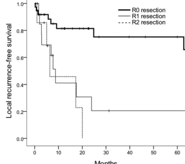
Figure 1. Local recurrence-free survival curves according to the resection margin status.
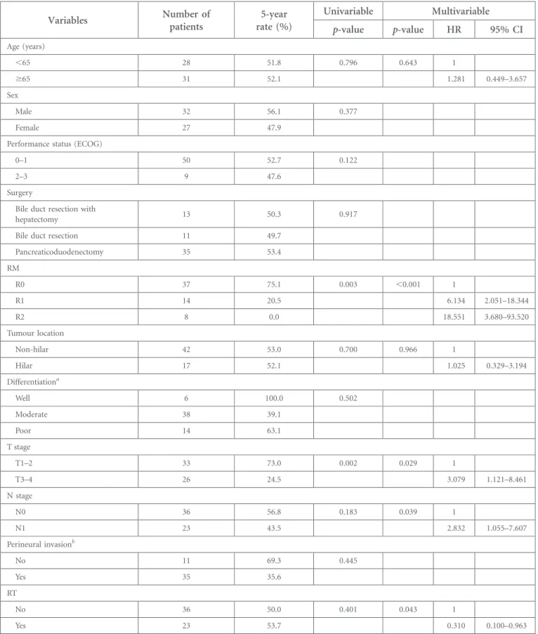
Table 2. Univariable and multivariable analyses for local recurrence-free survival
Variables Number ofpatients rate (%)5-year Univariable Multivariable
p-value p-value HR 95% CI Age (years) ,65 28 51.8 0.796 0.643 1 $65 31 52.1 1.281 0.449–3.657 Sex Male 32 56.1 0.377 Female 27 47.9
Performance status (ECOG)
0–1 50 52.7 0.122
2–3 9 47.6
Surgery
Bile duct resection with
hepatectomy 13 50.3 0.917
Bile duct resection 11 49.7
Pancreaticoduodenectomy 35 53.4 RM R0 37 75.1 0.003 ,0.001 1 R1 14 20.5 6.134 2.051–18.344 R2 8 0.0 18.551 3.680–93.520 Tumour location Non-hilar 42 53.0 0.700 0.966 1 Hilar 17 52.1 1.025 0.329–3.194 Differentiationa Well 6 100.0 0.502 Moderate 38 39.1 Poor 14 63.1 T stage T1–2 33 73.0 0.002 0.029 1 T3–4 26 24.5 3.079 1.121–8.461 N stage N0 36 56.8 0.183 0.039 1 N1 23 43.5 2.832 1.055–7.607 Perineural invasionb No 11 69.3 0.445 Yes 35 35.6 RT No 36 50.0 0.401 0.043 1 Yes 23 53.7 0.310 0.100–0.963
CI, confidence interval; ECOG, Eastern Cooperative Oncology Group; HR, hazard ratio; RM, resection margin; RT, radiotherapy.
aIn one patient, information on differentiation was unavailable. bIn 13 patients, information on perineural invasion was unavailable.
v. 18.0 (IBM Corp., New York, NY; formerly SPSS Inc., Chicago, IL). A two-tailed p, 0.05 was considered statisti-cally significant.
RESULTS
Patient and treatment characteristics
Patient and tumour characteristics according to receipt of ad-juvant RT are summarized inTable 1. Median age of all patients was 65 years. Patients$65 years of age were more common in the observation group (67% vs 30%, p5 0.008).
13, 11 and 35 patients underwent bile duct resection with hepatectomy, bile duct resection and pancreaticoduodenectomy, respectively. Clear RM (R0) was obtained in 25 (69%) patients in the observation group and 12 (52%) patients in the RT group. R1 resection was observed in 9 (25%) patients and 5 (22%) patients, and R2 resection was observed in 2 (6%) patients and 6 (26%) patients in the observation and RT groups, respectively. Even though RM status was not statisti-cally different, more patients in the RT group underwent R2 resection (26% vs 6%).
In the RT group (n5 23), RT was delivered to the tumour bed and regional lymph nodes including the porta hepatis, peri-choledochal, retrocaval and aortocaval lymph nodes accord-ing to the location of the primary tumour. The total dose was from 45 Gy to 61 Gy (median, 50.4 Gy) in 1.8–2.0 Gy/fraction for the tumour bed and 45 Gy in 1.8 Gy/fraction for the gional lymph nodes. 15 (65%) patients in the RT group re-ceived 5-fluorouracil- or gemcitabine-based chemotherapy during RT (n5 7), during and after RT (n 5 6) or after RT (n5 2). Among eight patients who received adjuvant che-motherapy after RT, enteric-coated tegafur/uracil was given for 1 year after RT or until progression in four patients, 5-fluorouracil for four cycles in three patients and gemcitabine for six cycles in one patient.
Patients with hilar tumour were more likely to receive adjuvant RT than patients with more distally located tumour (65% vs 29%, p5 0.017). No differences were observed between the groups with respect to sex, T stage and N stage.
Treatment outcome
Median follow-up was 19 months (range, 1–222 months) for all patients and 43 months (range, 11–178 months) for survivors. The median LFRS and 5-year LRFS rate were 63 months and 52%, respectively. On univariate analysis for LRFS, RM status and T stage were significant prognostic factors (p5 0.003 and 0.002, respectively). When age, RM status, tumour location, T stage, N stage and RT were in-corporated into a multivariable analysis, RM status was still correlated with LRFS; the 5-year LRFS rates of R0, R1 and R2 resection were 75%, 21% and 0%, respectively [HR for R1 6.134, 95% confidence interval (CI) 2.051–18.344; and HR for R2 18.551, 95% CI 3.680–93.520; p , 0.001] (Figure 1).
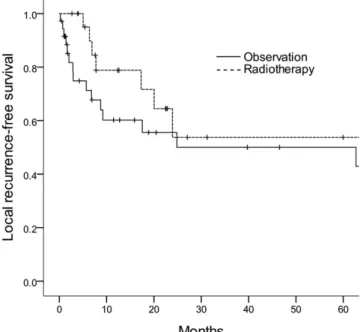
LRFS rates of the observation and RT groups were 50% and 54%, respectively (HR 0.310, 95% CI 0.100–0.963; p 5 0.043) (Table 2) (Figure 2).
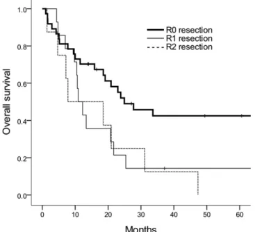
The median OS of all patients was 21 months and the 5-year OS rate was 29%. High T stage was a poor prognostic factor for OS in univariate analysis (p5 0.013). Adjuvant RT (HR 0.491, 95% CI 0.291–1.102; p 5 0.085), RM status (HR for R1 1.816, 95% CI 0.853–3.867; and HR for R2 3.564, 95% CI 1.175–10.809; p5 0.054) and N stage (HR 1.956, 95% CI 0.983–3.892; p5 0.056) showed non-significant trends for OS on multivari-able analysis incorporating age, RM status, tumour location, T stage, N stage and adjuvant RT (Table 3). The 5-year OS rates of R0, R1 and R2 resection were 42%, 14% and 0%, respectively (Figure 3). The 5-year OS rates were 29% for the observation group and 27% for the RT group (Figure 4).
Treatment failures were observed in 32 patients. As thefirst site failure, LR with or without distant metastasis was observed in 16 (44%) patients in the observation group and in 4 (17%) patients in the RT group (p5 0.021) (Table 4).
Treatment toxicity
Grade of toxicity was scored according to the Common Terminology Criteria for Adverse Events v. 4.03. Among 23 patients receiving adjuvant RT, acute complications of Grade$3 were observed in 2 (9%) patients; 1 patient (not receiving concomitant chemotherapy) experienced severe nausea and vomiting, and the other patient (receiving con-comitant weekly gemcitabine) had decreased platelet and white blood cell count.
DISCUSSION
In this study, adjuvant RT showed an increase in LRFS as well as a non-significant improvement in OS of patients with EHBD
Figure 2. Local recurrence-free survival curves according to the receipt of radiotherapy.
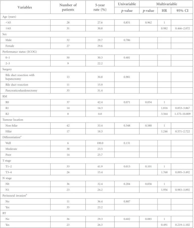
Table 3. Univariable and multivariable analyses for overall survival
Variables Number ofpatients rate (%)5-year Univariable Multivariable
p-value p-value HR 95% CI Age (years) ,65 28 27.6 0.851 0.962 1 $65 31 30.8 0.982 0.466–2.072 Sex Male 32 29.7 0.786 Female 27 29.6
Performance status (ECOG)
0–1 50 30.3 0.481
2–3 9 22.2
Surgery
Bile duct resection with
hepatectomy 13 30.8 0.981
Bile duct resection 11 15.9
Pancreaticoduodenectomy 35 31.4 RM R0 37 42.4 0.071 0.054 1 R1 14 14.3 1.816 0.853–3.867 R2 8 0.0 3.564 1.175–10.809 Tumour location Non-hilar 42 33.4 0.348 0.580 1 Hilar 17 18.3 1.246 0.571–2.722 Differentiationa Well 6 100.0 0.131 Moderate 38 23.5 Poor 14 25.7 T stage T1–2 33 41.9 0.013 0.101 1 T3–4 26 15.4 1.768 0.895–3.492 N stage N0 36 32.4 0.204 0.056 1 N1 23 24.2 1.956 0.983–3.892 Perineural invasionb No 11 36.4 0.887 Yes 35 23.2 RT No 36 29.3 0.602 0.085 1 Yes 23 26.3 0.491 0.219–1.102
CI, confidence interval; ECOG, Eastern Cooperative Oncology Group; HR, hazard ratio; RM, resection margin; RT, radiotherapy.
aIn one patient, information on differentiation was unavailable. bIn 13 patients, information on perineural invasion was unavailable.
cancer. Positive RM was associated with poor local control rate and adverse OS. LR was the predominant pattern of failure in the observation group, while distant metastasis was the domi-nant pattern in the RT group.
Poor prognosis of EHBD cancer was repeatedly observed in our study with a 5-year OS rate of 29%. It is noticeable that a treatment factor (i.e. RM status) had a greater impact on prognosis than any other patient or tumour characteristic. Many studies demonstrated that positive RM decreased median OS from 24–74 months (R0) to 12–13 months (R1 or R2).9,15,24
In
our study, the 5-year OS rates according to RM status were 42% in R0 resection, 14% in R1 resection and 0% in R2 resection, suggesting that positive RM would drastically decrease the chance of cure by surgical treatment. Moreover, LR was the major pattern of failure without receiving adjuvant RT in the first site of failure pattern analysis. Also, the 5-year LRFS rate of R0 resection was 75% compared with 21% and 0% of R1 and R2 resections, respectively (p, 0.001). These findings suggest that more effective strategies for local control are mandatory to improve OS rate.
Additional resection of positive RM could be a reasonable op-tion.25 Ribero et al26 demonstrated that reresection of positive proximal bile duct margin to gain secondary R0 resection in patients with hilar cholangiocarcinoma improved median OS to a level similar to that of patients who underwent primary R0 resection. However, the success rate of obtaining secondary R0 resection is limited because of procedural difficulties.9–15
Moreover, another author noted that additional resection of positive proximal bile duct RM in hilar cholangiocarcinoma did not improve OS, suggesting that further studies are indispens-able to evaluate the role of additional resection of positive RM.7 Several studies investigated the benefit of adjuvant RT to countervail the adverse effect of positive RM.14,17–19,27–30Small retrospective studies demonstrated that there was no significant OS difference between negative RM group without adjuvant RT and positive RM group with adjuvant RT.14,18 Gwak et al17 reported that patients with R1 resection who received adjuvant RT had significantly higher median disease-free survival than those who received R1 resection alone.
In our study, adjuvant RT group was compared with non-RT group, retrospectively. The RT group had more adverse features than its counterpart; among eight patients who had R2 re-section, six patients were included in the RT group (26% of the RT group), whereas the remaining two patients were in the observation group (6% of the observation group). After
in-Figure 3. Overall survival curves according to the resection margin status.
Figure 4. Overall survival curves according to the receipt of radiotherapy.
Table 4. Patterns of the first treatment failure
Site of failure Number of patients/total failures p-value Observation (n 5 36) (n 5 23)RT LR with or without DM No 4/20 8/12 0.021 Yes 16/20 4/12 DM with or without LR No 10/20 2/12 0.075 Yes 10/20 10/12
In spite of the high LR rate of EHBD cancer, RT has not yet been a well-established adjuvant treatment.31An analysis of data from the Surveillance, Epidemiology, and End Results program demonstrated that only 32% of patients with EHBD cancer underwent adjuvant RT.2 Because of the rarity of this disease, only small retrospective studies were available in many institu-tions, resulting in limited statistical powers. One meta-analysis, however, revealed that adjuvant RT derived survival benefit in R1 resection (odds ratio 0.33), although this improvement was not observed in R0 resection.19
In the present study, median age for the observation group and RT group was 71 years and 61 years, respectively. In the elderly patient, adjuvant RT was less likely to be administered because of concerns regarding RT-related morbidity. On univariate and multivariable analyses, however, age was not a significantly poor prognostic factor for survival. Toxicity of adjuvant RT was
minor; only 2 (9%) patients in RT group presented Grade$3 side effects. Therefore, more assertive utility of adjuvant RT might improve treatment outcomes in elderly patients with good performance status.
This study is a retrospective study with a small sample size in a single institution. While no patients received chemotherapy in the observation group, a major portion of patients in the RT group received chemotherapy with various regimens. Chemotherapy regimens and treatment durations were too heterogeneous to analyze. Benefits of adjuvant RT and che-motherapy could not be evaluated separately in this small study.
In conclusion, adjuvant RT improved LRFS for patients with localized EHBD cancer. RM status was a significant prognos-tic factor.
REFERENCES
1. Jung KW, Won YJ, Oh CM, Kong HJ, Cho H, Lee JK, et al. Prediction of cancer incidence and mortality in Korea, 2016. Cancer Res Treat 2016;48: 451–7. doi:https://doi.org/ 10.4143/crt.2016.092
2. Vern-Gross TZ, Shivnani AT, Chen K, Lee CM, Tward JD, MacDonald OK, et al. Survival outcomes in resected extrahepatic cholangiocarcinoma: effect of adjuvant ra-diotherapy in a surveillance, epidemiology, and end results analysis. Int J Radiat Oncol Biol Phys 2011;81: 189–98. doi:https://doi. org/10.1016/j.ijrobp.2010.05.001
3. Nakeeb A, Pitt HA, Sohn TA, Coleman J, Abrams RA, Piantadosi S, et al. Cholangio-carcinoma. A spectrum of intrahepatic, perihilar, and distal tumors. Ann Surg 1996; 224: 463. doi:https://doi.org/10.1097/ 00000658-199610000-00005
4. Tsao JI, Nimura Y, Kamiya J, Hayakawa N, Kondo S, Nagino M, et al. Management of hilar cholangiocarcinoma: comparison of an American and a Japanese experience. Ann Surg 2000;232: 166–74. doi:https://doi.org/ 10.1097/00000658-200008000-00003
5. Nagorney DM, Donohue JH, Farnell MB, Schleck CD, Ilstrup DM. Outcomes after curative resections of cholangiocarcinoma. Arch Surg 1993;128: 871–8. doi:https://doi. org/10.1001/archsurg.1993.01420200045008
6. Launois B, Terblanche J, Lakehal M, Catheline JM, Bardaxoglou E, Landen S, et al. Proximal bile duct cancer: high resectability rate and 5-year survival. Ann Surg 1999;230: 266. doi: https://doi.org/10.1097/00000658-199908000-00018
7. Shingu Y, Ebata T, Nishio H, Igami T, Shimoyama Y, Nagino M. Clinical
value of additional resection of a margin-positive proximal bile duct in hilar cholangiocarcinoma. Surgery 2010;147: 49–56. doi:https://doi.org/10.1016/j. surg.2009.06.030
8. Launois B, Reding R, Lebeau G, Buard JL. Surgery for hilar cholangiocarcinoma: French experience in a collective survey of 552 extrahepatic bile duct cancers. J Hepatobiliary Pancreat Surg 2000;7: 128–34. doi:https:// doi.org/10.1007/s005340050166
9. Rocha FG, Matsuo K, Blumgart LH, Jarnagin WR. Hilar cholangiocarcinoma: the Memo-rial Sloan-Kettering Cancer Center experi-ence. J Hepatobiliary Pancreat Sci 2010;17: 490–6. doi: https://doi.org/10.1007/s00534-009-0205-4
10. Kim K, Chie EK, Jang JY, Kim SW, Han SW, Oh DY, et al. Adjuvant chemoradiotherapy after curative resection for extrahepatic bile duct cancer: a long-term single center experience. Am J Clin Oncol 2012;35: 136–40. doi:https://doi.org/10.1097/ COC.0b013e318209aa29
11. Sakamoto Y, Kosuge T, Shimada K, Sano T, Ojima H, Yamamoto J, et al. Prognostic factors of surgical resection in middle and distal bile duct cancer: an analysis of 55 patients concerning the significance of ductal and radial margins. Surgery 2005;137: 396–402. doi:https://doi.org/10.1016/j. surg.2004.10.008
12. Kim S, Kim SW, Bang YJ, Heo DS, Ha SW. Role of postoperative radiotherapy in the management of extrahepatic bile duct cancer. Int J Radiat Oncol Biol Phys 2002;54: 414–9. doi:https://doi.org/10.1016/S0360-3016(02) 02952-8
13. Pichlmayr R, Weimann A, Klempnauer J, Oldhafer KJ, Maschek H, Tusch G, et al. Surgical treatment in proximal bile duct cancer. A single-center experience. Ann Surg 1996;224: 628–38. doi:https://doi.org/ 10.1097/00000658-199611000-00007
14. Oh D, Lim DH, Heo JS, Choi SH, Choi DW, Ahn YC, et al. The role of adjuvant radiotherapy in microscopic tumor control after extrahepatic bile duct cancer surgery. Am J Clin Oncol 2007;30: 21–5. doi:https://doi.org/10.1097/01. coc.0000245467.97180.78
15. Ben-David MA, Griffith KA, Abu-Isa E, Lawrence TS, Knol J, Zalupski M, et al. External-beam radiotherapy for localized extrahepatic cholangiocarcinoma. Int J Radiat Oncol Biol Phys 2006;66: 772–9. doi:
https://doi.org/10.1016/j.ijrobp.2006.05.061
16. Higuchi R, Ota T, Araida T, Kobayashi M, Furukawa T, Yamamoto M. Prognostic rele-vance of ductal margins in operative re-section of bile duct cancer. Surgery 2010;148: 7–14. doi:https://doi.org/10.1016/j. surg.2009.11.018
17. Gwak HK, Kim WC, Kim HJ, Park JH. Extrahepatic bile duct cancers: surgery alone versus surgery plus postoperative radiation therapy. Int J Radiat Oncol Biol Phys 2010;78: 194–8. doi:https://doi.org/10.1016/j. ijrobp.2009.07.003
18. Borghero Y, Crane CH, Szklaruk J, Oyarzo M, Curley S, Pisters PW, et al. Extrahepatic bile duct adenocarcinoma: patients at high-risk for local recurrence treated with surgery and adjuvant chemoradiation have an equivalent overall survival to patients with standard-risk treated with surgery alone. Ann
Surg Oncol 2008;15: 3147–56. doi:https:// doi.org/10.1245/s10434-008-9998-7
19. Horgan AM, Amir E, Walter T, Knox JJ. Adjuvant therapy in the treatment of biliary tract cancer: a systematic review and meta-analysis. J Clin Oncol 2012;30: 1934–40. doi:
https://doi.org/10.1200/JCO.2011.40.5381
20. Pitt HA, Nakeeb A, Abrams RA, Coleman J, Piantadosi S, Yeo CJ, et al. Perihilar chol-angiocarcinoma. Postoperative radiotherapy does not improve survival. Ann Surg 1995; 221: 788. doi:https://doi.org/10.1097/ 00000658-199506000-00017
21. Todoroki T, Ohara K, Kawamoto T, Koike N, Yoshida S, Kashiwagi H, et al. Benefits of adjuvant radiotherapy after radical resection of locally advanced main hepatic duct carcinoma. Int J Radiat Oncol Biol Phys 2000; 46: 581–7. doi:https://doi.org/10.1016/ S0360-3016(99)00472-1
22. Hejna M, Pruckmayer M, Raderer M. The role of chemotherapy and radiation in the management of biliary cancer: a review of the literature. Eur J Cancer 1998;34: 977–86. doi:
https://doi.org/10.1016/S0959-8049(97) 10166-6
23. Edge SB, Compton CC. The American Joint Committee on Cancer: the 7th edition of the
AJCC cancer staging manual and the future of TNM. Ann Surg Oncol 2010;17: 1471–4. doi: https://doi.org/10.1245/s10434-010-0985-4
24. Sasaki R, Takeda Y, Funato O, Nitta H, Kawamura H, Uesugi N, et al. Significance of ductal margin status in patients undergoing surgical resection for extrahepatic cholan-giocarcinoma. World J Surg 2007;31: 1788–96. doi:https://doi.org/10.1007/ s00268-007-9102-7
25. Neuhaus P, Jonas S, Bechstein WO, Lohmann R, Radke C, Kling N, et al. Extended resections for hilar cholangiocarcinoma. Ann Surg 1999;230: 808. doi:https://doi.org/ 10.1097/00000658-199912000-00010
26. Ribero D, Amisano M, Lo Tesoriere R, Rosso S, Ferrero A, Capussotti L. Additional re-section of an intraoperative margin-positive proximal bile duct improves survival in patients with hilar cholangiocarcinoma. Ann Surg 2011;254: 776–81; discussion 781–3. doi:https://doi.org/10.1097/
SLA.0b013e3182368f85
27. Itoh H, Nishijima K, Kurosaka Y, Takegawa S, Kiriyama M, Dohba S, et al. Magnitude of combination therapy of radical resection and external beam radiotherapy for patients with
carcinomas of the extrahepatic bile duct and gallbladder. Dig Dis Sci 2005;50: 2231–42. doi: https://doi.org/10.1007/s10620-005-3040-8
28. Urego M, Flickinger JC, Carr BI. Radiotherapy and multimodality management of cholangiocarcinoma. Int J Radiat Oncol Biol Phys 1999;44: 121–6. doi:https://doi.org/10.1016/S0360-3016 (98)00509-4
29. Stein DE, Heron DE, Rosato EL, Ann´e PR, Topham AK. Positive microscopic margins alter outcome in lymph node-negative chol-angiocarcinoma when resection is combined with adjuvant radiotherapy. Am J Clin Oncol 2005;28: 21–3. doi:https://doi.org/10.1097/ 01.coc.0000139017.90599.f5
30. Kim MY, Kim JH, Kim Y, Byun SJ. Post-operative radiotherapy appeared to improve the disease free survival rate of patients with extrahepatic bile duct cancer at high risk of loco-regional recurrence. Radiat Oncol J 2016;34: 297–304. doi:https://doi.org/ 10.3857/roj.2016.01879
31. Khan SA, Thomas HC, Davidson BR, Taylor-Robinson SD. Cholangiocarcinoma. Lancet 2005;366: 1303–14. doi:https://doi.org/ 10.1016/S0140-6736(05)67530-7