저작자표시-비영리-동일조건변경허락 2.0 대한민국 이용자는 아래의 조건을 따르는 경우에 한하여 자유롭게 l 이 저작물을 복제, 배포, 전송, 전시, 공연 및 방송할 수 있습니다. l 이차적 저작물을 작성할 수 있습니다. 다음과 같은 조건을 따라야 합니다: l 귀하는, 이 저작물의 재이용이나 배포의 경우, 이 저작물에 적용된 이용허락조건 을 명확하게 나타내어야 합니다. l 저작권자로부터 별도의 허가를 받으면 이러한 조건들은 적용되지 않습니다. 저작권법에 따른 이용자의 권리는 위의 내용에 의하여 영향을 받지 않습니다. 이것은 이용허락규약(Legal Code)을 이해하기 쉽게 요약한 것입니다. Disclaimer 저작자표시. 귀하는 원저작자를 표시하여야 합니다. 비영리. 귀하는 이 저작물을 영리 목적으로 이용할 수 없습니다. 동일조건변경허락. 귀하가 이 저작물을 개작, 변형 또는 가공했을 경우 에는, 이 저작물과 동일한 이용허락조건하에서만 배포할 수 있습니다.
Clinical and Pathologic Characteristics of
Hepatocellular Carcinoma following Resection
according to the Tumor Size
by
Seong Yeon Cho
Major in Medicine
Department of Medicine
Clinical and Pathologic Characteristics of
Hepatocellular Carcinoma following Resection
according to the Tumor Size
by
Seong Yeon Cho
A Dissertation Submitted to The Graduate School of Ajou University
in Partial Fulfillment of the Requirements for the Degree of
Master of Medicine
Supervised by
Hee-Jung Wang, M.D., Ph.D.
Major in Surgery
Department of Medicine
The Graduate School, Ajou University
February, 2010
This certifies that the dissertation
of Seong Yeon Cho is approved.
SUPERVISORY COMMITTEE
Hee-Jung Wang
Jae-Youn Cheong
Sung-Won Cho
The Graduate School, Ajou University
December, 21st, 2009
i
- ABSTRACT -
Clinical and Pathologic Characteristics of Hepatocellular
Carcinoma Following Resection According to the Tumor Size
A tumor size of hepatocellular carcinoma (HCC) has been known to be an important prognostic factor after resection and it is considered to be closely correlated with vascular invasion of HCC. We aimed this study to elucidate the outcomes according to some previously studied size criteria and the relationship between the size criteria and clinicopathologic parameters after surgical resection in a single center. Between Apr 2001 and Apr 2008, 307 patients with pure HCC (no combined intrahepatic cholangiocarcinoma) underwent first hepatic resection in a single center without preoperative transcatheter arterial chemoembolization (TACE). The 307 patients’ clinicopathologic data were retrospectively investigated. The tumor size criteria (2cm, 5cm and 10cm) were predictive factors for survival in univariate analysis, but not in multivariate analysis. Vascular invasion, tumor necrosis, and absence of tumor capsule were proved to be independent significant factors for overall survival. Among them, vascular invasion and tumor necrosis showed significant correlation with tumor size. On the other hand, tumor capsule did not show any correlation with tumor size. Tumor size should be further subdivided for T-classification of the AJCC/UICC staging system to predict prognosis more accurately. Considering that the AJCC/UICC staging system is a histopathologic staging system and tumor capsule was an independent significant prognostic factor irrespective of tumor size, tumor capsule should be included in T-classification of the AJCC/UICC staging system for more accurate prediction of survival.Key words: hepatocellular carcinoma, hepatectomy, tumor size, prognostic factor, vascular invasion, spontaneous tumor necrosis, tumor capsule.
ii
TABLE OF CONTENTS
ABSTRACT ··· i
TABLE OF CONTENTS ··· ii
LIST OF FIGURES ··· iii
LIST OF TABLES ··· iv
ABBREVIATION ··· v
Ⅰ. INTRODUCTION ··· 1
Ⅱ. MATERIALS AND METHODS ··· 2
A. MATERIALS ··· 2 B. METHODS ··· 2 Ⅲ. RESULTS ··· 3 Ⅳ. DISCUSSION ··· 10 Ⅴ. CONCLUSION ··· 12 REFERENCES··· 13 국문요약 ··· 18
iii
LIST OF FIGURES
Fig. 1. Overall survival curves according to the factors comprising the sixth AJCC/UICC T-classification system (Kaplan-Meier method). ··· 4 Fig. 2. Overall survival curves according to the tumor size (Kaplan-Meier method). ··· 5 Fig. 3. Adjusted overall survival curves of the independent prognostic factors (Cox model). ··· 7
iv
LIST OF TABLES
Table 1. Modalities of hepatic resection. ··· 3 Table 2. Clinicopathologic factors for survival (univariate and multivariate analyses). ··· 6 Table 3. The correlation between tumor size and independent prognostic factors. ··· 8
1
I. INTRODUCTION
Traditionally, a tumor size of hepatocellular carcinoma (HCC) has been known to be an important prognostic factor after resection and it is considered to be closely correlated with vascular invasion of HCC. (Matsuda et al., 2007; Sakata et al., 2008)
In the fifth AJCC/UICC staging system, T-classification was determined by tumor size (diameter) criterion of 2cm, vascular invasion and multiplicity. (Fleming et al., 1997) However, with the work of Vauthey et al., in the sixth AJCC/UICC, T-classification system changed as follows; a single tumor without vascular invasion regardless of size, that is, if the single tumor has no vascular invasion, it is T1. If a single tumor, not more than 5cm, has vascular invasion, it belongs to T2. (Vauthey et al., 2002; AJCC, 2002) It means that tumor size itself is not an independent factor; rather it seems related to some important prognostic factors such as vascular invasion or histologic differentiation. However, in the Liver Cancer Study Group of Japan (LCSGJ), still a single tumor not more than 2cm is considered to be T1. (LCSGJ, 2003)
Some authors investigated about HCC greater than 10cm in diameter and called it a ‘huge HCC’. Among those investigations, some demonstrated it poses worse prognosis compared with smaller one. (Chen et al., 2006; Pandey et al., 2007; Shah et al., 2007a)
Therefore, the debates about the prognostic significance of tumor size criteria of HCC still exist and to the best of our knowledge, there are few reports considering the clinicopathologic significance of each size criterion (2cm, 5cm and 10cm) of HCC in the same population. We aimed this study to elucidate the outcomes according to aforementioned each size criterion and the relationship between clinicopathologic parameters and the size criteria after surgical resection in a single center.
2
II. MATERIALS AND METHODS
MATERIALS
We investigated the patients underwent the first hepatic resection at the National Cancer Center of Korea between Apr 2001 and Apr 2008. Among them, 552 patients were proved to have HCC by histopathologic examination of the first hepatic resection specimens. We excluded the patients with multiple tumors (n=33) or with combined hepatocellular-cholangiocellular carcinoma (n = 24) and the patients undergone transcatheter arterial chemoembolization (TACE) before surgery (n = 188) from this study for the validity of histopathologic and clinical analyses. The remaining 307 patients’ clinicopathologic data were retrospectively investigated.
The 307 patients were divided into four groups according to the primary tumor size; group A: tumor size ≤ 2cm, group B: 2cm < tumor size ≤ 5cm, group C: 5cm < tumor size ≤ 10cm, group D: tumor size > 10cm. We analyzed the clinicopathologic data to elucidate outcomes and prognostic factors of HCC in patients undergoing hepatic resection. Comparative analyses of clinicopathologic factors and prognosis according to the size criteria and groups were conducted.
METHODS
For those comparative analyses, the following factors were analyzed: age, gender, hepatitis viral markers, tumor marker, blood transfusion, tumor size, vascular invasion, histologic grade, spontaneous tumor necrosis, tumor capsule (absence vs. partial/complete), bile duct invasion, cirrhosis, and resection margin. The cumulative survival rate was calculated by the Kaplan-Meier method. Univariate correlations between factors and cumulative survivals were examined by the log-rank test. Multivariate correlations between factors were made by the Cox-regression model. Comparisons of nominal variables between groups were made using the chi-square test. A P-value of less than 0.05 was considered statistically significant.
3
III. RESULTS
Clinicopathologic profile
In the 307 patients, median age was 54.5 years (range: 26-83) and gender ratio was 251:56 (81.8:18.2%). Viral hepatitis was detected as follows; HBV in 220 (71.7%), HCV in 17 (5.5%), simultaneous HBV and HCV in 2 (0.7%) and no hepatitis virus in 68 (22.1%).
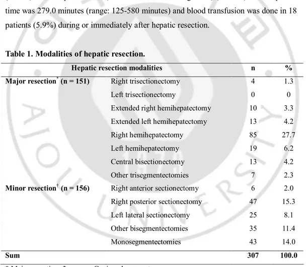
There were 151 (49.2%) major hepatectomies (3 or more Couinaud segments) and 156 (50.8%) minor hepatectomies (less than 3 Couinaud segments) (Table 1). Mean operation time was 279.0 minutes (range: 125-580 minutes) and blood transfusion was done in 18 patients (5.9%) during or immediately after hepatic resection.
Table 1. Modalities of hepatic resection.
Hepatic resection modalities n %
Right trisectionectomy 4 1.3
Left trisectionectomy 0 0
Extended right hemihepatectomy 10 3.3
Extended left hemihepatectomy 13 4.2
Right hemihepatectomy 85 27.7
Left hemihepatectomy 19 6.2
Central bisectionectomy 13 4.2
Major resection* (n = 151)
Other trisegmentectomies 7 2.3
Right anterior sectionectomy 6 2.0
Right posterior sectionectomy 47 15.3
Left lateral sectionectomy 25 8.1
Other bisegmentectomies 35 11.4
Minor resection† (n = 156)
Monosegmentectomies 43 14.0
Sum 307 100.0
* Major resection: 3 or more Couinaud segments. † Minor resection: less than 3 Couinaud segments.
4
The median tumor size was 4.0cm (range: 1.3-18.5cm). We divided the patients into four groups according to the primary tumor size with 2cm, 5cm and 10cm criteria. Vascular invasion was detected by histopathologic examination in 135 (44.0%) among the 307 patients. As a result, the sixth AJCC/UICC T-classification of the 307 patients with single tumor were as follows; T1 in 172 (56.0%), T2 in 119 (38.8%), T3 in11 (3.6%) and T4 in 5 (1.6%).
Spontaneous tumor necrosis was observed in 143 patients (46.6%) without preoperative TACE; median ratio of estimated necrotic volume was 10% (range: 1-99). Tumor capsule formation was observed in 240 patients (78.2%); complete tumor capsule in 135 (44.0%) and partial tumor capsule in 105 (34.2%).
Overall survival
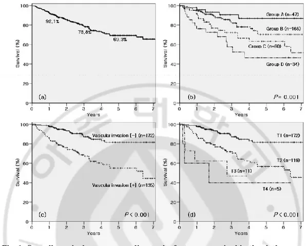
For the 307 patients, 1-, 3- and 5-year overall survival rates were 92.1%, 78.8% and 69.3%, respectively (Fig. 1 a).
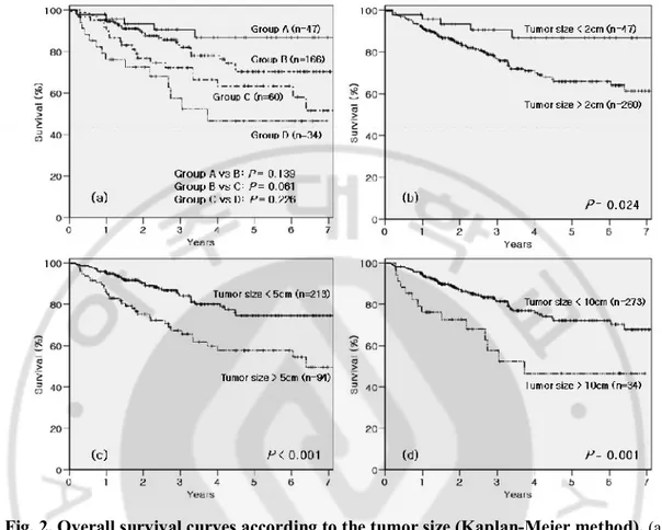
Overall survival was significantly different between the size groups (P = 0.001) (Fig. 1 b) and whether vascular invasion was presents or not (P < 0.001) (Fig. 1 c). Overall survival according to the sixth AJCC/UICC T-classification system also showed significant
difference (P < 0.001) (Fig. 1 d). In intergroup analyses for each neighboring paired size groups, none of neighboring groups gained statistical significance (group A vs. B, P = 0.139; group B vs. C, P = 0.061; group C vs. D, P = 0.226) (Fig. 2a). However, all the size criteria of the 307 patients showed significant differences (2cm-criterion, P = 0.024; 5cm-criterion, P < 0.001; 10cm-criterion, P = 0.001) (Fig. 2 b, c and d).
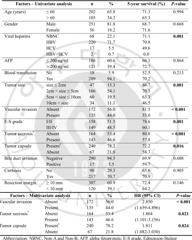
The clinicopathologic factors possibly affecting prognosis following resection of HCC were analyzed. Among them, HCV carrier, tumor size > 2cm, > 5cm and > 10cm,
respectively, vascular invasion, high Edmonson-Steiner grade, spontaneous tumor necrosis and absence of tumor capsule were poor prognostic factors for overall survival in univariate analysis (Table 2).
5
Fig. 1. Overall survival curves according to the factors comprised in the sixth
AJCC/UICC T-classification system (Kaplan-Meier method). (a) Overall survival curve of
307 patients. (b) Tumor size groups. (c) Vascular invasion. (d) T-classification. Group A: tumor size ≤ 2cm, Group B: 2cm < tumor size ≤ 5cm, Group C: 5cm < tumor size ≤ 10cm, Group D: tumor size > 10cm.
6
Fig. 2. Overall survival curves according to the tumor size (Kaplan-Meier method). (a)
Four tumor size groups with comparison of intergroup differences. (b) Tumor size criterion, 2cm. (c) Tumor size criterion, 5cm. (d) Tumor size criterion, 10cm. Group A: tumor size ≤ 2cm, Group B: 2cm < tumor size ≤ 5cm, Group C: 5cm < tumor size ≤ 10cm, Group D: tumor size > 10cm.
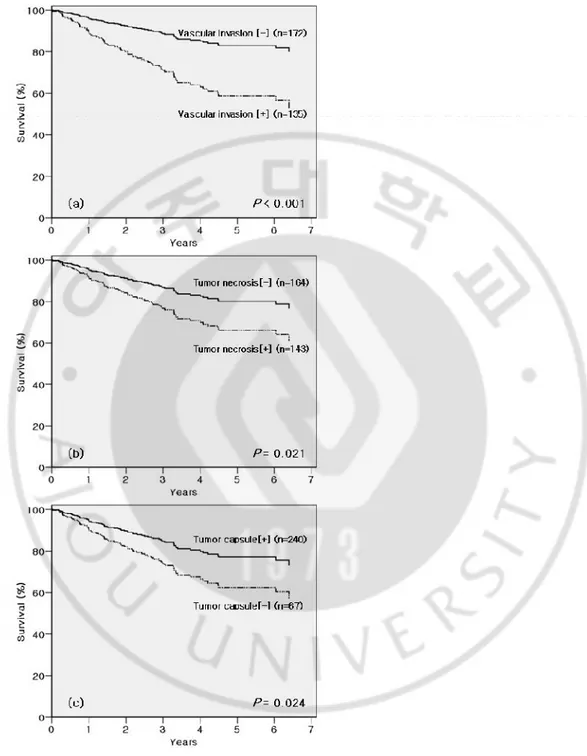
Three size criteria (2cm-, 5cm- and 10cm-borders) were included respectively for the each Cox-regression analysis; however, none of them gained statistical significance in multivariate analyses. By these multivariate analyses vascular invasion (P < 0.001), tumor necrosis (P = 0.021), and absence of tumor capsule (P = 0.024) were proved to be
7
Table 2. Clinicopathologic factors for survival (univariate and multivariate analyses).
Factors – Univariate analysis n % 5-year survival (%) P-value
Age (years) ≤ 60 > 60 202 105 65.8 34.2 71.3 65.3 0.994 Gender Male Female 251 56 81.8 18.2 68.7 71.6 0.668 Viral hepatitis NBNC HBV HCV HBV+HCV 68 220 17 2 22.1 71.7 5.5 0.7 71.1 70.8 49.6 0.0 0.001 AFP ≤ 200 ng/ml > 200 ng/ml 186 121 60.6 39.4 66.1 72.7 0.864 Blood transfusion No Yes 18 289 5.9 94.1 52.5 70.2 0.213
Tumor size size ≤ 2cm
2cm < size ≤ 5cm 5cm < size ≤ 10cm 10cm < size 47 166 60 34 15.3 54.1 19.5 11.1 86.7 70.3 63.3 46.5 0.001
Vascular invasion Absent
Present 172 135 56.0 44.0 81.5 55.0 < 0.001
E-S grade I/II
III/IV 158 149 51.5 48.5 78.6 60.1 0.001
Tumor necrosis* Absent
Present 164 143 53.4 46.6 80.8 57.8 < 0.001
Tumor capsule Present†
Absent 240 67 78.2 21.8 72.2 58.7 0.016
Bile duct invasion Negative Positive 290 17 94.5 5.5 69.9 59.7 0.608 Cirrhosis No Yes 90 217 29.3 70.7 65.6 70.9 0.905 Resection margin ≥ 10 mm < 10 mm 307 120 60.9 39.1 72.4 64.2 0.146
Factors – Multivariate analysis n % HR (95% CI) P-value
Vascular invasion Absent
Present 172 135 56.0 44.0 2.850 (1.659-4.896) < 0.001
Tumor necrosis* Absent
Present 164 143 53.4 46.6 1.864 (1.101-3.156) 0.021
Tumor capsule Present†
Absent 240 67 78.2 21.8 1.811 (1.082-3.030) 0.024
Abbreviation: NBNC, Non-A and Non-B; AFP, alpha-fetoprotein; E-S grade, Edmonson-Steiner grade; HR, Hazard ratio; CI, Confidence interval.
* Spontaneous tumor necrosis. † Partial or complete capsule.
8
Fig. 3. Adjusted overall survival curves of the independent prognostic factors (Cox model). (a) Vascular invasion. (b) Tumor necrosis. (c) Tumor capsule.
9
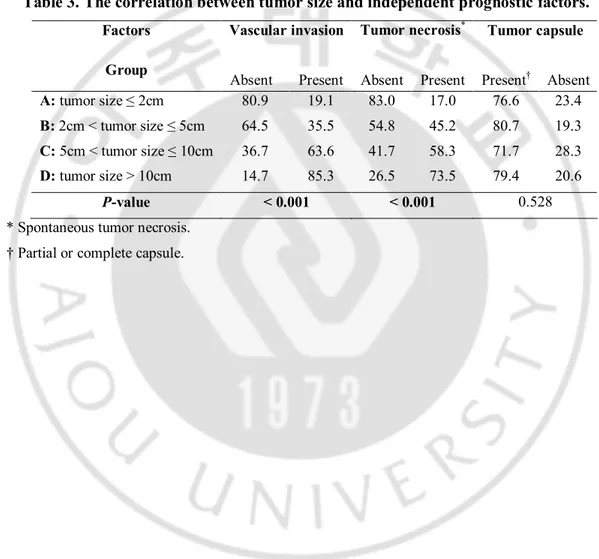
The relationship between tumor size and independent prognostic factors
We analyzed the relationship between the tumor sizes and the three independent prognostic factors (Table 3). Among them, vascular invasion (P < 0.001) and tumor necrosis (P < 0.001) showed significant correlation with tumor size. On the other hand, tumor capsule did not show any correlation with tumor size (P = 0.528).
Table 3. The correlation between tumor size and independent prognostic factors.
Vascular invasion Tumor necrosis* Tumor capsule Factors
Group
Absent Present Absent Present Present† Absent
A: tumor size ≤ 2cm 80.9 19.1 83.0 17.0 76.6 23.4
B: 2cm < tumor size ≤ 5cm 64.5 35.5 54.8 45.2 80.7 19.3
C: 5cm < tumor size ≤ 10cm 36.7 63.6 41.7 58.3 71.7 28.3
D: tumor size > 10cm 14.7 85.3 26.5 73.5 79.4 20.6
P-value < 0.001 < 0.001 0.528
* Spontaneous tumor necrosis. † Partial or complete capsule.
10
IV. DISCUSSION
HCC is a major cancer in Korea. (Park et al., 2004; Cheon et al., 2004; Korean Liver Cancer Study Group and National Cancer Center of Korea, 2009) HCC is generally known to be a highly malignant tumor with a poor prognosis. Unlike other primary cancers, the prognosis of HCC is not solely determined by cancer itself, since most of HCC occur in the background liver with cirrhosis and/or chronic viral hepatitis. Considerable portion of patients with HCC die of liver failure due to underlying liver disease. As a result, several staging systems which include liver function parameters have been proposed. Recently, liver transplantation has become so-called ‘rising sun’ for the primary treatment modality of both HCC and underlying liver cirrhosis simultaneously. However, because of organ shortage, it is limited to some lucky patients. As a result, hepatic resection is still the most common and curative measure for HCC.
Despite of all, as for many other cancers, the AJCC/UICC staging system is most commonly used worldwide after surgical resection for HCC. In the sixthAJCC/UICC TNM staging system, T-classification is determined by tumor size, vascular invasion, multiplicity of tumor and direct invasion of adjacent organ.
Multiplicity of HCC is considered to be a poor prognostic factor. (Chen et al., 2003) HCC with multiple tumors is allocated to T2 or more in the sixth AJCC/UICC system. However, in the present study, patients with multiple tumors were observed only in 33 (9.7%) among the 340 patients who underwent first hepatic resection without preoperative TACE and they were excluded from this study.
The tumor size criterion is defined only in 5cm in the sixth AJCC/UICC system. However, other staging system such as LCSGJ, suggests the tumor size criterion as 2cm for T-classification. Besides, HCC larger than 10cm is classified as so-called ‘huge HCC’ and is considered to possess worse prognosis than smaller one in some previous reports. (Chen et al., 2006; Pandey et al., 2007; Shah et al., 2007b; Lee et al., 2007) In the present study, the patients were divided into four groups according to the aforementioned size criteria (2cm, 5cm and 10cm). Patients with tumor size between 2cm and 5cm were most common (54.1%) among the four size groups, and those with tumor size more than 10cm were 11.1%. The
11
survival rate comparisons in all the three size criteria showed significant differences. The tumor size proved to be a significant prognostic factor in univariate analysis, but not in multivariate analysis.
Vascular invasion is an established risk factor following surgery as well as a component of the sixth AJCC/UICC T-classification system. (Kaibori et al., 2009; Nathan et al., 2009; Shah et al., 2007) Hematogenous route is considered to be important for both intrahepatic and extrahepatic metastases. (Mitsunobu et al., 1996; Toyosaka et al., 1996; Sawabe et al., 1987) In our study, vascular invasion is proved to be a significant prognostic factor not only in univariate analysis but also in multivariate analysis. Moreover, it is the strongest independent prognostic factor in Cox-regression model of our study.
While some studies suggested bile duct invasion is a poor prognostic factor like the LCSGJ T-classification system, (Ikenaga et al., 2009; Kojiro et al., 1982; Minagawa et al., 2007; Yeh et al., 2004) other studies demonstrated the opposite results. (Lau et al., 1997; Satoh et al., 2000; Shiomi et al., 2001) In our study, bile duct invasion did not attain statistical significance.
In this study, among these factors of T-classification, survival differences were observed in tumor size and vascular invasion by univariate analysis. However, by multivariate analysis, only vascular invasion was proved to be an independent significant factor among these factors. Other independent significant factors were spontaneous tumor necrosis and absence of tumor capsule, in addition to vascular invasion.
We investigated the relationship between the tumor size and three independent prognostic factors, respectively. Among them, tumor size showed statistically significant correlation with spontaneous tumor necrosis and vascular invasion, but not with absence of tumor capsule. Spontaneous tumor regression is rare in HCC. However, generally, a tumor is too large to be fed with enough blood supply to the entire tumor tissue, ischemic necrosis may happen. So, larger tumor tends to accompany spontaneous ischemic necrosis more frequently even without therapeutic embolization, as in our study. Some previous studies demonstrated the correlation between tumor size and vascular invasion. (Esnaola et al., 2002; Pawlik et al., 2005) The authors of these studies suggested that tumor size is regarded as a predictor of microvascular invasion for liver transplantation candidates.
Our study demonstrated that all the respective size criteria (2cm, 5cm and 10cm) were predictive factors for survival in univariate analysis, but not in multivariate analysis. Tumor
12
size showed significant correlation with some of independent prognostic factors, except for absence of tumor capsule.
13
V. CONCLUSION
In summary, all the tumor size criteria were valuable predictors for survival in patients with single HCC following resection. Tumor size should be subdivided further for T-classification to predict prognosis more accurately. Though tumor size itself was not an independent prognostic factor, it represents some of independent prognostic factors, but not all of them. Considering that the AJCC/UICC staging system is a histopathologic staging system rather than preoperative parameter such as the Milan criteria and tumor capsule was an independent significant prognostic factor irrespective of tumor size, tumor capsule should be included in T-classification of the AJCC/UICC staging system for more accurate prediction of survival.
14
REFERENCES
1. AJCC: AJCC Cancer Staging Manual, 6th ed. New York: Springer, 2002
2. Chen MF, Tsai HP, Jeng LB, Lee WC, Yeh CN, Yu MC, Hung CM: Prognostic factors after resection for hepatocellular carcinoma in noncirrhotic livers: univariate and multivariate analysis. World J Surg 27: 443-447, 2003
3. Chen XP, Qiu FZ, Wu ZD, Zhang BX: Hepatectomy for huge hepatocellular carcinoma in 634 cases. World J Gastroenterol 12: 4652-4655, 2006
4. Cheon JH, Park JW, Park KW, Kim YI, Kim SH, Lee WJ, Park HS, Park SJ, Hong EK, Kim CM: The clinical report of 1,078 cases of hepatocellular carcinomas: National Cancer Center experience. Korean J Hepatol 10: 288-297, 2004
5. Esnaola NF, Lauwers GY, Mirza NQ, Nagorney DM, Doherty D, Ikai I, Yamaoka Y, Regimbeau JM, Belghiti J, Curley SA, Ellis LM, Vauthey JN: Predictors of microvascular invasion in patients with hepatocellular carcinoma who are candidates for orthotopic liver transplantation. J Gastrointest Surg 6: 224-232, 2002
6. Fleming ID, Cooper JS, Henson DE, Hutter RVP, Kennedy BJ, Murhpy GP, O'Sullivan B, Sobin LH, Yarbro JW: Liver AJCC Cancer Staging Manual (ed 5). Philadelphia, PA, Lippincott-Raven, pp.98-126, 1997
7. Ikenaga N, Chijiiwa K, Otani K, Ohuchida J, Uchiyama S, Kondo K: Clinicopathologic characteristics of hepatocellular carcinoma with bile duct invasion. J Gastrointest Surg 13: 492-497, 2009
8. Kaibori M, Ishizaki M, Saito T, Matsui K, Kwon AH, Kamiyama Y: Risk factors and outcome of early recurrence after resection of small hepatocellular carcinomas. Am J Surg 198: 39-45, 2009
15
9. Kojiro M, Kawabata K, Kawano Y, Shirai F, Takemoto N, Nakashima T: Hepatocellular carcinoma presenting as intrabile duct tumor growth: a clinicopathologic study of 24 cases. Cancer 49: 2144-2147, 1982
10. Korean Liver Cancer Study Group and National Cancer Center of Korea: Practice guidelines for management of hepatocellular carcinoma 2009. Korean J Hepatol 15: 391-423, 2009
11. Lau W, Leung K, Leung TW, Liew CT, Chan MS, Yu SC, Li AK: A logical approach to hepatocellular carcinoma presenting with jaundice. Ann Surg 225: 281-285, 1997
12. LCSGJ: General Rules for the Clinical and Pathological Study of Primary Liver Cancer, 2nd English edition. Tokyo: Kanehara. 2003
13. Lee SG, Hwang S, Jung JP, Lee YJ, Kim KH, Ahn CS: Outcome of patients with huge hepatocellular carcinoma after primary resection and treatment of recurrent lesions. Br J Surg 94: 320-326, 2007
14. Matsuda M, Suzuki T, Kono H, Fujii H: Predictors of hepatic venous trunk invasion and prognostic factors in patients with hepatocellular carcinomas that had come into contact with the trunk of major hepatic veins. J Hepatobiliary Pancreat Surg 14: 289-296, 2007 15. Minagawa M, Ikai I, Matsuyama Y, Yamaoka Y, Makuuchi M: Staging of hepatocellular
carcinoma: assessment of the Japanese TNM and AJCC/UICC TNM systems in a cohort of 13,772 patients in Japan. Ann Surg 245: 909-922, 2007
16. Mitsunobu M, Toyosaka A, Oriyama T, Okamoto E, Nakao N: Intrahepatic metastases in hepatocellular carcinoma: the role of the portal vein as an efferent vessel. Clin Exp Metastasis 14: 520-529, 1996
17. Nathan H, Schulick RD, Choti MA, Pawlik TM: Predictors of survival after resection of early hepatocellular carcinoma. Ann Surg 249: 799-805, 2009
16
18. Pandey D, Lee KH, Wai CT, Wagholikar G, Tan KC: Long term outcome and prognostic factors for large hepatocellular carcinoma (10 cm or more) after surgical resection. Ann Surg Oncol 14: 2817-2823, 2007
19. Park JW: Practice guideline for diagnosis and treatment of hepatocellular carcinoma. Korean J Hepatol 10: 88-98, 2004
20. Pawlik TM, Delman KA, Vauthey JN, Nagorney DM, Ng IO, Ikai I, Yamaoka Y, Belghiti J, Lauwers GY, Poon RT, Abdalla EK: Tumor size predicts vascular invasion and histologic grade: Implications for selection of surgical treatment for hepatocellular carcinoma. Liver Transpl 11: 1086-1092, 2005
21. Sakata J, Shirai Y, Wakai T, Kaneko K, Nagahashi M, Hatakeyama K: Preoperative predictors of vascular invasion in hepatocellular carcinoma. Eur J Surg Oncol 34: 900-905, 2008
22. Satoh S, Ikai I, Honda G, Okabe H, Takeyama O, Yamamoto Y, Yamamoto N, Iimuro Y, Shimahara Y, Yamaoka Y: Clinicopathologic evaluation of hepatocellular carcinoma with bile duct thrombi. Surgery 128: 779-783, 2000
23. Sawabe M, Nakamura T, Kanno J, Kasuga T: Analysis of morphological factors of hepatocellular carcinoma in 98 autopsy cases with respect to pulmonary metastasis. Acta Pathol Jpn 37: 1389-1404, 1987
24. Shah SA, Cleary SP, Wei AC, Yang I, Taylor BR, Hemming AW, Langer B, Grant DR, Greig PD, Gallinger S: Recurrence after liver resection for hepatocellular carcinoma: risk factors, treatment, and outcomes. Surgery 141: 330-339, 2007a
25. Shah SA, Wei AC, Cleary SP, Yang I, McGilvray ID, Gallinger S, Grant DR, Greig PD: Prognosis and results after resection of very large (>or=10 cm) hepatocellular carcinoma. J Gastrointest Surg 11: 589-595, 2007b
17
26. Shiomi M, Kamiya J, Nagino M, Uesaka K, Sano T, Hayakawa N, Kanai M, Yamamoto H, Nimura Y: Hepatocellular carcinoma with biliary tumor thrombi: aggressive operative approach after appropriate preoperative management. Surgery 129: 692-698, 2001
27. Toyosaka A, Okamoto E, Mitsunobu M, Oriyama T, Nakao N, Miura K: Intrahepatic metastases in hepatocellular carcinoma: evidence for spread via the portal vein as an efferent vessel. Am J Gastroenterol 91: 1610-1615, 1996
28. Vauthey JN, Lauwers GY, Esnaola NF, Do KA, Belghiti J, Mirza N, Curley SA, Ellis LM, Regimbeau JM, Rashid A, Cleary KR, Nagorney DM: Simplified staging for hepatocellular carcinoma. J Clin Oncol 20: 1527-1536, 2002
29. Yeh CN, Jan YY, Lee WC, Chen MF: Hepatic resection for hepatocellular carcinoma with obstructive jaundice due to biliary tumor thrombi. World J Surg 28: 471-475, 2004
18 - 국문요약 -