Introduction
The combination of stem cells and biomaterials has provided potential for regenerative medicine. The key point of the ap-proach is to differentiate stem cells into specific cell lineages or tissues using biomaterials (1). This tissue-engineered approach requires an understanding of the characteristics about both of the stem cells and the biomaterials and the interaction between them. In the case of tissue-engineered applications and con-ventional in vitro cell experiments, a 2D culture system provides advantages of easily supplementing oxygen and nutrients (2). Investigating the cell responses on the surface of the implant using the 2D culture system, studies on the interaction of stem
cells and surface topography have been conducted.
Mesenchymal stem cells (MSCs) have been focused because they can be easily harvested from the human body and have capability of differentiating into various cells or tissues for sur-gical procedures. Many efforts to investigate and simulate stem cell niche exist, as it is known that cell responses are affected by the surrounding microenvironment and sensing at the same time (3). An external local microenvironment of stem cell, called stem cell niche, affects stem cell behavior such as adhesion, proliferation, and differentiation. The above-mentioned surface topography corresponds to the physical component among the three components (chemical, physical, and cellular components) that make up the stem cell niche (4). Therefore, observing the Young-Shik Yun1,2, Eun-Hye Kang3, In Sik Yun3, Yong Oock Kim3, and Jong-Souk Yeo1,2
1School of Integrated Technology, Yonsei University, Incheon, Korea
2Yonsei Institute of Convergence Technology, Yonsei University, Incheon, Korea
3Department of Plastic & Reconstructive Surgery, College of Medicine, Yonsei University, Seoul, Korea
In the field of tissue engineering, researches have been actively conducted to regulate stem cell fate by understanding the inter-action between cell and materials. This approach is expected as a promising therapeutic method in the future medicine by uti-lizing differentiation of stem cells into desired cells or tissues using biomaterial. For this regenerative medicine, there exist lots of attempts to construct optimized structures of various shapes and sizes that can regulate the stem cell fate. In this review, we will empathize the topographic effect as stem cell niche on the mesenchymal stem cell (MSC) response (cell attachment, prolif-eration, and differentiation) according to the shape and size of the structure of the substrates, and comprehensively analyze the importance and the effect of shape and size of the surface topography.
Key WordsZZTissue engineering ㆍRegenerative medicine ㆍMesenchymal stem cells ㆍTopographic effect. Received: May 20, 2017 / Revised: May 22, 2017 / Accepted: May 24, 2017
Address for correspondence: Jong-Souk Yeo
Yonsei Institute of Convergence Technology, Yonsei University, 85 Songdo-gwahak-ro, Yeonsu-gu, Incheon 21983, Korea
Tel: 82-32-749-5838, Fax: 82-32-818-5801, E-mail: [email protected]
This is an Open Access article distributed under the terms of the Creative Commons Attribution Non-Commercial License (http://creativecommons.org/licenses/ by-nc/4.0/) which permits unrestricted non-commercial use, distribution, and reproduction in any medium, provided the original work is properly cited.
response of stem cells on various surface topography is crucial for understanding their interactions, thus developing tissue-engineered applications (Fig. 1).
There have been many attempts to regulate stem cell fate based on the understanding the interaction of stem cells and the microenvironment (5). Previous researches demonstrate that stem cell fate is mostly influenced by biochemical compo-nents such as growth factor, protein, and hormones and also by physical components such as topography and stiffness (1, 6-8). Furthermore, it has been shown that topographic effect alone without biochemical factors can also affect stem cell fate (9, 10). As nanotechnology develops further, it has become possible to create nanostructures that are much smaller and more precise than ever before. Electron beam lithography is a typical meth-od and the nanostructures down to below 10 nm can be pat-terned on the surface by this method (11, 12). Additional nano-fabrication techniques include top-down and bottom-up processes to fabricate nanoscale and microscale structures. By using combinations of different fabrication techniques, it is possible to produce various nanostructures of desired shape and specific size. Those techniques form a basis for defining topography that effectively controls the cell responses.
We review the researches about the response of MSCs to sur-face topography and the related fabrication processes with respect to tissue engineering. In order to understand the mechanism of regulating stem cell fate induced by topographical effect, size and shape of topography is emphasized. Then, we consider the remaining challenges in using MSCs for regenerative medicine.
Mesenchymal Stem Cells (MSCs)
in Tissue Engineering
Stem cells refer to the cells that are capable of differentiating
into various cell lineages and proliferating while keeping un-differentiated state at the same time. The characteristics of stem cells can be clearly observed in embryonic stem cells. The plu-ripotency and self-renewal ability of embryonic stem cells can be used in combination with tissue engineering to show high potential for regenerative medicine (13). However, embryonic stem cells also have ethical issues as well as potential problems of differentiating into cancer cells (14).
MSCs are attracting attention as one of the alternatives to embryonic stem cells. The MSC was first found from bone mar-row in the early 1960s (15). Since MSCs are capable of differenti-ating into muscle tissue, fat, cartilage, and bone tissue, and can be easily harvested from the patient’s body, studies on their tis-sue engineered application have been actively carried out (16, 17). There are three requirements for the use of stem cells in tissue engineering (18). A large number of cells should be collected and tissue engineering products from them should be safe when they are used on the human body. They should also maintain undifferentiated state until it is used for tissue engineering. It should be noted that some challenges remain as the amount that can be collected from the human body is limited, and the state of the MSCs is affected by the age and physical condition of the patient (19, 20). To meet those requirements, researchers have attempted to regulate stem cell fate based on the under-standing of the functions of stem cell niche in the tissue engi-neering.
Fabrication Techniques
for Surface Topography
With the development of nanotechnology, it has become pos-sible to fabricate various structures that are comparable in the size with extracellular matrix (ECM). The most commonly Fig. 1. Multipotent of mesenchymal stem cell and physical component of stem cell niche in tissue engineering.
fabrication is an additive process where atoms and molecules are assembled to make structures. Self-assembled nanostruc-tures, bottom-up synthesis such as nanowires and nanodots are the representative methods for bottom-up process.
The top-down fabrication benefits from the precise control of energetic beam such as photon, electron, and ion to put the desired patterns in exact location. While it is widely used in mi-croelectronics manufacturing, its resolution is limited by the very nature of beam control process such as a mask used for a photon beam that does not have electric charge and an
electro-development of hybrid nanofabrication techniques such as na-noimprinting and soft lithography where nanoscale structures are fabricated by slow and costly top-down process such as elec-tron beam lithography (EBL) or low cost bottom-up fabrication process such as colloidal self-assembly and then replicated by transferring the nanopatterns with soft lithography process.
Among the top-down processes, the direct milling can be used to cut the surface of a substrate in a desired structure. Typ-ical methods include focused ion beam and laser milling. Pho-tolithography is a core technique of semiconductor production
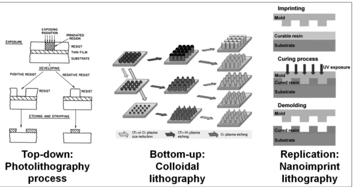
Fig. 2. Representative schematic of fabrication techniques (Top-down: Photolithography process) The schematic diagram shows the process of photolithography including exposure through mask after resin coating, development, and post process of etching and stripping. The residual resin pattern after development is used to fabricate surface topography as mask (21). Reprinted with permission from Ref. 21. Copyright 1983, American Chemical Society.(Bottom-up: Colloidal lithography and post processes for the fabrica-tion of nanopillars) At the left side, nanoparticles are aligned as hexagonal closed packed monolayer by self-assembly. From the left, the underlying substrate is modified to pillar array using the monolayer as an etch mask. Size reduction of nanoparticle with post pro-cesses to control the size of mask makes the altered shape of nanopillars (23). Reproduced from Ref. 23 with permission of The Royal Society of Chemistry. (Replication: Pattern transfer process using UV-Nanoimprint Lithography) The schematic diagram shows the process of ultraviolet (UV) nanoimprint lithography (NIL) including resist coating, imprinting mold into the resin, UV curing and demold-ing process.
method. The process of photolithography includes photoresist coating on Si wafer, UV exposure, development, and post pro-cess (21). The photoresist made of a polymer resin is spin-coat-ed onto the Si wafer, then exposspin-coat-ed through the mask pattern to react under UV light. The resin dissolves selectively in a devel-oper because the solubility of resin changes by UV exposure. The development of semiconductor fabrication technology has led to the optimized process so any types of structure can be generated in microscale with relative ease.
However, when going down to the nanoscale, there is a reso-lution limit due to the diffraction of light depending on the wave-length of the UV used as the light source. To overcome the reso-lution limit of photolithography, the methods of using shorter wavelength sources such as electron beam and extreme ultra violet (EUV) have been investigated. Electron beam lithography (EBL), which is used in the prototyping for many nanoscale ex-periments, can form a nanoscale pattern down to 10 nm or less on a substrate (11, 22). In contrast to conventional photolithog-raphy where exposure through a mask defines a pattern in the process, EBL enables a direct patterning without using a mask by utilizing a computer aided design (CAD) tool to design a pat-tern and then by using a X, Y axis controller or an electron beam blanker attached to scanning electron microscope to provide positioning capability of the electron beam. Patterns made us-ing these lithography processes can be used directly or in
com-bination with the other methods of post processing such as de-position or etching.
These post processes can be used to introduce additional to-pography on the patterns fabricated with the methods de-scribed previously. The deposition methods include chemical vapor deposition (CVD) and physical vapor deposition (PVD). The CVD method is a technique of forming a thin film by react-ing at the surface of a substrate in ambient source gas usreact-ing ex-ternal energy such as heat, plasma, and light. PVD uses vapor-izing or ionvapor-izing a metal or a dielectric material in a vacuum to form a thin film on the surface of a substrate. Depending on how materials are transferred from target material to a sub-strate, the techniques are called an evaporation coating if it in-volves vaporization of source material, a sputtering if it utilize ion bombardment to sputter species off from the source, or an ion plating if glow discharge plasma is initiated to enhance the energy of the species for better deposition. Etching is catego-rized into wet etch and dry etch. Wet etch is immersed in a re-active solution as the name implies, and dry etch is a method of selectively removing the area that is not shielded with a mask by using a plasma process.
Self-assembly is a fundamental method for the bottom-up process and has the advantage of high resolution by directly using molecules or nanoparticles. Colloidal lithography forms a pattern through the self-assembly of colloidal polystyrene Fig. 3. Representative result of topographic effect. SEM images of nanopit arrays from ordered to disordered arrangement (upper side), fluorescence microscopy images of actin (red) and osteopontin (OPN)/osteocalcin (OCN) (green) as bone-specific ECM protein (bottom side): nanoscale disordered pit array induced MSCs to produce bone mineral without biochemical environment (9) Reprinted by per-mission from Macmillan Publishers Ltd: Nature Materials (ref. 9), copyright 2007.
nanospheres and then uses the bottom-up monolayer arrange-ment as a mask to define the nanoscale topography. The mono-layer can be in the form of a hexagonal close packed (HCP) struc-ture. Using this monolayer as a mask, a structure of nanopillar or nanopit array can be generated through the post process of etching (23).
In addition to the exposure or self-assembly based methods, nanoimprint lithography (NIL) is relatively new and provides a hybrid approach of taking advantages from both methods to form nanopatterns (24). Nanoscale patterns meticulously pre-pared by electron beam lithography or self-assembly based process can be replicated by transferring the patterns by stamp-ing onto the UV curable resin coated on the surface of silicon wafers. Soft lithography leverages similar principle and surface chemistry to control the transfer of the nanostructures from a mold onto polymeric resin materials (25). Capillary force li-thography is one of the representative methods. (26, 27). A few drops of UV resin are applied to the surface of the nanostruc-ture. The resin fills the gap of the nanostructure by a capillary force and then it is cured and separated to produce replicated structure. Materials such as Polydimethylsiloxane (PDMS) can be used and cured by heat and then separated to obtain a repli-ca of nanostructures.
Surface Topographic Effect
Shape and size of structures on the substrate have been wide-ly known as factors of topographic effect (6, 9, 28-30). Natural
ECM structures are composed of complex topography of en-tangled fiber and porous structure in nanoscale (31) and the com-monly used forms such as a roughened surface, pillar struc-tures, and ridge/groove are inspired from the natural structures. These types of topography from various fabrication techniques affect the regulation of stem cell fates (Table 1). The first observed behavior of MSCs is morphological change. MSCs are elongat-ed or spread on the surface along with the shape and size of underlying topography. The morphological changes generally accompany cytoskeletal reorganization and stress. The cyto-skeleton tension and reorganization with focal adhesion are the key factors of topographic effect investigated so far (32). We classify the previous studies according to the type of surface structure and classify the effect of target cell reaction and struc-ture type. In the case of patterning, anisotropic and isotropic structures are considered to reflect the response of MSCs. Anisotropic pattern has ridge/groove structures, and isotropic pattern has structures such as pit, pillar, and nanotube. Roughened surface
In recent experiments using micropatterning to observe the reaction of human mesenchymal stem cells (hMSCs) from bone marrow on a substrate with a planar surface and a roughened surface, MSCs has shown better adhesion on planar surfaces than roughened surfaces (33). This research described inhibi-tion of focal adhesion on nanoroughened area and suggested the random walk model for analysis of organization of cells on the micropatterned surface.
(nano) ~20 nm tip Wet etching osteogenisis.
Nanotube 15, 20, 30, 50, 70, 100 nm
diameter Anodization Smaller nanotubes enhance MSC responses of adhesion, proliferation, and osteogenic differentiation
(30)
30, 50, 70, 100 nm
diameter Anodization Small nanotube promotes cell adhesion and large nanotube enhances elongation and osteogenic differetation
Roughened surfaces have been used since the early stages of the study on surface morphology and cell response as the rough-ened surface is relatively easier to fabricate. Roughness on the surface is isotropic and randomly disordered with its charac-teristic typically defined as the mean value of the height along the z-axis. The cell response on the roughened surface focuses on the area where the surface is defined and measured. Ran-domly roughened surfaces have different sharpness and smooth-ness of the protrusions on the surface, and height and spacing of the protrusions, which makes clear conclusions rather diffi-cult as the roughness values (Ra) are averaged over the surface. Ridge/groove
The effect of ridge/groove structure on cell activity is mainly classified according to the distance between the ridges and the size of the ridges. Such a structure can be made either directly by beam based machining methods such as ion or laser micro-machining or indirectly by transferring a pattern of a mold formed by lithography and etching onto a biocompatible polymer. Abagnale et al. analyzed osteogenic and adipogenic differentia-tion of MSCs isolated from lipoaspirate using various sizes of ridge/groove structures (28). The research has shown that a specific size in microscale promoted the differentiation of stem cells. In microscale, osteogenic differentiation was enhanced on 2 μm ridge and adipogenic differentiation was enhanced on 15 μm ridge. In nanoscale, both osteogenic and adipogenic dif-ferentiations were amplified on the ridge/groove structures of 650 nm pitch.
Since ridge/groove is an anisotropic structure, it has a direction-al effect on cell attachment and maturation. It has direction-also been used for experiments requiring specific orientation and shape of cell arrays such as differentiation of muscle cells and nerve cells. Yim et al. induced hMSCs from bone marrow to differ-entiate into neuronal-like cells on the nanograting structure with 350 nm width (34). They demonstrated neuronal differ-entiation of MSCs on the nanograting associated with morpho-logical changes and emphasized the significance of nanotop-ography.
Pit array
According to Dalby et al.’s results (Fig. 3), not only the size but also the arrangement of nanostructures are important for os-teogenic differentiation of stem cells (9). The surface morphol-ogy of the substrate used in the experiment was changed by varying the diameter and depth of the nanopit on the surface so that the resulting osteogenesis was compared. This study is considered as a representative example showing the importance of topographic effects as it demonstrated that osteogenesis can
be induced in the specific patterns of square array and disorder levels even in the absence of specific growth factors.
Pillar array
Brammer et al. demonstrated the role of nanopillar that affects osteogenic differentiation of MSCs from rat bone marrow (35). They found that nanopillar increased cell attachment, growth and cell aggregation. Furthermore, the cell aggregation induced on the nanopillar enhances osteogenic mineralization. Recent-ly, researchers have reported that controlling the distance of the pillar array can regulate the spread of the filopodia to pro-mote neuronal differentiation of adipose-derived stem cells (10). In order to fabricate the PDMS nanopillar array used in this experiment, a nanopit array mold was fabricated using EBL and etching process, and a pattern was transferred through soft lithography to fabricate the nanopillar structure. In this case, the nanopillar was utilized to induce cell elongation and this morphological change was shown to regulate the stem cell fate. Nanotube
Tubular arrays have been extensively studied as a means to make porous oxide film on the surface of a Ti alloy, a material frequently used as an implant. For the in vitro studies, titani-um oxide nanotube is formed through the anodization process or etching with a porous anodized aluminum oxide (AAO) as a template. In experiments using TiO2 nanotubes, the response of MSCs from bone marrow to various sizes was shown to be different (29, 30). Park et al. observed that adhesion, prolifera-tion, and osteogenic differentiation of MSCs were enhanced in nanotubes of 15 nm diameter (30). However, the results of Oh et al. using nanotubes indicated that osteogenic differentiation was enhanced for larger diameters (29). The two experimental results were not exactly consistent and it would be hard to keep the detailed environment of each experiment same, but these results suggest the necessity of systematic approaches in the development and design for tissue engineered application.
Summary and Conclusion
The development of bio-alternative materials for regenera-tive medicine through the combination of stem cells and bio-materials lays the foundation for future medical technology development. This requires a deeper understanding of the in-teraction between stem cells and biomaterials. Activities such as differentiation and attachment of MSCs are influenced by the local microenvironment called stem cell niche. The fabri-cation of nanostructures opens up the possibility for systematic approaches in mimicking extracellular environments. It has
For regenerative medicine, it is necessary to systematically approach the effects of stem cell niche, especially for the physi-cal components that can be controlled by various fabrication technologies. Many experiments have been conducted in this regard, but there are many cases in which the experimental re-sults are not sufficient to draw conclusive explanation on the topographic effect. In order to understand the regulatory mech-anisms of stem cells by topographic effect, we need to analyze the features of each nanostructure as well as the function of stem cells, and apply the systematic approach to control the experi-mental environment clearly.
Acknowledgements
This research was supported by the “Mid-career Researcher Pro-gram” (NRF-2016R1A2B2014612) supervised by the NRF (National Research Foundation). This study was also supported by a grant of Ministry of Trade, Industry & Energy, Korea (Grant No. 10045651) and Basic Science Research Program through the National Re-search Foundation of Korea (NRF) funded by the Ministry of Edu-cation, Science and Technology (2015R1D1A1A01060361).
References
1. Bianco P, Robey PG. Stem cells in tissue engineering. Nature 2001; 414:118-121
2. Russell AJ, Bertram T, Lanza R, Langer R, Vacanti J. Moving into the clinic. Principles of Tissue Engineering 2007:15-32
3. Geiger B, Spatz JP, Bershadsky AD. Environmental sensing through focal adhesions. Nat Rev Mol Cell Bio 2009;10:21-33
4. Samadikuchaksaraei A, Lecht S, Lelkes PI, Mantalaris A, Polak JM. Stem cells as building blocks. Principles of tissue engineering 2014 5. Kshitiz, Park J, Kim P, Helen W, Engler AJ, Levchenko A, et al.
Control of stem cell fate and function by engineering physical mi-croenvironments. Integr Biol-Uk 2012;4:1008-1018
6. Engler AJ, Sen S, Sweeney HL, Discher DE. Matrix elasticity directs stem cell lineage specification. Cell 2006;126:677-689
7. Li Z, Gong YW, Sun SJ, Du Y, Lu DY, Liu XF, et al. Differential regulation of stiffness, topography, and dimension of substrates in rat mesenchymal stem cells. Biomaterials 2013;34:7616-7625 8. Griffin MF, Butler PE, Seifalian AM, Kalaskar DM. Control of stem
cell fate by engineering their micro and nanoenvironment. World J
15. Friedenstein AJ, Piatetzky S, II, Petrakova KV. Osteogenesis in trans-plants of bone marrow cells. J Embryol Exp Morphol 1966;16:381-390
16. Hass R, Kasper C, Bohm S, Jacobs R. Different populations and sources of human mesenchymal stem cells (msc): A comparison of adult and neonatal tissue-derived msc. Cell Commun Signal 2011;9 17. Ullah I, Subbarao RB, Rho GJ. Human mesenchymal stem cells -
current trends and future prospective. Bioscience Rep 2015;35 18. Obokata H, Vacanti CA. Chapter 31 - stem cells in tissue engineering.
InPrinciples of tissue engineering (fourth edition). Boston: Academ-ic Press, 2014:595-608
19. Rodriguez JP, Garat S, Gajardo H, Pino AM, Seitz G. Abnormal os-teogenesis in osteoporotic patients is reflected by altered mesenchy-mal stem cells dynamics. Journal of Cellular Biochemistry 1999; 75:414-423
20. Quarto R, Thomas D, Liang CT. Bone progenitor-cell deficits and the age-associated decline in bone repair capacity. Calcified Tissue Int 1995;56:123-129
21. Thompson LF. An introduction to lithography. InIntroduction to mi-crolithography: American Chemical Society, 1983:1-13.
22. Wagner C, Harned N. Euv lithography lithography gets extreme. Nat Photonics 2010;4:24-26
23. Ji S, Park J, Lim H. Improved antireflection properties of moth eye mimicking nanopillars on transparent glass: Flat antireflection and color tuning. Nanoscale 2012;4:4603-4610
24. Zhou W. Nanoimprint lithography process. InNanoimprint lithogra-phy: An enabling process for nanofabrication. Berlin, Heidelberg: Springer Berlin Heidelberg, 2013:111-146
25. Rogers JA, Nuzzo RG. Recent progress in soft lithography. Mater Today 2005;8:50-56
26. Ho D, Zou J, Zdyrko B, Iyer KS, Luzinov I. Capillary force lithogra-phy: The versatility of this facile approach in developing nanoscale applications. Nanoscale 2015;7:401-414
27. Jeong HE, Kwak R, Khademhosseini A, Suh KY. Uv-assisted capil-lary force lithography for engineering biomimetic multiscale hier-archical structures: From lotus leaf to gecko foot hairs. Nanoscale 2009; 1:331-338
28. Abagnale G, Steger M, Nguyen VH, Hersch N, Sechi A, Joussen S, et al. Surface topography enhances differentiation of mesenchymal stem cells towards osteogenic and adipogenic lineages. Biomaterials 2015;61:316-326
29. Oh S, Brammer KS, Li YS, Teng D, Engler AJ, Chien S, et al. Stem cell fate dictated solely by altered nanotube dimension. Proc Natl Acad Sci U S A 2009;106:2130-2135
30. Park J, Bauer S, von der Mark K, Schmuki P. Nanosize and vitality: Tio2 nanotube diameter directs cell fate. Nano Lett 2007;7:1686-1691
31. Abrams GA, Goodman SL, Nealey PF, Franco M, Murphy CJ. Na-noscale topography of the basement membrane underlying the
cor-neal epithelium of the rhesus macaque. Cell Tissue Res 2000;299: 39-46
32. Yao X, Peng R, Ding JD. Effects of aspect ratios of stem cells on lin-eage commitments with and without induction media. Biomaterials 2013;34:930-939
33. Perez DG, Quijorna EP, Sanz R, Torres-Costa V, Ruiz JPG, Silvan MM. Nanotopography enhanced mobility determines mesenchy-mal stem cell distribution on micropatterned semiconductors
bear-ing nanorough areas. Colloid Surface B 2015;126:146-153 34. Yim EKF, Pang SW, Leong KW. Synthetic nanostructures inducing
differentiation of human mesenchymal stem cells into neuronal lineage. Exp Cell Res 2007;313:1820-1829
35. Brammer KS, Choi C, Frandsen CJ, Oh S, Jin S. Hydrophobic nano-pillars initiate mesenchymal stem cell aggregation and osteo-dif-ferentiation. Acta Biomaterialia 2011;7:683-690