의학
의학
의학
의학 박사학위
박사학위
박사학위
박사학위 논문
논문
논문
논문
A Study on the Regulation of
Microglial Inflammatory Responses
by Gangliosides and Oxidized Low
Density Lipoprotein
아
아
아
아 주
주
주
주 대
대
대
대 학
학
학
학 교
교 대
교
교
대
대
대 학
학
학
학 원
원
원
원
의
의
의
의 학
학
학 과
학
과
과
과
김
김
김
김 온
온
온 순
온
순
순
순
A Study on the Regulation of Microglial
Inflammatory Responses by Gangliosides and
Oxidized Low Density Lipoprotein
by
Ohn Soon Kim
A Dissertation Submitted to The Graduate School of Ajou University
in Partial Fulfillment of the Requirements for the Degree of
DOCTOR OF PHILOSOPHY
Supervised by
Ilo Jou, M.D., Ph.D.
Department of Medical Sciences
The Graduate School, Ajou University
김온순의
김온순의
김온순의
김온순의 의학
의학
의학
의학 박사학위
박사학위
박사학위 논문을
박사학위
논문을
논문을 인준함
논문을
인준함
인준함
인준함....
심 사 위 원 장
심 사 위 원 장
심 사 위 원 장
심 사 위 원 장
백
백
백
백 은
은 주
은
은
주
주
주
인
인
인
인
심
심
심
심 사
사 위
사
사
위
위
위 원
원
원
원
주
주 일
주
주
일
일 로
일
로
로
로
인
인
인
인
심
심
심
심 사
사 위
사
사
위
위
위 원
원
원
원
조
조 은
조
조
은
은 혜
은
혜
혜
혜
인
인
인
인
심
심
심
심 사
사 위
사
사
위
위
위 원
원
원
원
김
김 완
김
김
완
완 기
완
기
기
기
인
인
인
인
심
심
심
심 사
사
사 위
사
위
위
위 원
원
원
원
박
박
박 은
박
은
은 미
은
미
미
미
인
인
인
인
아
아
아
아 주
주
주
주 대
대
대
대 학
학
학
학 교
교 대
교
교
대
대
대 학
학
학
학 원
원
원
원
2005
2005
2005
2005년
년
년
년 12
12
12
12월
월
월
월 22
22
22일
22
일
일
일
-ABSTRACT-
A Study on the Regulation of Microglial Inflammatory Responses
by Gangliosides and Oxidized Low Density Lipoprotein
The brain is abundant in lipids, and lipid metabolism is clearly related to brain functions such as synaptogenesis, neuronal survival and signal transduction. Microglia are one type of glial cells in the brain and play a role as major immune effector cells. Because microglia-mediated brain inflammation participate in the initiation or progression of neurodegenerative disorders, it is important to understand the regulation of microglial inflammatory responses. In this study, I investigated whether lipid derivatives, gangliosides and oxidized low density lipoprotein (oxLDL), could modulate microglia-mediated brain inflammation.
In part 1, I examined whether gangliosides activate JAK-STAT pathway, an essential inflammatory signaling pathway in microglia. Neuronal cell membranes are particularly rich in gangliosides, which play important roles in brain physiology and pathology. In this study, I provide evidence that JAK-STAT inflammatory signaling mediates gangliosides-stimulated microglial activation. Both in rat primary microglia and murine BV2 microglial cells, gangliosides stimulated nuclear factor binding to GAS/ISRE elements, which are known to be STAT-binding sites. Consistent with this, gangliosides rapidly activated JAK1 and JAK2 and induced phosphorylation of STAT1 and STAT3. In addition, gangliosides increased
transcription of the inflammation-associated genes inducible nitric oxide synthase (iNOS), intracellular adhesion molecule-1 (ICAM-1), and monocyte chemoattractant protein-1 (MCP-1), which are reported to contain STAT-binding elements in their promoter regions. AG490, a JAK inhibitor, reduced induction of these genes, nuclear factor binding activity, and activation of STAT1 and STAT3 in gangliosides-treated microglia. AG490 also inhibited gangliosides-induced release of NO, an inflammation hallmark. Furthermore, AG490 markedly reduced activation of ERK1/2 MAPK, indicating that ERKs act downstream of JAK-STAT signaling during microglial activation. However, AG490 did not affect activation of p38 MAPK. I also reported that the sialic acid present on gangliosides may be one of the essential components in activation of JAK-STAT signaling. These results indicate that JAK-STAT signaling is an early event in gangliosides-induced brain inflammatory responses.
In part 2, I studied the effects of oxLDL on microglia-mediated brain inflammation. LDL is readily oxidized under certain conditions, resulting in the formation of oxLDL. Despite numerous in vitro reports that reveal the pathogenic role of oxidative stress, anti-oxidative strategies have underperformed in the clinic. In this study, I examined the role of oxLDL in brain inflammatory responses using cultured rat brain microglia. I demonstrate that oxLDL inhibits lipopolysaccharide (LPS)-induced inflammatory responses in these cells. It decreases LPS-induced expression of iNOS and production of NO, and reduces LPS-induced secretion of tumor necrosis factor-alpha (TNFα) and MCP-1. Oxysterols, known components of
oxLDL, can simulate the inhibitory effects of oxLDL in LPS-activated microglia. In addition, their inhibitory effects were mimicked by liver X receptor (LXR) agonists and potentiated by a retinoid X receptor (RXR) agonist, suggesting these molecules heterodimerize to function as oxysterol receptors. Taken together, these results demonstrate that oxLDL inhibits LPS-induced inflammatory responses in brain microglia and that these inhibitory effects are mediated by oxysterols and, at least in part, by the nuclear receptor LXR, suggesting an additional mechanism of action for oxidative stress that acts indirectly via modulation of inflammatory responses. Although further studies are needed, these results answer in part the question of why anti-oxidative strategies have not been successful in clinical situations. Moreover, as brain inflammation participates in the initiation and progression of several neurodegenerative disorders, the present data provide information that should prove useful for designing therapeutic strategies to combat oxidative brain diseases.
In summary, lipid derivates, gangliosides and oxLDL, could regulate microglial-mediated brain inflammation. Gangliosides induce microglial activation through JAK-STAT pathway and oxLDL suppress LPS-induced microglial activation.
Key words: Microglia, Neuroinflammation, Gangliosides, OxLDL, Oxysterol, JAK-STAT, LXR
TABLE OF CONTENTS
ABSTRACT ··· i
TABLE OF CONTENTS ··· iv
LIST OF FIGURES ··· vii
LIST OF TABLES ··· x
ABBREVIATION ··· xi
I. INTRODUCTION ··· 1
A. Brain inflammation ··· 1
B. Gangliosides and oxidized low density lipoprotein (oxLDL) ··· 2
1. Gangliosides ··· 2
2. OxLDL ··· 3
C. Inflammatory signaling pathways ··· 7
1. JAK-STAT pathway ··· 7
2. Lipopolysaccharide (LPS) –induced signaling pathways ··· 8
3. Liver X receptor (LXR) pathway as anti-inflammatory signaling Pathway ··· 9
D. Aims of study ··· 10
II. MATERIALS AND METHODS ··· 11
A. Reagents ··· 11
B. Cell culture ··· 12
D. Reverse transcriptase-polymerase chain reaction (RT-PCR) ··· 13
E. Western blot analysis ··· 15
F. Determination of NO release ··· 15
G. Enzymatic digestion of sialic acid ··· 16
H. Oxidation of LDL ··· 16
I. Relative electrophoretic mobility (REM) assay ··· 16
J. Thiobarbituric acid reacting substances (TBARS) assay ··· 17
K. Lipid hydroperoxide (LPO) assay ··· 17
L. Enzyme-linked immunosorbent assay (ELISA) ··· 17
M. Data analysis ··· 18
III. RESULTS ··· 19
A. JAK-STAT Signaling Mediates Gangliosides-induced Inflammatory Responses in Brain Microglial Cells ··· 19
1. Gangliosides Induce Nuclear Factor Binding to GAS/ISRE Elements ··· 19
2. Gangliosides Induce the Phosphorylation of STAT1 and STAT3 ·· 23
3. Gangliosides Induce Phosphorylation and Activation of JAK1 and JAK2 ··· 25
4. Gangliosides Stimulate STAT-responsive Inflammatory Gene Expression ··· 28
5. AG490 Reduces Gangliosides-induced Release of NO ··· 28
7. Sialic Acid Residues Are Important for Gangliosides-induced
Phosphorylation of STAT ··· 32
B. Oxidized Low Density Lipoprotein (oxLDL) Suppresses Lipopolysaccharide (LPS)–induced Inflammatory Responses in Microglia ··· 38
1. OxLDL Potently Inhibits LPS-induced Expression of iNOS and COX2 in Primary Microglia Cultured from Rat Brain ··· 38
2. OxLDL Suppresses the Expression and Release of Inflammatory Mediators in an Oxidation-dependent Manner ··· 38
3. Inhibitory Effects of OxLDL are Mediated by Interference with LPS-induced NFκB Activation, IFNβ Release, and STAT1 activation in Brain Microglia ··· 49
4. Oxysterol/LXR Agonists Mimic Anti-inflammatory Effects of OxLDL on LPS-activated Microglia ··· 54
IV. DISCUSSION ··· 64
A. JAK-STAT Signaling Mediates Gangliosides-induced Inflammatory Responses in Brain Microglial Cells ··· 64
B. Oxidized Low Density Lipoprotein (oxLDL) Suppresses LPS–induced Inflammatory Responses in Microglia ··· 71
V. CONCLUSION ··· 76
REFERENCES ··· 77
LIST OF FIGURES
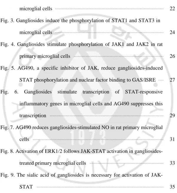
Fig. 1. Gangliosides stimulate iNOS transcription and nuclear factor binding to GAS/ISRE elements in microglial cells ··· 20 Fig. 2. Gel shift assay using anti-STAT1 and anti-STAT3 in BV2
microglial cells ··· 22 Fig. 3. Gangliosides induce the phosphorylation of STAT1 and STAT3 in
microglial cells ··· 24 Fig. 4. Gangliosides stimulate phosphorylation of JAK1 and JAK2 in rat
primary microglial cells ··· 26 Fig. 5. AG490, a specific inhibitor of JAK, reduce gangliosides-induced
STAT phosphorylation and nuclear factor binding to GAS/ISRE ···· 27 Fig. 6. Gangliosides stimulate transcription of STAT-responsive
inflammatory genes in microglial cells and AG490 suppresses this transcription ··· 29 Fig. 7. AG490 reduces gangliosides-stimulated NO in rat primary microglial
cells ··· 31 Fig. 8. Activation of ERK1/2 follows JAK-STAT activation in
gangliosides-treated primary microglial cells ··· 33 Fig. 9. The sialic acid of gangliosides is necessary for activation of
JAK-STAT ··· 35 Fig. 10. Asialo-GM1 does not activate the JAK-STAT signaling in primary
microglial cells ··· 36 Fig. 11. OxLDL suppressed iNOS and COX2 expression in LPS-activated
microglia ··· 39 Fig. 12. Effects of other modified LDLs on LPS-induced iNOS and COX2
expression in microglia ··· 40 Fig. 13. Relative agarose gel electromobility of modified LDLs ··· 42 Fig. 14. OxLDLs suppress LPS-induced iNOS expression and NO
production in an oxidation-dependent manner ··· 45 Fig. 15. OxLDLs suppress LPS-induced TNFα and MCP-1 secretion in an
oxidation-dependent manner ··· 47 Fig. 16. The inhibitory effects of oxLDL are not due to the displacement of
LPS from their cellular receptor ··· 50 Fig. 17. OxLDL inhibit LPS-induced intracellular signaling pathway in
microglia ··· 52 Fig. 18. OxLDL dose not inhibit LPS-induced MAPK pathway in microglia·· 53 Fig. 19. Individual components of oxLDL for their anti-inflammatory
effects ··· 56 Fig. 20. LXR agonists mimicked anti-inflammatory effect of oxLDL··· 58 Fig. 21. Oxysterols/LXR agonists suppress LPS-induced iNOS expression
and NO production in microglia··· 59 Fig. 22. Oxysterols/LXR agonists suppress LPS-induced TNFα and MCP-1
Fig. 23. RXR agonist potentiates anti-inflammatory effects of oxLDL on LPS-induced iNOS expression and NO production in microglia ··· 61 Fig. 24. RXR agonist potentiates anti-inflammatory effects of oxLDL on
LIST OF TABLES
Table 1. Primer sequences for PCR ··· 14 Table 2. Thiobarbituric acid-reacting substances and lipid hydroperoxide
ABBREVIATION
AAPH: 2,2’-Azobis(2-amidimopropane) Dihydrochloride AcLDL: Acetylated LDL
COX: Cyclooxygenase
ERK: Extracellular Signal-regulated Kinase GAS: Gamma Interferon Activated Site Gmix: Gangliosides Mixture
HC: Hydroxycholesterol
ICAM: Intercellular Adhesion Molecule IFN: Interferon
iNOS: Inducible NO Synthase
ISRE: Interferon-Stimulated Regulatory Element JAK: Janus Kinase
JNK: c-Jun N-terminal Kinase KC: Ketocholesterol
LDL: Low Density Lipoprotien LPO: Lipid Hydroperoxide LPS: Lipopolysaccharide LXR: Liver X Receptor MA: Methoprene Acid
oxLDL: Oxidized LDL
REM: Relative Electrophoretic Mobility RXR: Retinoid X Receptor
STAT: Signal Transducer and Activator of Transcription TBARS: Thiobarbituric Acid Reacting Substance
I. INTRODUCTION
A. Brain inflammation
Until recently, it has been thought that the brain is an immune-privileged organ, but there have been increasing numbers of reports of immune reactions in the brain. Glial cells, which include microglia and astrocytes, mainly play a role in immune reactions in the central nervous system (CNS). Microglia are a class of brain mononuclear phagocytes and are thought to be the principal immune cell resident to the CNS. Microglia have functions similar to those of other tissue macrophages, including phagocytosis, antigen presentation and production of cytokines, eicosanoids, complement components, excitatory amino acid (glutamate), proteinase and nitric oxide (Gehrmann et al., 1995; Streit et al., 1999). The inflammatory mediators from activated microglia protect brain from bacterial and viral infection. When the CNS is injured, microglia rapidly proliferate and migrate to the injured sites where they secrete inflammatory mediators. In this way, microglia are thought to protect neurons from external injuries. Of course, microglial activation can occur as a result of brain injury, and there is substantial evidence that microglial activation can aggravate brain injuries, resulting in neurodegenerative diseases. Activated microglia are observed in the brains of patients of Alzheimer’s disease (AD), multiple sclerosis, stroke and other neurodegenerative diseases (Benveniste, 1997; Breitner, 1996; Danton and Dietrich, 2003; Nelson et al., 2002). Because
microglia-mediated brain inflammation can participate in the initiation or progression of neurodegenerative disorders, it is important to understand not only their mechanisms of activation but also their functions.
B. Gangliosides and oxidized low density lipoprotein (oxLDL)
1. Gangliosides
Gangliosides are sialic acid-containing glycosphingolipids that are constituents of mammalian cell membrane. Gangliosides are particularly abundant in neuronal cell membrane and participate in various cellular events of nervous system (Riboni et al., 1997; Tettamanti and Riboni, 1994). The major types of gangliosides in the brain are GM1, GD1a, GD1b, GT1b and GQ1b, which differ in their profiles of sialic acid residues and carbohydrate moieties (Dreyfus et al., 1997; Tettamanti, 2004). Exogenously added gangliosides exert neuritogenic, neurotrophic and neuroprotective effects on a variety of cell systems of neural origin (Byrne et al., 1983; Facci et al., 1984). Several lines of evidence point to the importance of the brain-derived gangliosides in immune responses and pathogenesis of brain disease. There are reports that brain injury can cause release of gangliosides from damaged neuronal cells into the extracellular space, which may lead to pathophysiological conditions (Blennow et al., 1991; Gisslen et al., 1997; Michikawa et al., 2001). Gangliosides have been also reported to interact with β-amyloid (Aβ), suggesting
associated with Alzheimer’s disease (AD). In addition, gangliosides regulate the production of various inflammatory mediators, such as cytokines and inducible nitric oxide synthase (iNOS) (Ding et al., 1998; Oderfeld-Nowak and Zaremba, 1998). Despite the evidence of a role for gangliosides in brain pathology, little appears known regarding how gangliosides act.
2. OxLDL
(A) LDL in brain
Lipoproteins are macromolecular complex containing an envelope of phospholipids, some free cholesterol and a core of triglycerides or cholesteryl esters (Steinberg, 2002). The lipoproteins vary in origin, size, density in an aqueous environment, lipid and apolipoprotein content. The brain contains almost 25% of total body cholesterol, and lipoproteins also are present in the brain (Dietschy and Turley, 2001). LDL is present in the brain, largely as the result of cholesterol metabolism (Pitas et al., 1987). Although the cell types that produce and release lipoproteins in the brain has not yet been determined, there have been increasing numbers of reports of possibilities of astrocyte and microglia (Fujita et al., 1999; Mori et al., 2004; Saura et al., 2003; Xu et al., 2000). LDL in the brain is also supplied by cellular uptake from the circulation as well as in peripheral tissues. Although, the blood-brain barrier prevents diffusion of large molecules at the level of tight junctional attachments between adjacent capillary endothelial cells, surprisingly,
it has been shown that brain endothelial cells have the potential to take up LDL cholesterol through luminal LDL receptors and translocation of LDL across the cell (Dehouck et al., 1994; Dehouck et al., 1997).
(B) Oxidative modification of LDL
LDL is rapidly oxidized under certain conditions, which results in the formation of oxLDL. Although the oxidants responsible for in vivo oxidationof LDL are unknown at present, there are several candidates for oxidants including 15-lipoxygenase (Kuhn et al., 1994), myeloperoxidase (Daugherty et al., 1994), endothelial nitric oxide synthase (White et al., 1994), or transition metals (Balla et al., 1991; Ehrenwald and Fox, 1996; Heinecke et al., 1984). LDL within the brain is highly vulnerable to oxidative modification. CNS is particularly sensitive to oxygen radical damage because of poor antioxidant defense and an abundant supply of transition metals (Coyle and Puttfarcken, 1993). There are reports that oxidative stress is associated with neurodegenerative disorders, including Parkinson’s disease (PD), AD and Amyotorphic laterlal sclerosis (ALS). Markers for lipid peroxidation, such as 4-hydroxynonal (4-HNE) and malondialdehyde (MDA), have been identified in the cortex and hippocampus of patients with AD (Butterfield et al., 2002), the substantia nigra of patients with PD (Dexter et al., 1989) and in spinal fluid from patients with ALS (Pedersen et al., 1998). In addition, ischemic injury to brain is associated with disruption of blood-brain barrier (BBB), increasing the possibility of exposing the CNS to plasma LDL or oxLDL (del Zoppo and Hallenbeck, 2000).
Actually Uno et al. have shown raised plasma oxLDL in acute cerebral infarction (Uno et al., 2003). Together, these findings suggest that oxLDL may contribute to pathophysiology of brain.
(C) Biological functions of oxLDL
OxLDL are believed to play a critical role in atherosclerosis and exert diverse biological effects on different cell types. OxLDL stimulates endothelial cells to produce MCP-1 which recruits circulating monocytes into the intima (Cushing et al., 1990). Macrophages in the intima bind and take up oxLDL, and subsequently change themselves into foam cells (Itabe, 2003). OxLDL also induces atherosclerosis by stimulating monocyte infiltration and smooth muscle cell migration and proliferation (Mertens and Holvoet, 2001).
In addition to its well-established role in atherosclerosis, oxLDL exerts complex effects on inflammation. OxLDL has been demonstrated to modulate the expression of inflammatory cytokines in cell types present in the vascular wall and this activity is believed to be an important mechanistic feature of its pathophysiological action. It is interesting to note that oxLDL can modulate inflammatory gene expression in both positive and negative fashions. On the one hand, oxLDL induces the expression of several inflammatory mediators (Cushing et al., 1990; Lin et al., 2003; Terkeltaub et al., 1994). On the other hand, oxLDL antagonizes certain inflammatory gene expression initiated after LPS stimulation (Chung et al., 2000; Fong et al., 1991; Hamilton et al., 1990). Among the variables
that determine the nature of the effect are the cell type and the magnitude of LDL oxidation.
(D) Active components of oxLDL
OxLDL contains a complex, variable, incompletely characterized mixture of oxidation products. Both the diversity of product formation and observed cellular responses depend on the methodand time course of oxidation (Navab et al., 1996). OxLDL contains several lipid-derived bioactive molecules such as lysophosphatidylcholine (LPC), platelet-activating factor (PAF)-like bioactive lipids and oxysterols (Hajjar and Haberland, 1997; Heery et al., 1995; Huang et al., 1999). One well studied, stable end product includes LPC, which is generated by phospholipase A2 (PLA2) hydrolysis duringcellular oxidative modification of LDL (Quinn et al., 1988). LPC has been demonstrated to be a chemoattractant to monocytes and T lymphocytes (McMurray et al., 1993). PAF-like bioactive lipids have previously been implicated to play roles in vascular cell activation. Binding of PAF by theG protein-coupled PAF receptor leads to activation of heterotrimericG proteins linked to phosphoinositide–phospholipase C (PI-PLC) andtriggers a broad array of biological actions. PAF receptor antagonists blockthe ability of PAF-like phospholipids extracted from highly oxLDL to induce mitogenesis of smooth muscle cells (Heery et al., 1995). The cholesterol oxidation products, termed oxysterols, that are found in oxLDL include 7-ketocholesterol (Jialal et al., 1991) and 22 (R)-hydroxycholesterol (Fowler et al., 2003). Oxysterols may induce apoptosis in a wide
variety of cells, making them resonable candidates for the apoptotic activity of oxLDL. Recent studies suggest that oxysterols possess anti-inflammatory properties (Fowler et al., 2003; Liu et al., 1998).
C. Inflammatory signaling pathways
1. JAK-STAT pathway
JAK-STAT (Janus kinase-signal transducers and activators of transcription) signaling pathways have been reported to be involved not only in the immune response of numerous cytokines but also in the actions of primarily non-immune mediators such as growth factors and hormones. Specific subtypes of JAK and STAT molecules are activated by different signals, resulting in specificity of response (Igaz et al., 2001; Kishimoto et al., 1994). The binding of ligand to its receptor induces assembly of an active receptor complex and consequent phosphorylation of the receptor-associated JAKs (JAK1, JAK2, JAK3, TYK2). Phosphorylated JAKs lead to the activation of neighboring JAKs, receptor subunits and several other substrates. Phosphorylation of JAKs provides the docking sites for STATs, which in turn become phosphorylated on tyrosine and serine residues; phosphorylation of both amino acid species being required for full STAT activity. Phosphorylated STATs are released from the receptor complex and form dimers. These dimers translocate to the nucleus where they directly bind to IFNγ-activated sites (GAS) of specific target genes, thus regulating transcription of these genes such as ICAM-1 and MCP-1,
which are involved in immune responses (Ramana et al., 2000; Tessitore et al., 1998; Zhou et al., 2001). JAK-STAT signaling can specifically mediate the inflammatory pathways activated by various stimulators in the brain, and appropriate regulation of JAK-STAT intensity and duration can protect against inflammation-induced brain injury.
2. Lipopolysaccharide (LPS)–induced signaling pathways
LPS is an integral cell wall component of gram-negative bacteria that can provoke life-threatening inflammatory conditions through the sequential activation of intracellular signaling molecules (Aderem and Ulevitch, 2000; Ulevitch and Tobias, 1995). LPS binds to LPS-binding protein (LBP) in plasma and is delivered to the surface receptor CD14. Next, LPS is transferred to the transmembrane signaling receptor, toll-like receptor 4 (TLR4) and its accessory protein MD2. LPS stimulation activates several intracellular signaling pathways that include the transcription factor NF- B pathway and three mitogen-activated protein kinase (MAPK) pathways: extracellular signal-regulated kinases (ERK) 1 and 2, c-Jun N-terminal kinase (JNK) and p38 (Guha et al., 2001; Guha and Mackman, 2001). Recently, LPS has been found to activate the STAT inflammatory signaling cascade in macrophages and glial cells, leading to the massive production of inflammatory cytokines and causing multiple organ system failure and death (Dell'Albani et al., 2001; Jacobs and Ignarro, 2001). LPS activates NF-κB, leading to the induction and secretion of interferon-β (IFNβ). The secreted IFNβ then bind to interferon receptor, thereby leading to
phosphorylation of STAT1. The activated STAT1 then bind to the promoter of inflammation-associated genes, leading to the secretion of newly synthesized cytokines and nitric oxide (NO) and the induction of various inflammatory responses.
3. Liver X receptor (LXR) pathway as anti-inflammatory signaling pathway LXRs (LXRα and LXRβ) are members of the nuclear receptor superfamily and are activated by oxysterols and intermediates in the cholesterol synthetic pathway. LXRs form obligate heterodimers with retinoid X receptors (RXRs), which are members of the nuclear receptor superfamily that is regulated by 9-cis-retinoic acid (9cRA). Although originally identified as liver enriched transcription factors, the LXRs are now being intensely studied in various tissues as well as in liver. LXRs are also expressed in the brain, but their roles in tissue remain to be clarified. Although both LXR subtypes are expressed in the brain, LXRβ, in particular, is broadly expressed in the developing and adult rodent brain (Kainu et al., 1996). LXRs are able to regulate the expression of a number of genes involved in cholesterol metabolism (Mangelsdorf and Evans, 1995; Peet et al., 1998; Schultz et al., 2000). A recent report shows that LXRs have an important function in lipid homeostasis in the brain, and that loss of these receptors results in neurodegenerative diseases (Whitney et al., 2002). In addition to its importance in lipid metabolism, LXR activation has recently been demonstrated to regulate immune processes and to inhibit inflammatory gene expression in macrophage (Castrillo et al., 2003; Joseph et al., 2003). Also, Synthetic LXR agonists have been demonstrated to prevent
atherosclerosis in murine models and to inhibit inflammation (Joseph et al., 2003; Joseph et al., 2002).
D. Aims of study
The brain is abundant in lipids, and lipid metabolism is clearly related to brain functions such as synaptogenesis (Mauch et al., 2001), neuronal survival (Xu et al., 2000) and signal transduction (Brown and London, 1998). In this study, I investigated whether lipid derivatives, gangliosides and oxLDL, could modulate microglia-mediated brain inflammation. Because microglia-mediated brain inflammation participates in the initiation or progression of neurodegenerative disorders, it is important to understand their mechanism of activation. In part 1, I tested whether gangliosides activate JAK-STAT pathway, an essential inflammatory signaling pathway in microglia. In part 2, I studied the effects of oxLDL on microglia-mediated brain inflammation.
II. MATERIALS AND METHODS
A. Reagents
Bovine brain gangliosides mixture, GM1 and GD1a, was purchased from Matreya (Pleasant Gap, PA). Asialogangliosides GM1 wasfrom Sigma-Aldrich (St. Louis, MO). Human LDL was purchased from Calbiochem (La Jolla, CA) and Cu-oxLDL was purchased from Biomedical Technologies Inc. (Stoughton, MA). Rat IFN-γ, α-cyano-(3,4-dihydroxy)-N-benzylcinnamide (AG490), and PD98059 were from Calbiochem. Anthrobacter ureafaciens neuraminidase was purchased from Sigma. Salmonella typhimurium LPS and 7-ketocholesterol were purchased from Sigma-Aldrich. 22(R)-hydroxycholesterol and TO901317 were purchased from Cayman (Ann Arbor, MI). Methoprene acid was purchased from BIOMOL (Plymouth Meeting, PA). GW3965 was kindly provided by GlaxoSmithKline High Throughput Chemistry (Stevenage SG1 2NY, UK). Antibodiesagainst STAT1, Tyr-701-phosphorylated STAT1, Ser-727-phosphorylated STAT1, and Tyr-705-phosphorylated STAT3 were from Cell Signaling Technology (Beverly, MA). Antibodies against phosphorylated ERK, phosphorylated JNK and phosphorylated p38 were from Calbiochem. Antibodies againstphosphorylated JAK1 and -2 were from Affinity Bioreagents (Denver, CO). Antibodies (Abs) against inducible NO synthase (iNOS) was purchased from Upstate Biotechnology (Lake Placid, NY), and Abs against cyclooxygenase-2 (COX2) and actin were purchased from Santa Cruz
Biotechnology (Santa Cruz, CA).
B. Cell Culture
Primary microglia were cultured from the cerebral cortices of 1- to 3-day-old Sprague-Dawley rats as described previously (Pyo et al., 1999).Briefly, the cortices were triturated into single cells in minimal essential medium (MEM, Invitrogen, Carlsbad, CA) containing 10% fetal bovine serum (FBS, HyClone, Logan,UT) and plated in 75-cm2 T-flasks (0.5 hemisphere/flask) for 2-3 weeks. Microglia werethen detached from the flasks by mild shaking and filtered through a nylon mesh to remove astrocytes. Cells were plated in 6-well plates (7 × 10⁴ cells/well), 60-mm dishes (5 × 10⁵ cells/dish), or 100-mm dishes (106 cells/well). One hour later, the cells were washed to removeunattached cells before being used in experiments. BV2 immortalizedmurine microglia cells were from Dr. E. J. Choi. The BV2 cellline was grown in Dulbecco's modified Eagle's medium (DMEM, Invitrogen) and supplemented with 5% FBS. Cells were serum-starved overnightbefore treatment withgangliosides.
C. Electrophoretic Mobility Shift Assay (EMSA)
Cells were harvested and suspended in 9 times packaged cell volume of a hypotonic solution (10 mM HEPES, pH 7.9, 10 mM KCl,0.1 mM EDTA, 0.1 mM EGTA, 1 mM dithiothreitol (DTT), 0.5 mM phenylmethylsulfonylfluoride (PMSF)) including 0.5% Nonidet P-40. Cells were centrifugedat 500 × g for 10 min at 4 °C,
and the pellet (nuclear fraction)was saved. The nuclear fractions were resuspended in a buffercontaining 20 mM HEPES, pH 7.9, 20% glycerol, 0.4 M NaCl, 1 mM EDTA, 1 mM EGTA, 1 mM DTT, and 1 mM PMSF, incubated on ice for 60 min with occasional gentle shaking,and centrifuged at 12,000 × g for 20 min. The crude nuclear proteinsin the supernatant were collected and stored at 70 °C until used. EMSA was performed for 30 min on ice in a volume of 20 µl, containing4 µg of nuclear protein extract in a reaction buffer containing8.5 mM EDTA, 8.5 mM EGTA, 8% glycerol, 0.1 mM ZnSO4, 50 µg/mlpoly (dI-dC), 1 mM DTT, 0.3 mg/ml bovine serum albumin,6 mM MgCl2, and γ-32P-radiolabeled oligonucleotide probe (3 × 10⁴ cpm), with or without 20-50-fold excess unlabeled probe. In supershiftexperiments, protein extracts were incubated with 0.2-0.5 µg ofSTAT1 and STAT3 antibodies (Santa Cruz Biotechnology) for 30 minprior to the addition of 32P-labeled probe. DNA-protein complexes were separated on 6% polyacrylamidegels in Tris/glycine buffer. The dried gels were exposed to x-rayfilm. The following double-stranded oligonucleotide was used inthese studies: GAS/ISRE, 5'-AAG TAC TTT CAG TTT CAT ATT ACT CTA-3', 27bp (Santa Cruz Biotechnology, sc-2537). 5'-end-labeled probeswere prepared with 40 µCi of [γ-32P] ATP using T4 polynucleotide kinase (Promega, Madison, WI) and were purifiedon Quick Spin Columns Sephadex G-25 (Roche MolecularBiochemicals, Mannheim, Germany).
D. Reverse transcriptase-polymerase chain reaction (RT-PCR)
and cDNA was prepared using avian reverse transcriptase (Takara, Shiga, Japan), according to the manufacturer'sinstructions. PCR was performed with 30 cycles of sequential reactionsas follows: 94 °C for 60 s, 55 °C for 30 s, and 72 °C for 90 s. Oligonucleotide primers were purchased from Bioneer (Seoul, Korea). The sequences of PCR primers were shown on Table 1. PCR products were separated by electrophoresis on a 1.5% agarose gel and detected under UV light.
E. Western Blot Analysis
Cells were washed twice with cold phosphate-buffered saline and then lysed in ice-cold modified RIPA buffer (50 mM Tris-HCl, pH 7.4, 1% Nonidet P-40, 0.25% sodium deoxycholate, 150 mM NaCl, 1 mM Na3VO4, and 1 mM NaF) containing protease inhibitors (2 mM PMSF, 100 µg/ml leupeptin, 10 µg/ml pepstatin, 1 µg/ml aprotinin, and 2 mM EDTA). The lysates were centrifuged for 10 min at 12,000 × g at 4 °C, and the supernatant was collected. Proteins were separated by SDS-PAGE and transferred to a nitrocellulose membrane. The membrane was incubated with primary antibodies and peroxidase-conjugated secondary antibodies (Vector Laboratories, Burlingame, CA) and then visualized using an enhanced chemiluminescencesystem(Sigma-Aldrich).
F. Determination of NO Release
Media nitrite concentration was measured as an indication of NO release. Following the indicated cell incubations, 50 µl ofculture medium was removed and
mixed with an equal volume of Griessreagent (0.1% naphthylethylenediamine, 1% sulfanilamide, 2.5%H3PO4), and absorbance of the mixture at 540 nm wasmeasured.
G. Enzymatic Digestion of Sialic Acid
Neuraminidase derived from A. ureafaciens was used for cleaving sialic acids residues from gangliosides. Gangliosides were dissolved in 10 mM sodium acetate buffer, pH 5.0, containing 1µg of sodium cholate per µl and were incubated with A. ureafaciensneuraminidase (Sigma-Aldrich) at 37 °C for 2h.
H. Oxidation of LDL
I was prepared oxLDL using a standard method of 2,2’-azobis(2-amidimopropane) dihydrochloride (AAPH; Sigma-Aldrich)-mediated oxidation (Neuzil et al., 1998; Shie et al., 2004). Oxidation of LDL was performed at 37 °C under the following conditions to obtain different degrees of oxidation. First, LDL was oxidized by 10 mM AAPH for 2, 6, or 18 h. Second, oxidation of LDL was performed for 18 h at AAPH concentrations of 1, 5, or 10 mM. Produced oxLDLs were represented as “oxLDL-concentration of AAPH (mM): oxidation period (h)”.
I. Relative electrophoretic mobility (REM) assay
Electrophoretic mobility relative to LDL was measured by agarose gel (0.8% agarose in 0.08 mol/L Tris-HCl buffer, pH 8.3) electrophoresis and Coomassie Brilliant Blue R250 staining. This allows detection of changes in electric charge
induced by oxidation (Napoli et al., 1999; Sparks and Phillips, 1992).
J. Thiobarbituric acid reacting substances (TBARS) assay
200 µl of LDL/oxLDL (100 µg) was added to a test tube containing 200 µl SDS (8%, w/v), 400 µl acetic acid (20%, w/v), and 400 µl of thiobarbituric acid (0.8%, w/v). The mixture was vortexed well and boiled for 1 h. After cooling, the specimens were centrifuged (13,000 rpm, 10 min) and the absorbance of the supernatant was determined at 540 nm using a spectrophotometer. The amount of TBARS was determined by comparison to a standard of malondialdehyde (MDA) equivalents prepared using 1,1,3,3-tetraethoypropane (Sigma-Aldrich).
K. Lipid hydroperoxide (LPO) assay
The LPO level in oxLDL was determined by a LPO assay kit provided by Cayman. 100 µg LDL/oxLDL were used for LPO measurements. The absorbance at 500 nm was measured using a spectrophotometer (Amersham Pharmacia Biotech, San Francisco, CA).
L. Enzyme-linked immunosorbent assay (ELISA)
TNFα and MCP-1 levels in cell culture media were determined by ELISA as described by the manufacturer (OptEIA Sets, Pharmingen, San Diego, CA). TNFα and MCP-1 concentrations in the media were determined by spectrophotometer and calibrated from standards containing known concentrations of the cytokines.
M. Data analysis
Data were expressed as mean ± S.E.M. Analysis of variance followed by Dunnett’s multiple comparison tests were used for statistical comparisons.
III. RESULTS
A. JAK-STAT Signaling Mediates Gangliosides-induced Inflammatory Responses in Brain Microglial Cells
1. Gangliosides Induce Nuclear Factor Binding to GAS/ISRE Elements
Functional GAS/ISRE elements are found in the promoter regions of several inflammation-related genes, such as iNOS, and theseelements are known to bind the phosphorylated STAT dimer. In an attempt to explore the molecular mechanism of gangliosides on microglial activation, I investigated whether STATs could be involved in gangliosides-induced activation of microglia. I first examined the transcript level of iNOS in gangliosides-treatedrat primary microglia. Gangliosides markedly induced iNOS mRNAwithin 1 h (Fig. 1A, a), suggesting that gangliosides directly regulate NOproduction at the level of transcription. This observation was subsequently evaluated by EMSA using a γ-32P-labeled consensus GAS/ISRE oligonucleotides probe. After the cells were treated with 50 µ g/ml brain-derived gangliosides mixturefor the indicated times, nuclear extracts were prepared and then analyzed by EMSA. The specific binding complex was detected innuclear extracts from gangliosides-treated rat primary and murineBV2 microglia (Fig. 1, A(b) and B). Time course analysis showed thatgangliosides rapidly induced the nuclear factor binding within5 min and that the binding activity was decreased to basal levelsafter
Fig. 1. Gangliosides stimulate iNOS transcription and nuclear factor binding to GAS/ISRE elements in microglial cells. (A) Rat primary microglial cells were treated with or without 50 µg/ml brain gangliosides mixture (Gmix) for 1 h, after which total RNA was isolated, and levels of iNOS mRNA were measured using an RT-PCR-based assay. The transcription of GAPDH was measured for normalization (a). Cells were treated with Gmix for 5 min, after which nuclear extracts were prepared and assayed for the amount of binding activity to GAS/ISRE oligonucleotides using EMSA (b). (B) BV2 cells were treated with 50 µg/ml Gmix for the indicated periods. Nuclear extracts were prepared, and binding activity to GAS/ISRE oligonucleotides was determined by EMSA.
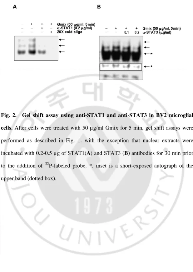
Fig. 2. Gel shift assay using anti-STAT1 and anti-STAT3 in BV2 microglial cells. After cells were treated with 50 µ g/ml Gmix for 5 min, gel shift assays were performed as described in Fig. 1. with the exception that nuclear extracts were incubated with 0.2-0.5 µ g of STAT1(A) and STAT3 (B) antibodies for 30 min prior to the addition of 32P-labeled probe. *, inset is a short-exposed autograph of the upper band (dotted box).
30 min in both microglial cell types (Fig. 1B). The specificity of the shifted bands was confirmed by competition assay using excess amounts of unlabeled oligonucleotides(cold oligo). In addition, gel shift assay showed that the binding complex was diminished by addition of anti-STAT1 and anti-STAT3,indicating that both STAT1 and STAT3 are constituents of the nuclearfactor binding complex (Fig. 2). These results show that functional GAS/ISRE elements may be involved in gangliosides-induced activationof microglia.
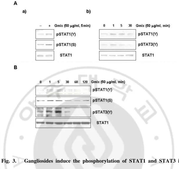
2. Gangliosides Induce the Phosphorylation of STAT1 and STAT3
Essential roles for STAT signaling in brain inflammatory response have emerged. Because gangliosides rapidly induced the GAS/ISRE-nuclear factor binding, I examined whethergangliosides indeed caused phosphorylation of STAT proteins. Primarymicroglial cells were stimulated with 50 µg/ml gangliosides forthe indicated times, and the levels of phosphorylated STAT1 were determined by Western blot analysis using antibodies against Tyr-701-STAT1and Ser-727-STAT1. Both phosphorylations of STAT1 occurred within5 min of gangliosides addition (Fig. 3A, a). Similar patterns of phosphorylation were observed in lysatesfrom murine BV2 microglial cells, where incubation of cells withgangliosides resulted in STAT1 phosphorylation on tyrosine and serine residues, with phosphorylation levels returning to basalat 30 min (Fig. 3B). In addition to phosphorylation of STAT1,I detected gangliosides-induced phosphorylation of STAT3 in both microglial cell types. The pattern of STAT3 tyrosine phosphorylationappeared similar to that of
Fig. 3. Gangliosides induce the phosphorylation of STAT1 and STAT3 in microglial cells. Rat primary microglial cells (A) and mouse BV2 microglial cells (B) were serum-starved for 12 h and then stimulated with 50 µg/ml Gmix for the indicated times. Cell lysates were separated by 10% SDS-PAGE and Western blots probed with anti-pSTAT1 (Y), anti-pSTAT1 (S), or pSTAT3 (Y). The membrane was then stripped and analyzed with anti-STAT1 antibody to determine loading.
STAT1 phosphorylation (Fig. 3, A andB). The Western blotting data show that gangliosides trigger rapidphosphorylation of STAT1 and STAT3, suggesting their involvement in gangliosides-induced microglial activation. The phosphorylation patterns of both STAT1 and STAT3 determined by Western blottingcorrelate with the binding activity results from EMSA.
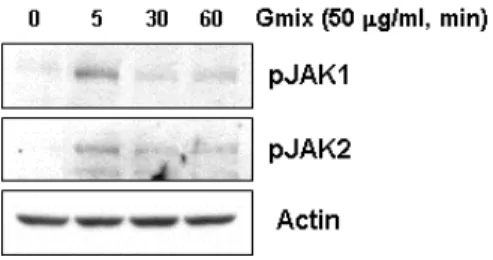
3. Gangliosides Induce Phosphorylation and Activation of JAK1 and JAK2 Phosphorylation of STATs depends on the activation of JAKs. JAKs both functionally and physically associate with cytokinesignaling. In particular, activation of JAK1 and JAK2 providesa molecular explanation for cellular actions of a broad rangeof cytokines. Thus, I investigated whether JAK1and JAK2 could be involved in gangliosides-induced STAT phosphorylation. Primary rat microglial cells were stimulated with 50 µg/ml gangliosidesfor the indicated times, and cell lysates were Western blottedusing antibodies directed against phosphorylated JAK1 and JAK2. The data presented in Fig. 4 show that following addition ofgangliosides to cells, phosphorylation of both JAK1 and JAK2 occurred within 5 min, after which phosphorylation levels returned to basallevels by 30 min. The involvement of JAK signaling in gangliosides-induced microglial activation was also shown using a second, independent approach. The pharmacological agent AG490 is known to inhibitthe phosphorylation of both JAK1 and JAK2. I found thatpretreatment of rat primary microglial cells with AG490 effectively reduced gangliosides-induced phosphorylation of STAT1 and STAT3(Fig. 5A). In addition, AG490 inhibited the
Fig. 4. Gangliosides stimulate phosphorylation of JAK1 and JAK2 in rat primary microglial cells. Cells were serum-starved for 12 h and then stimulated with 50 µg/ml Gmix for 5 min. The phosphorylation of JAK1 and JAK2 was determined by Western blot analysis using antibodies specific for phosphoJAK1 or -JAK2.
Fig. 5. AG490, a specific inhibitor of JAK, reduce gangliosides-induced STAT phosphorylation and nuclear factor binding to GAS/ISRE. (A) Cells were pretreated with 10 µM AG490 for 1 h and then stimulated with 50 µg/ml Gmix for 2 min. Western blots were probed with anti-pSTAT1 (Tyr-701) and pSTAT3 (Tyr-705). The membrane was subsequently stripped and probed with anti-STAT1 and STAT3 antibodies. (B) Cells were pretreated with 10 µM AG490 for 1 h and then stimulated with 50 µg/ml Gmix for 5 min. Nuclear extracts were prepared, and binding activity to GAS/ISRE oligonucleotides was determined by EMSA. *, inset is a short-exposed autograph of the upper band (dotted box).
nuclear factor bindingto GAS/ISRE nucleotides in gangliosides-treated microglial cells(Fig. 5B). These results indicate that gangliosides induce phosphorylationand activation of STAT1 and STAT3 through phosphorylation andactivation of JAK1 and JAK2.
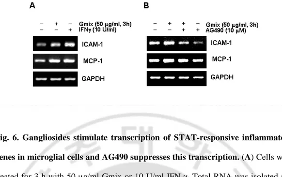
4. Gangliosides Stimulate STAT-responsive Inflammatory Gene Expression Brain inflammatory responses are coordinated by the production of cytokines, chemokines, and reactive oxygen species. The above data indicate that gangliosides-induced microglial activationmay be mediated, at least in part, by JAK-STAT-dependent transcriptionalresponses. Therefore, I examined the transcript level of genesthat have been reported previously to have functionalGAS elements and act as mediators of inflammation, namely MCP-1 and ICAM-1. Rat primary microglial cells and BV2 cells were stimulatedwith 50 µg/ml gangliosides for 3 h, and total RNA was extractedfor RT-PCR analysis. Addition of gangliosides rapidly increased the mRNA levels of both MCP-1 and ICAM-1, as did IFN-γ, whichwas included as a positive control (Fig. 6A). Pretreatment with AG490 significantly inhibited gangliosides-induced transcription of both genes (Fig. 6B). These findings demonstrate that gangliosidestrigger STAT-dependent transcriptional activation of inflammatorygenes in microglia.
5. AG490 Reduces Gangliosides-induced Release of NO
Fig. 6. Gangliosides stimulate transcription of STAT-responsive inflammatory genes in microglial cells and AG490 suppresses this transcription. (A) Cells were treated for 3 h with 50 µg/ml Gmix or 10 U/ml IFN-γ. Total RNA was isolated and analyzed for levels of MCP-1 and ICAM-1 mRNA using an RT-PCR-based assay. The transcription of GAPDH was measured for normalization. (B) Cells were pretreated with 10 µM AG490 for 1 h and then stimulated with 50 µg/ml Gmix for 3 h. mRNA expression of ICAM-1 and MCP-1 was detected using an RT-PCR-based assay.
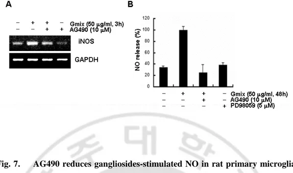
Aberrant iNOS expression and excessive NO production are observed in various pathophysiological conditions. Previously, I showed that gangliosides-induced microglialactivation was accompanied by induction of NO release. Thus, Itested whether gangliosides induced NO release via JAK-STAT signaling.First, I examined the effect of JAK inhibition on gangliosides-inducedtranscription of iNOS in rat primary microglial cells. RT-PCRanalysis showed that the inhibitor AG490 reduced mRNA levels ofiNOS (Fig. 7A). Second, I investigated the effect of AG490 onNO release. In these studies, the ERK inhibitor, PD98059, wasalso used since I have shown previously that it reducedgangliosides-induced NO release (Pyo et al., 1999). In the presence of AG490, microglialcells were treated with 50 µg/ml gangliosides for 48 h, and theamount of NO produced was determined by measuring the amount of nitrite converted from NO in the media. AG490 significantly reduced gangliosides-enhanced NO release, as did PD98059 (Fig. 7B). Comparedwith cells treated with gangliosides alone, NO release was reduced to 38.6 ± 4.3 and 25.2 ± 14% in cells co-treated with PD98059and AG490, respectively. These results are consistent with theresults shown in Figs. 5 and 6. The findings indicate that JAK-STAT signaling is required for NO release and provide evidence of the critical functional involvement of JAK-STAT signaling in gangliosides-inducedmicroglial activation.
6. ERK Activity Appears to be Regulated by JAK Activation
Fig. 7. AG490 reduces gangliosides-stimulated NO in rat primary microglial cells. (A) Cells were pretreated with 10 µM AG490 for 1 h and then stimulated with 50 µg/ml Gmix for 3 h. mRNA expression of iNOS was detected by RT-PCR analysis. The transcription of GAPDH was measured for normalization. (B) Cells were treated with 50 µ g/ml Gmix for 48 h in the presence or absence of AG490 or PD98059. The amount of NO was determined by measuring the amount of nitrite in the media, as described under "Experimental Procedures.".
is regulated through mitogen-activated protein kinases (MAPKs). MAPKs are considered as common intracellular signaling molecules involved in microglial activation. Previous reports by others and us showed that gangliosides induced activation of MAPKs in microglia. In the present study, I used pharmacological inhibitors to examine possible cross-talk between the JAK-STAT and MAPKs signaling pathways. When primaryrat microglial cells were pretreated for 2 h with the JAK inhibitor AG490, gangliosides-induced activation of ERK1/2 was significantlyreduced compared with controls with no AG490 (Fig. 8). In contrast,no significant suppression of p38 was observed under this condition.However, in the presence of PD98059, an ERK inhibitor, not onlyERK but also p38 activation was completely inhibited. These results indicate that gangliosides-stimulated JAK activation leads to activation of ERK in microglial cells. These pharmacological studiesalso indicate that gangliosides-stimulated activation of p38 maynot be due to activation of ERK by JAK.
7. Sialic Acid Residues Are Important for Gangliosides-induced Phosphorylation of STAT
The major types of gangliosides in brain are GM1, GD1a, GD1b, GT1b, and GQ1b. These gangliosides differ with respect to thenumber and position of sialic acid residues attached to the carbohydrates. The approximate percentages of each gangliosides presentin the brain gangliosides mixture used in the current study are 18% GM1, 55% GD1a, 15% GD1b, 10% GT1b, and 2% others. To addresswhether
Fig. 8. Activation of ERK1/2 follows JAK-STAT activation in gangliosides-treated primary microglial cells. Primary microglial cells were pregangliosides-treated with AG490 or PD98059 for 1 h and then treated with 50 µg/ml Gmix for 30 min. Cell lysates were separated by 10% SDS-PAGE and Western blots probed with anti-phospho-ERK and anti-phospho p38, respectively. The membrane was then stripped and probed with anti-ERK antibody. At least four experiments were independently performed, and representative data are shown in this figure.
the structural diversity of gangliosides affected activationof STAT, I compared the effect of GM1, which has one moleculeof sialic acid, with GD1a, which has two molecules of sialic acid, on phosphorylation of STAT1. Primary microglial cells were treated with GM1 or GD1a for 2 min, and levels of phosphorylated STAT1 were determined by Western blot analysis using antibodies againstTyr-701-STAT1. The data in Fig. 9 show both GM1 and GD1a stimulatedphosphorylation of STAT1 within 2 min. The level of STAT1 phosphorylation stimulated by either GM1 or GD1a was similar to that caused bythe gangliosides mixture, suggesting that the number of sialic acid residues per gangliosides molecule has little effect on the phosphorylation of STAT1 in microglial cells (Fig. 9A). Becausesialic acid residues are characteristic of gangliosides, I examined whether sialic acid residues were important for gangliosides-stimulated STAT phosphorylation. Gangliosides were preincubated with either550 or 1000 units/ml A. ureafaciens neuraminidase, which is known to release sialic acid attached to an internal galactose in anygangliosides including GM1. Primary microglia cells were stimulated with gangliosides or neuraminidase-treated gangliosides(desialylated gangliosides) for 2 min, and levels of phosphorylated STAT1 were determined by Western blot analysis. The data presented in Fig. 9, B and C show a dose-dependent inhibitory effect of neuraminidasetreatment on phosphorylation of STAT1, indicating that sialic acid residues are required for stimulation of JAK-STAT signaling. To rule out the possibility that these reductions are due to contaminatingsialic acid or neuraminidase, I compared the effect of GM1 and asialo-GM1 (Sigma) on phosphorylation of
Fig. 9. The sialic acid of gangliosides is necessary for activation of JAK-STAT signaling. (A) Primary microglial cells were treated with 20 µ g/ml GM1 or GD1a for 2 min. Cell lysates were subjected to Western blot analysis, and levels of phosphorylated STAT1 were determined using anti-pSTAT1 (Tyr-701). (B and C) To remove the sialic acid residue, gangliosides were preincubated with 550 or 1000 units/ml A. ureafaciens neuraminidase as described under "Experimental Procedures." Cells were treated with the indicated gangliosides or the desialylated gangliosides for 2 min, after which cell lysates were prepared, separated by 10% SDS-PAGE, Western blotted, and probed using anti-pSTAT1 (Tyr-701). The membrane was then stripped and probed with anti-STAT1 antibody.
Fig. 10. Asialo-GM1 does not activate the JAK-STAT signaling in primary microglial cells. (A) Primary microglial cells were treated with 20 µ g/ml of GM1 or asialo-GM1 for 2 min. Cell lysates were subjected to Western blot analysis, and levels of phosphorylated STAT were determined using pSTAT1 and anti-pSTAT3. (B) Total RNA was isolated and analyzed for levels of iNOS, MCP-1, and ICAM-1 mRNA using an RT-PCR-based assay.
STAT and transcriptionof STAT-responsive genes. Consistent with Fig. 9, not only the phosphorylation of STAT1 and STAT3 but also the transcriptions of iNOS, MCP-1, and ICAM-1 were not induced in asialo-GM1-treated primary microglial cells (Fig. 10). Taken together, these resultssuggest that the presence of sialic acid residues is importantfor gangliosides-stimulated JAK-STAT signaling, although the number of sialic residues per gangliosides molecule may not influence phosphorylation.
B. Oxidized Low Density Lipoprotein (oxLDL) Suppresses Lipopolysaccharide (LPS)–induced Inflammatory Responses in Microglia.
1. OxLDL Potently Inhibits LPS-induced Expression of iNOS and COX2 in Primary Microglia Cultured from Rat Brain.
To investigate the role of oxLDL on neuroinflammation, I first examined the effectof LDL or modified LDLs on the expression of iNOS and COX2, key enzymes in inflammatory processes in activated microglia. For the experiment, I prepared oxLDL using a standard method of AAPH-mediated oxidation (Neuzil et al., 1998; Shie et al., 2004). LDL was oxidized under 10 mM AAPH for 18 hr at 37℃. LDL or modified LDLs treatment for 24hr itself had no effect on iNOS and COX2 expression in rat primary microglia. But pretreatment of oxLDL for 1hr, but not LDL and AcLDL, markedly suppressed LPS-induced iNOS and COX2 expression in microglia (Fig. 11A and Fig 12). These anti-inflammatory effects of oxLDL were dose dependent (Fig. 11B). The inhibition effect of oxLDL was also observed when microglia was activated by gangliosides (Fig. 11C). Although oxLDL has been reported to show cytotoxic effects in other cells(Han and Pak, 1999; Sugawa et al., 1997), 50 µg/ml or below oxLDL had little effect oncell viability in our experiments. Thus, I used 50 µg/ml oxLDL for subsequent experiments.
2. OxLDL Suppresses the Expression and Release of Inflammatory Mediators in an Oxidation-dependent Manner.
Fig. 11. OxLDL suppressed iNOS and COX2 expression in LPS-activated microglia. Rat primary microglia were first incubated with LDL or oxLDL (50
µg/ml) for 1h and then activated with LPS (10 ng/ml) (A) or Gmix (50 µg/ml) (C) for further 24h. (B) Microglia were pretreated with oxLDLs at indicated doses for 1h, and stimulated with LPS (10 ng/ml) for 24h. Then, cell lysates were separated using 10% SDS-PAGE, and Western blot analysis was performed using antibodies against iNOS and COX2. Actin was used as a loading control. Data shown are representative of three independent experiments.
Fig. 12. Effects of other modified LDLs on LPS-induced iNOS and COX2 expression in microglia. Microglia were pretreated with indicated LDLs for 1h, and stimulated with LPS (10 ng/ml) for 24h. Then, cell lysates were separated using 10% SDS-PAGE, and Western blot analysis was performed using antibodies against iNOS and COX2. Actin was used as a loading control. Data shown are representative of three independent experiments. Ac-LDL: acetylated LDL; Cu-oxLDL: oxidized LDL by Cu2+; AAPH-oxLDL: oxidized LDL by AAPH.
To better define the suppressive effect of oxLDL on brain inflammation, I tested the effects of oxLDLs with different degree of oxidation. LDLs were oxidized under various conditions to achieve different levels of oxidation, and I characterized the oxidation content of oxLDL using several methods. First, the oxidative modification of LDL was determined by REM assay. Alteration of electrophoretic mobility on the agarose gel reflects the increase of negative charge in LDL particle after oxidation. OxLDL not only migrates faster but is also less visible with Commassie Blue stain because of partial degradation of LDL protein. As the REM data in Fig. 13B show, the increase in electrophoretic mobility corresponded with the oxidation period (1.1, 1.4 and 2.3) and AAPH concentration (1.1, 1.7 and 2.3) in oxLDLs. Traditionally, lipid peroxidation is quantified by measuring malondialdehyde (MDA), the degradation products of polyunsaturated fatty acids (PUFAs) hydroperoxides (Esterbauer et al., 1991; Janero, 1990). As oxidation progressed, MDA content of oxLDL preparations increased gradually with oxidation period- and AAPH concentration-dependent manner as measured by TBARS assay (Table 2). The better parameters for LDL oxidation were the content of lipid hydroperoxides. Because TBARS assays use by-products as indicators of lipid peroxidation, I performed direct measurement of lipid hydroperoxide (Mihaljevic et al., 1996). As Table 2 show, LPO content of oxLDL preparations increased gradually with oxidation period- and AAPH concentration-dependent manner as measured by direct LPO assay.
Fig. 13. Relative agarose gel electromobility of modified LDLs. LDLs were oxidized with various concentrations of AAPH for indicated time periods. After incubation, 50 µg of LDLs were loaded onto 0.8 % agarose gel for electrophoresis. The gel was stained with Coomassie brilliant blue R-250 (a), and the migration distance was measured (b). The stained gel is representative of three independent oxLDL preparations.
levels of oxidation on LPS-induced inflammatory responses. First, LDLs were oxidized for three different time periods (2, 6 and 18 hr) in 10 mM AAPH (Fig. 14, left panel). As shown in Fig. 14A, oxLDL reduced LPS-induced iNOS and COX2 expression with oxidation period-dependent pattern. Also, LPS-mediated NO production was inhibited by oxLDLs pretreatment in oxidation period-dependent manner. The NO inhibition content of oxLDL-2hr, -6hr and -18hr were roughly 10, 50 and 70%, respectively (Fig. 14B). Next, I tested effects of oxLDLs on LPS-induced cytokine and chemokine secretion. As the ELISA data in Fig. 15A and 15B show, the LPS-induced secretion of both TNFα and MCP-1 also were reduced in the oxLDL pretreated microglia with oxidation period-dependent manner. LPS-induced TNFα secretions were inhibited by 13.7, 26.8 and 48.3%, respectively by the pretreatment of oxLDL-2hr, -6hr and -18hr (Fig. 15A). In the case of MCP-1, the inhibition content of oxLDL-2hr, -6hr and -18hr were 13.2, 37.1 and 67.5, respectively (Fig. 15B).
Second, I performed experiments as mentioned above using oxLDLs that oxidized for 18 hr in three different concentration of AAPH (1, 5 and 10 mM) (Fig. 14, right panel). OxLDLs suppressed LPS-induced iNOS and COX2 expression with AAPH concentration-dependent pattern (Fig. 14A). Also, NO production was inhibited by oxLDLs pretreatment in AAPH concentration-dependent manner. The NO inhibition content of oxLDL-1mM, oxLDL-5mM and oxLDL-10mM were roughly 20, 60 and 70%, respectively (Fig. 14B). LPS-induced TNFα secretions were inhibited by 9.6, 20.8 and 60.2%, respectively by the pretreatment of oxLDL-1mM, -
Fig. 14. OxLDLs suppress LPS-induced iNOS expression and NO production in an oxidation-dependent manner. Left panel, Rat primary microglia were pretreated with oxLDLs (50 µg/ml) that were oxidized with AAPH for indicated times and then stimulated with LPS (10 ng/ml) for 24h. Right panel, microglia were pretreated with oxLDLs (50 µg/ml) that were oxidized with indicated doses of AAPH for 18h and stimulated with LPS (10 ng/ml) for 24h. (A) Cell lysate was isolated and analyzed for levels of iNOS and COX2 protein using a Western blot analysis. (B) Media nitrite concentration was measured as an indication of NO release, using Griess reagent. Data represent the mean ± S.E.M. of three independent experiments. Significantly different from the LPS group by Dunnett’s multiple range test, *P<0.05; **P<0.01.
Fig. 15. OxLDLs suppress LPS-induced TNFαααα and MCP-1 secretion in an oxidation-dependent manner. Left panel, Rat primary microglia were pretreated with oxLDLs (50 µg/ml) that were oxidized with AAPH for indicated times and then stimulated with LPS (10 ng/ml) for 24h. Right panel, microglia were pretreated with oxLDLs (50 µg/ml) that were oxidized with indicated doses of AAPH for 18h and stimulated with LPS (10 ng/ml) for 24h. Release of TNFα (A) and MCP-1 (B) were determined by ELISA. Data represent the mean ± S.E.M. of three independent experiments. Significantly different from the LPS group by Dunnett’s multiple range test, *P<0.05; **P<0.01.
5mM and -10mM (Fig 15A). In the case of MCP-1, the inhibition content of oxLDL-1mM, -5mM and -10mM were 14.8, 60.2 and 68.9%, respectively (Fig.15B).
REM assay revealed 3-fold increase in electrophoretic mobility in AcLDL than in LDL (Fig. 13A). LPO production level was similar in AcLDL and LDL, although MDA production was found to be higher (about 6-fold) in AcLDL in TBARS assay (Table 2). Cu-oxLDL, another modified LDL which did not have a suppressive effect against the LPS-induced iNOS expression, showed no differences in electronegativity (Fig. 13A) and LPO content, while showed significant up-regulation of MDA content as shown in Table 2. The MDA content of Cu-oxLDL was 18 µM/mg, which was similar with oxLDL5:18 having sufficient suppressive effect. Based on these results, suppressive effect of AAPH-oxLDL is considered to be elicited by oxidation (especially on LPO content) increased in proportion to oxidation time and AAPH concentration.
3. Inhibitory Effects of OxLDL are Mediated by Interference with LPS-induced NFκB Activation, IFNβ Release, and STAT1 activation in Brain Microglia.
The next question I addressed was how oxLDL pretreatment inhibited LPS-induced inflammatory responses. The first possibility was that oxLDL simply blocked LPS binding on their receptor.This possibility was tested by adding oxLDL at times aftertreatment of LPS.I added oxLDL into cellsat various time points, from 1 hr pretreatment to 1 hr aftertreatment of LPS, and incubated for indicated times. The suppressive effects of oxLDL on expression of iNOS and COX2 were
Fig. 16. Theinhibitory effects of oxLDL are not due to the displacementof LPS from their cellular receptor. (A) OxLDL (50 µg/ml) was added to microglia 1h before, simultaneously with or after (0.5 or 1h) LPS (10 ng/ml). iNOS, COX2 and Actin were determined in microglia lysates by Western blot analysis. (B) Rat primary microglia were stimulated with LDL or oxLDL (50 µg/ml) for 8 hrs. Total RNA was isolated and analyzed for levels of genes of oxLDL receptors using an RT-PCR assay.