2011년도 추계학술발표회 논문요약집
대한방사선방어학회
212
_http://www.karp.or.kr
The potential impact of low dose ionizing ɤ-radiation on
immune response activity up-regulated by Ikaros in IM-9 B
lymphocytes
Sung Jin Kim, Seon-A Jang, Kwang Hee Yang*, Ji-Young Kim, Cha Soon Kim,
Seon Young Nam, Meeseon Jeong, Young-Woo Jin
Radiation Health Research Institute, Korea Hydro & Nuclear Power Co., LTD, Seoul, Korea
E-mail: [email protected]
Key words : Low dose ionizing radiation (LDIR), Ikaros, Autotaxin (ATX), Immune activation, Lysophosphatidylcholine
(LPC), Lysophosphatidic acid (LPA)
Abstract
The biological effects of low dose ionizing radiation (LDIR) remain
insufficiently understood. We examined for the scientific evidence to
show the biological effects of LDIR using radiation-sensitive immune
cells. We found that Ikaros protein was responsed to low
dose-dependent effects of gamma radiation in IM-9 B lymphocytes.
Ikaros encodes zinc finger transcription factors that is important
regulators of a hematopoietic stem cells (HSCs) progression to the B
lymphoid lineage development, differentiation and proliferation. In this
study, we observed that cell proliferation was enhanced from 10% to
20% by LDIR (0.05 Gy) in IM-9 B lymphocytes. The Ikaros protein
was phosphorylated in its serine/threonine (S/T) region and decreased
its DNA binding activity in the cells exposed to LDIR.
We found that Ikaros phosphorylation was up-regulated by
CK2/AKT pathway and the residues of ser-304 and ser-306 in Ikaros
was phosphorylated by LDIR. We also observed that Ikaros protein
was localized from the nucleus to the cytoplasm after LDIR and
bound with Autotaxin (ENPP2, ATX) protein, stimulating proliferation,
migration and survival of immune cells. In addition, we found that the
lysoPLD activity of ATX was dependent on Ikaros-ATX binding
activity.
These results indicate that the Ikaros is an important regulator of
immune activation. Therefore, we suggest that low dose ionizing
radiation can be considered as a beneficial effects, stimulating the
activation of immune cells.
Materials and Methods
Human B lymphoblastoid cell line IM-9 and human embryonic
kidney cell line 293T were used. The cells were then uniformly
irradiated at room temperature with various doses of a
137Cs γ-source
(dose rate of 5.41 Gy/min or 0.01 Gy/hour). siRNA of target genes
were used by Amaxa nucleofector kit to knock down a transcription
level in IM-9 lymphocytes. Cell proliferation was investigated using
MTT, XTT and BrdU assay. Ikaros-DNA interactions were examined
by Electrophoretic Mobility Shift Assays (EMSA). Expression of target
protein was determined using western blot. ATX/lyso-PLD activity
(convert LPC into LPA) in the conditioned medium was measured
using FS-3, a fluorescent LPC analogue, as substrate.
Results
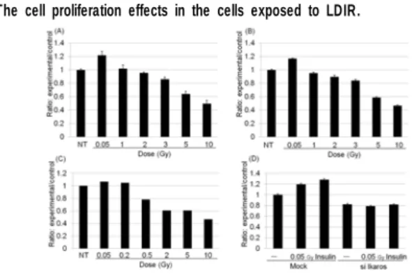
The cell proliferation effects in the cells exposed to LDIR.
Fig. 1. The effects of cell proliferation by LDIR in IM-9 B lymphocytes.
IM-9 B lymphocytes exposed to various doses of radiation were cultured for 24 hours. And then the cells were evaluated for cell proliferative capacity using by (A) MTT assay, (B) BrdU assay and (C) Nucleo counter assay. (D) IM-9 B lymphocytes were transfected with control siRNA or Ikaros siRNA for 9 hours and then the cell were exposed to LDIR. Insulin was used as a positive control. Cell proliferation was determined by BrdU.
The regulation of Ikaros by phosphorylation in the cells exposed to
LDIR.
Fig. 2. The alteration of Ikaros phosphorylation, Ikaros DNA binding activity and localization of Ikaros in IM-9 B lymphocytes exposed to LDIR.
(A) The Ikaros was phosphorylated by chronic LDIR (0.01 Gy, Dose rate: 0.01 Gy/hour) in IM-9 B lymphocytes. Cells were irradiated with chronic 0.01 Gy, incubated for various times, and the phosphorylation of Ikaros was detected via Ikaros immunoprecipitation (IP) and then Western blotting was conducted with p-S/T antibody. (B) The DNA-binding activity of Ikaros determined by EMSA in IM-9 B lymphocytes exposed to chronic LDIR (0.01Gy). Position of the Ikaros-DNA complexe in the EMSA experiment is indicated by arrow. (C and D) Ikaros is translocated from the nucleus to the cytoplasm by acute LDIR (dose rate of 5.41 Gy/min) in IM-9 B lymphocytes. (C) IM-9 B lymphocytes were fractionated into their nuclear and cytosolic components and subjected to western blotting analysis using an anti-Ikaros antibody. Cell fractionation was confirmed by western blot using anti-Lamin A/C antibody and anti-GAPDH antibody. (D) Cells were fixed, permeabilized, and immunostained with anti-Ikaros primary antibody and FITC-conjugated secondary antibody. Nuclei were visualized with DAPI.
2011년도 추계학술발표회 논문요약집
대한방사선방어학회
제5분과(의료 및 생물)_
213
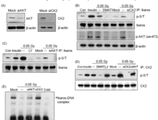
The CK2/AKT/Ikaros signaling pathway in the cells exposed to LDIR.
Fig. 3. Up-regulation of Ikaros activation pathway by CK2, AKT/PKB and recovery of Ikaros DNA binding activity by AKT/PKB and CK2 siRNA transfection in IM-9 B lymphocytes exposed to LDIR.
(A) After the transfection of IM-9 lymphocytes with AKT siRNA and CK2 siRNA, the effects of AKT and CK2 knock-down were identified via Western blotting. (B, C and D) The phosphorylation of Ikaros was detected via Ikaros immunoprecipitation (IP) and then western blotting was conducted with p-S/T antibody and the CK2 specific inhibitor, 20 μM DMAT and CK2 siRNA were used for the inhibition of the CK2 signaling pathway. 200 nM insulin was used as a positive control. The 20 μM LY294002 (PI3K-AKT/PKB inhibitor) and AKT siRNA were used for the blockage of the AKT/PKB signaling pathway. (E) Ikaros-DNA binding activity determined by EMSA.
Site-specific phosphorylation of Ikaros in the cells exposed to LDIR.
Fig. 4. Site-specific phosphorylation of Ikaros by LDIR sensitivity in 293T cells.
(A) Characterization of the major phosphorylated residues and site-specific phosphorylation mutant of Ikaros. Site-directed mutations (S304A, S306A and S304,S306>A) generated in the Ikaros were also shown. (B) Identification of specific Ikaros phosphorylation sites by LDIR. (C) EMSA analysis of p-Ikaros mutant-DNA binding activity in 293T cells. (WT: Using site directed mutagenesis, but not mutation) (D) In vitro kinase assay of the phosphorylation of recombinant Ikaros by activated AKT. (E) The cell proliferation was determined by measuring MTT assay in cells transfected with p-Ikaros mutant (S304,S306>A) exposed LDIR. Abbreviation Key: S/TRR=S/T rich region; AD=activation domain; DBD=DNA-binding domain; DD=dimerization domain.
Binding activity between Ikaros and ATX in the cells exposed to
LDIR.
Fig. 5. Phosphorylation of Ikaros by LDIR determines its ability to bind with ATX. (A and B) The phosphorylation of Ikaros and ATX expression was detected via Ikaros immunoprecipitation and then western blotting was conducted with p-S/T antibody or ATX antibody in IM-9 B lymphocytes. (C) Time course induction of Ikaros protein phosphorylation and AKT-binding activity in IM-9 B lymphocytes exposed to LDIR. Immunoprecipitation of Ikaros and western blotting with anti-p-S/T antibody and anti-ATX antibody were conducted to analyze Ikaros ATX binding activity in IM-9 B lymphocytes. (D) 293T cells were transfected with p-Ikaros mutant constructs. The binding activity of Ikaros-ATX were detected via Ikaros immunoprecipitation and then western blotting was conducted with anti-ATX antibody.
The relationship between Ikaros and ATX activation in the cells
exposed to LDIR.
Fig. 6. The effects of cell proliferation by LPC in IM-9 B lymphocytes exposed to LDIR. IM-9 B lymphocytes were treated to vehicle alone or LPC at the indicated concentrations and then exposed to LDIR (0.05 Gy), grown for 24 hours. Cell proliferation was determined by measuring XTT assay (A) or MTT assay (B).
Fig. 7. The lysoPLD activity of ATX by Ikaros-ATX binding activity.
(A) Cells were treated to vehicle alone or LPA (18:1) at the indicated concentrations in IM-9 B lymphocytes. Cell proliferation was determined by measuring MTT assay. (B) After stimulation with LDIR (0.05 Gy or 0.1 Gy) for 24 hours, the lysoPLD activity in concentrated (45-fold) conditioned serum-free culture medium of IM-9 lymphocytes were analyzed using FS-3 as substrate. (C and D) The lysoPLD activity in 293T cells. (E) mRNA expression of LPA receptors (LPA1, LPA2 and LPA3) in IM-9 B lymphocytes.
Conclusions
▪ The cell proliferation is enhanced by LDIR in B lymphoblast cell line. ▪ Ikaros DNA binding activity is decreased by LDIR in B lymphoblast cell line. ▪ Ikaros phosphorylation is up-regulated by CK2/AKT pathway.
▪ The residues of S304 and S306 in Ikaros is phosphorylated by LDIR. ▪ Ikaros ATX binding activity is dependent on phosphorylation of Ikaros.
▪ The lysoPLD activity of ATX was dependent on Ikaros-ATX binding activity.
▪ Ikaros may be an important regulator of immune response in B lympho- blast cell line exposed to LDIR.
Fig. 8. Scheme of Ikaros in B lymphoblast cell line exposed to LDIR.
References
1. Georgopoulos K. 2002 Nat Rev Immunol 2:162-174. 2. Shu-Zheng L. 2003 Critical Reviews in Toxicology, 33:431-441. 3. Rebollo A. Schmitt C. 2003 Immunol. Cell Biol. 81:171-175. 4. Di Maira G. et al 2005 Cell Death and Differentiation 12:668-677. 5. Gurel Z. et al 2008 J Biol Chem 28;283(13):8291-300. 6. Ferguson CG. et al 2006 Org Lett. 8:2023–2026. 7. Satoh Y. et al. 2007 Eur J Haematol 78: 510–517.