Differentiation of Thyroid Nodules
With Macrocalcifications
Role of Suspicious Sonographic Findings
Min Jung Kim, MD, Eun-Kyung Kim, MD, Jin Young Kwak, MD, Cheong Soo Park, MD, Woong Youn Chung, MD, Kee-Hyun Nam, MD, Ji Hyun Youk, MDObjective. The purpose of this study was to determine which types of macrocalcifications are associ-ated with thyroid carcinoma and to assess the role of other suspicious sonographic findings in thyroid nodules with macrocalcifications. Methods. Our Institutional Review Board approved this retrospective study, and informed consent was not required. We reviewed sonographic findings of thyroid nodules in 722 patients that underwent thyroid surgery in our institution between March 2006 and August 2006. Sonographic results were grouped into 3 types of macrocalcifications. Each lesion was evaluat-ed on the basis of other suspicious sonographic criteria, including markevaluat-ed hypoechogenicity, irregular or microlobulated margins, and a taller-than-wide shape. Sensitivity and specificity based on sono-graphic criteria were calculated and compared among the subtypes of macrocalcifications. Results. One hundred seventy-four nodules showed macrocalcifications; 116 were malignant, and 58 were benign. Among the macrocalcification categories, solitary calcifications were more common in benign thyroid lesions, whereas coarse calcifications not otherwise specified were more common in malignant lesions (P < .05). Although the risk of malignancy was 17.2% in cases with no suspicious sonograph-ic findings, the risk of malignancy was up to 82.8% in cases with at least 1 of the sonographsonograph-ic crite-ria (P < .05). On the basis of the suspicious sonographic critecrite-ria, the overall sensitivity was 82.8%. There was no statistically significant difference in sensitivity among the macrocalcification subtypes (P > .05). Conclusions. Suspicious sonographic features such as marked hypoechogenicity, irregular or microlob-ulated margins, and a taller-than-wide shape can play important roles in differentiating benign and malignant thyroid nodules with macrocalcifications. Key words: calcifications; sonography; thyroid.
alcifications, often described in sonography reports, can be detected in both benign and malignant thyroid nodules. Although it is well known that microcalcifications are associated with thyroid malignancy,1–4the results for macrocalcifi-cations are controversial. Several recent reports have refocused attention on thyroid macrocalcifications.5–10 Wang et al5and Seiberling et al7reported that detection of macrocalcifications as well as microcalcifications should increase the clinical index of suspicion for thyroid carci-noma. Taki et al6classified macrocalcifications into 3 sub-types and reported that any type of sonographically detected calcifications represents a risk of malignancy in 33% to 52% of cases. Consorti et al10reported no correla-tion between the presence of calcificacorrela-tions and their
con-C
Article includes CME test
CME
Received November 7, 2007, from the Department of Radiology, Research Institute of Radiological Science (M.J.K., E.-K.K., J.Y.K.), and Department of Surgery (C.S.P., W.Y.C., K.-H.N.), Yonsei University Heath System, Seoul, Korea; and Department of Radiology, Bundang CHA General Hospital, Pochon CHA University, Seoul, Korea (J.H.Y.). Revision requested December 3, 2007. Revised manuscript accepted for publication March 26, 2008.
Address correspondence to Eun-Kyung Kim, MD, Department of Radiology, Yonsei University Heath System, 134 Shinchon-dong, Seodaemun-ku, Seoul 120-752, Korea.
E-mail: ekkim@yuhs.ac
Abbreviations
FNAB, fine-needle aspiration biopsy; NOS, not otherwise specified
sidered sonographic characteristics of nodules, including poorly defined margins, hypoe-chogenicity, and a solid structure, but they did not supply statistical data to support their con-clusion. None of these reports addressed the role of other sonographic findings in assessing the risk of malignancy in thyroid nodules with macrocalcifications.
The purpose of this study was to determine which types of macrocalcifications are associat-ed with thyroid carcinoma and to assess the role of other sonographic findings in diagnosing malignancy in thyroid nodules with macrocalci-fications.
Materials and Methods
Our Institutional Review Board approved this ret-rospective study, and informed consent was not required.
Between March and August 2006, 722 patients underwent thyroid surgery in our institution. They consisted of 616 women (85.3%) and 106 men (14.7%). Their ages ranged from 9 to 82 years (mean age, 46 years). The reasons for roid evaluation were as follows: a palpable thy-roid mass in 117 patients, a palpable lateral neck mass in 6 patients, hoarseness in 5 patients, and discomfort including dysphagia in 36 patients. The remaining 558 patients had incidentalomas. Preoperative sonograms obtained in our institu-tion were available for 697 patients.
Preoperative sonographic evaluation was per-formed with an HDI 3000 or HDI 5000 system (Philips Medical Systems, Bothell, WA) or an Acuson Sequoia 512 system (Siemens Medical Solutions, Mountain View, CA) for evaluation of the thyroid gland and neck. With the use of the HDI 5000 machine, compound imaging was performed in all cases. Preoperative real-time sonography was performed by 1 of 3 certificated radiologists with an average of 6.7 years (4, 6, and 10 years, respectively) of experi-ence in thyroid imaging. The time elapsed between sonography and surgery ranged from 27 to 63 days (median, 48 days).
One radiologist (M.J.K) blinded to the final pathologic diagnosis and the ratio of malignant to benign lesions retrospectively reviewed sono-graphic data for the 697 patients. We excluded 22
patients that underwent a second operation for tumor recurrence. Among the remaining 675 patients, each lesion was also evaluated for the presence of sonographically visible macrocalcifi-cations. When microcalcifications, which were defined as multiple punctate bright echoes of less than 2 mm with or without acoustic shadow-ing, were present, the lesions were excluded from statistical analysis because the purpose of this study was to assess the role of sonographic macrocalcifications in evaluating thyroid nod-ules, and an analysis including microcalcifica-tions would have detracted from the study. All calcifications that were not microcalcifications were regarded as macrocalcifications. If a thyroid nodule had a combination of microcalcifications and macrocalcifications, it was classified as a nodule with microcalcifications because it is well established that microcalcifications in thyroid lesions are more suspicious for malignancy than macrocalcifications.2,4,5,11
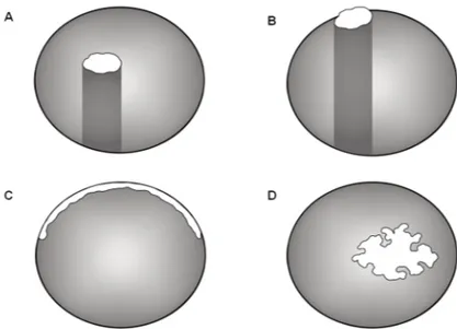
Among 675 patients, 174 nodules containing macrocalcifications constituted our study popu-lation. There were 116 malignant and 58 benign nodules. All of the malignant thyroid nodules were papillary carcinoma. Benign thyroid nod-ules included adenomatous hyperplasia in 56 nodules, follicular adenoma in 1 nodule, and lymphocytic thyroiditis in the remaining nodule. The macrocalcification patterns were classified into 3 categories: type 1, solitary calcifications (linear or round hyperechoic structure >2 mm with or without acoustic shadowing in the mid-dle of the nodule or along the margin of the nodule that encompassed less than 120° the cir-cumference; Figure 1, A and B); type 2, eggshell calcifications (curvilinear hyperechoic structure parallel to the margin of the nodule that encom-passed 120° or more of the circumference; Figure 1C); and type 3, all other coarse but not otherwise specified (NOS) calcifications (Figure 1D). If a patient had several thyroid nodules with macro-calcifications, each thyroid nodule with calcifica-tions was classified separately.
Each lesion with macrocalcifications was also evaluated for other suspicious sonographic fea-tures according to published data,1,2,12including marked hypoechogenicity (decreased echogenic-ity compared with the surrounding strap muscle), irregular or microlobulated margins, and a
taller-than-wide shape (greater in its anteroposterior dimension than its transverse dimension), except for microcalcifications. We refer to this combination as the triple criteria. Each lesion was classified by the number of malignant sono-graphic features present.
Pathologic results were reviewed from surgical records and pathology reports. Sonographic findings and the pathologic diagnosis were cor-related according to the size and location of the nodule and the calcification pattern described in the pathology report.
We determined the pattern and prevalence of macrocalcifications in resected thyroid lesions. The sensitivity and specificity were determined on the basis of the other sonographic features. We also compared the sensitivity and specificity according to the subtype of the calcifications. Statistical analyses were conducted with the χ2 test for nonparametric variables and the t test for parametric inference. P < .05 was considered statistically significant. A 95% confidence inter-val was calculated. Statistical analyses were performed with SAS version 9.1 software for Windows (SAS Institute Inc, Cary, NC).
Results
The size of the 174 nodules ranged from 5 to 50 mm (mean, 15 mm). There was no statistical difference between the benign and malignant nodules with regard to size (P > .05). There were solitary calcifications in 60 nodules (Figures 2 and 3), eggshell calcifications in 81 (Figure 4), and coarse NOS calcifications in 33 (Figure 5 and Table 1). Among these macrocalcification sub-types, solitary calcifications were more common in benign thyroid lesions, and coarse NOS calci-fications were more common in malignant thy-roid lesions (P < .05). Table 2 shows the number of combined suspicious sonographic features in thyroid nodules with calcifications according to the triple criteria (hypoechogenicity, irregular or microlobulated margins, and a taller-than-wide shape). Although the risk of malignancy was 34.5% in cases with no suspicious sonographic findings (Figures 2 and 4), it was as much as 82.8% in cases with at least 1 of the triple criteria (P < .05; Figures 3 and 5). On the basis of the number of combined suspicious sonographic
features among the triple criteria, the sensitivity was higher in the presence of 1 suspicious find-ing (82.8%) than in the presence of 2 or 3 suspi-cious findings (50% and 24.1%, respectively).
Figure 1. Schematic subtypes of calcifications on sonography. A and B, Type 1,
solitary calcification: hyperechoic structure larger than 2 mm in the middle of a nodule (A) or along the margin of a nodule (B). C, Type 2, eggshell calcification: curvilinear hyperechoic structure parallel to the margin of a nodule encompassing more than 120° of the circumference. D, Type III, coarse NOS calcification: irregu-lar macrocalcifications not specified in any other calcification subtype.
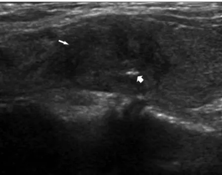
Figure 2. Transverse sonogram showing a round hyperechoic structure (>2 mm,
type 1 solitary calcification; arrow) along the margin of an oval circumscribed nod-ule. It has no suspicious findings of the triple criteria. The pathologic result was adenomatous hyperplasia.
On the basis of the macrocalcification subtype, there were no significant differences in the sensi-tivity and specificity in the presence of at least 1 of the triple criteria (P > .05; Table 3).
Discussion
Many authors have reported sonographic criteria that facilitate differentiation of benign and malignant lesions,2,8,13,14but thyroid sonography has not been thought to allow such a distinction despite its widely accepted usefulness.15,16 For accurate diagnosis, fine-needle aspiration biopsy (FNAB) with cytologic evaluation is the most important procedure and the most noninvasive and effective method for screening a thyroid nodule for cancer, especially with sonographic guidance.6,7,10However, the role of FNAB in thy-roid lesions with calcifications is controversial17 because one of the common causes of nondiag-nostic sampling is calcified lesions. In addition, Khoo et al8 reported that 25% of thyroid malig-nancies with calcifications in their study popula-tion had benign preoperative FNAB results. The importance of sonographic interpretation of thy-roid nodules with calcifications deserves further attention for follow-up and treatment.
Although microcalcifications have a well-known association with thyroid malignancy,2–4 a peripheral or eggshell calcification within a thyroid nodule is thought to be an indicator of benignity with just a few exceptions.18However, Taki et al6reported that 43% of the thyroid lesions with peripheral calcifications they examined were associated with cancer, and several recent investigations suggested that detection of macrocalcifications as well as microcalcifica-tions should increase the clinical index of suspi-cion for thyroid carcinoma.1,5–7,10These reports detailed the prevalence of calcifications (includ-ing macrocalcifications) in benign and malig-nant thyroid lesions and correlated calcifications and malignancy in terms of the calcification size, location, and patient age. In a study by Wang et al,5 microcalcifications were more significant than macrocalcifications (P = .001) for predicting thyroid malignancy, and the relative risk of malig-nancy was significantly higher in patients older than 45 years with macrocalcifications (P = .03). Moreover, Frates et al1 reported that coarse or rim calcifications doubled the risk of malignancy compared with a similar nodule without calcifi-cations, and the risk of malignancy increased when a nodule was solitary and solid. Although Consorti et al10reported no correlations between
Figure 3. Sagittal sonogram showing a hyperechoic structure (>2 mm, type 1
central calcification; thick arrow) in the middle of a microlobulated hypoechoic nodule. It has 1 suspicious finding (microlobulated margin; thin arrow) among the triple criteria. The pathologic result was papillary carcinoma.
Figure 4. Transverse sonogram showing a curvilinear hyperechoic structure (type
2 eggshell calcification; arrow), parallel to the margin of a nodule and encom-passing more than 120° of the circumference. It has no suspicious findings of the triple criteria. The pathologic result was adenomatous hyperplasia.
the presence of calcifications and the sono-graphic characteristics of nodules considered, they did not present statistical data to support their conclusion. To our knowledge, no study before ours addressed the role of other sono-graphic findings in thyroid nodules with macro-calcifications in assessment of the risk of malignancy.
Our results showed that 82.8% of malignant thy-roid nodules with macrocalcifications could be detected by the sonographic criteria outlined by Kim et al,2and the figures were relatively consis-tent across the macrocalcification subtypes. However, we found that the sensitivity of eggshell calcifications was comparatively lower than those of the other macrocalcification subtypes (solitary and coarse NOS), although it was not statistically significant. In some cases, it was difficult to apply these criteria to the thyroid nodules with eggshell calcifications because the calcification-lining margin of the nodule could obscure margin inter-pretation. Further modified criteria should be used to improve the sensitivity in thyroid nodules with eggshell calcifications.
One of the limitations of our study was the small percentage of benign nodules compared with pre-vious reports in the literature. We reviewed con-secutive surgical lesions over 6 months. Although we encountered benign calcified nodules fre-quently during FNAB, nodules referred for surgi-cal excision are more likely to be malignant.19 Considering the high sensitivity and specificity, FNAB should be recommended for nodules with macrocalcifications that have any suspicious sonographic findings. However, we did not assess how accurately FNAB could differentiate thyroid nodules with macrocalcifications prospectively, and further studies are warranted. The second limitation was that this study was retrospective. This could have had an effect on the results, but the degree of the effect on benign and malignant lesions seemed similar because the radiologist who reviewed the cases was blinded with respect to the final pathologic diagnosis and the ratio of malignant to benign lesions. A prospective analy-sis should be conducted. Last, we did not assess the role of macrocalcifications in evaluation of thyroid nodules in a case-control review, compar-ing macrocalcifications in nodules with noncalci-fied nodules and controlling for the presence of
the triple criteria. This would also be an informa-tive study of the importance of macrocalcifica-tions as a stand-alone predictor of malignancy. We wanted to determine whether we could differenti-ate between benign and malignant thyroid nod-ules with macrocalcifications on the basis of sonographic findings suspicious for malignancy, and the result was that the sonographic findings were useful for differentiation. Further study of thyroid nodules with no suspicious sonographic findings but with macrocalcifications is needed.
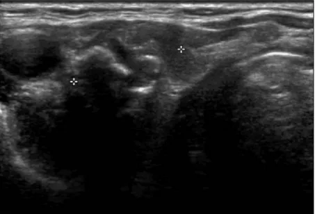
Figure 5. Transverse sonogram showing irregular macrocalcifications (type 3,
coarse NOS calcifications; calipers) in a hypoechoic nodule. This lesion has 2 sus-picious findings (microlobulated margin and a taller-than-wide shape) of the triple criteria. The pathologic result was papillary carcinoma.
Table 1. Macrocalcification Patterns and Prevalence of Malignancy in 174 Resected Thyroid Lesions With Macrocalcifications
Pathologic Type Solitary Eggshell Coarse NOS Total
Malignant 36 (60) 52 (64.2) 28 (84.8) 116 Benign 24 (40) 29 (35.8) 5 (15.2) 58
Total 60 81 33 174
Numbers in parentheses are percentages of the prevalence in the same calcification subtype.
Table 2. Triple Criteria Versus Pathologic Type
Suspicious Sonographic Features Malignant Benign
0 20 (17.2) 38 (65.5)
1 38 (32.8) 14 (24.1)
2 30 (25.9) 6 (10.4)
3 28 (24.1) 0 (0)
Total 116 (100) 58 (100)
In conclusion, our study indicates that other sonographic criteria can play an important role in differentiating benign and malignant thyroid nodules with macrocalcifications.
References
1. Frates MC, Benson CB, Doubilet PM, et al. Prevalence and distribution of carcinoma in patients with solitary and mul-tiple thyroid nodules on sonography. J Clin Endocrinol Metab 2006; 91:3411–3417.
2. Kim EK, Park CS, Chung WY, et al. New sonographic crite-ria for recommending fine-needle aspiration biopsy of non-palpable solid nodules of the thyroid. AJR Am J Roentgenol 2002; 178:687–691.
3. Holtz S, Powers WE. Calcification in papillary carcinoma of the thyroid. Am J Roentgenol Radium Ther Nucl Med 1958; 80:997–1000.
4. Frates MC, Benson CB, Charboneau JW, et al. Management of thyroid nodules detected at US: Society of Radiologists in Ultrasound consensus conference state-ment. Radiology 2005; 237:794–800.
5. Wang N, Xu Y, Ge C, Guo R, Guo K. Association of sono-graphically detected calcification with thyroid carcinoma. Head Neck 2006; 28:1077–1083.
6. Taki S, Terahata S, Yamashita R, et al. Thyroid calcifications: sonographic patterns and incidence of cancer. Clin Imaging 2004; 28:368–371.
7. Seiberling KA, Dutra JC, Grant T, Bajramovic S. Role of intrathyroidal calcifications detected on ultrasound as a marker of malignancy. Laryngoscope 2004; 114:1753– 1757.
8. Khoo ML, Asa SL, Witterick IJ, Freeman JL. Thyroid calcifi-cation and its association with thyroid carcinoma. Head Neck 2002; 24:651–655.
9. Kakkos SK, Scopa CD, Chalmoukis AK, et al. Relative risk of cancer in sonographically detected thyroid nodules with calcifications. J Clin Ultrasound 2000; 28:347–352. 10. Consorti F, Anello A, Benvenuti C, et al. Clinical value of
cal-cifications in thyroid carcinoma and multinodular goiter. Anticancer Res 2003; 23:3089–3092.
11. Takashima S, Fukuda H, Nomura N, Kishimoto H, Kim T, Kobayashi T. Thyroid nodules: re-evaluation with ultra-sound. J Clin Ultrasound 1995; 23:179–184.
12. Tae HJ, Lim DJ, Baek KH, et al. Diagnostic value of ultra-sonography to distinguish between benign and malignant lesions in the management of thyroid nodules. Thyroid 2007; 17:461–466.
13. Peccin S, de Castsro JA, Furlanetto TW, Furtado AP, Brasil BA, Czepielewski MA. Ultrasonography: is it useful in the diagnosis of cancer in thyroid nodules? J Endocrinol Invest 2002; 25:39–43.
14. Chan BK, Desser TS, McDougall IR, Weigel RJ, Jeffrey RB Jr. Common and uncommon sonographic features of papil-lary thyroid carcinoma. J Ultrasound Med 2003; 22:1083– 1090.
15. Hegedus L. Thyroid ultrasound. Endocrinol Metab Clin North Am 2001; 30:339–360, viii–ix.
16. Haber RS. Role of ultrasonography in the diagnosis and management of thyroid cancer. Endocr Pract 2000; 6: 396–400.
17. Belfiore A, La Rosa G. Fine-needle aspiration biopsy of the thyroid. Endocrinol Metab Clin North Am 2001; 30:361– 400.
18. Park CH, Rothermel FJ, Judge DM. Unusual calcification in mixed papillary and follicular carcinoma of the thyroid gland. Radiology 1976; 119:554.
19. Burgess JR, Tucker P. Incidence trends for papillary thyroid carcinoma and their correlation with thyroid surgery and thyroid fine-needle aspirate cytology. Thyroid 2006; 16:47– 53.
Table 3. Sensitivity and Specificity of Each Macrocalcification Pattern Based on Triple Criteria
Value Solitary (n = 60) Eggshell (n = 81) Coarse NOS (n = 33)
Sensitivity 31/36 (86.1) 40/52 (76.9) 5/28 (89.3) Specificity 16/24 (66.7) 21/29 (72.4) 1/5 (20) Numbers in parentheses are percentages.