V o l u m e 4 , Number 1 , A p r i l , 2 0 0 1
Transforming growth factor-β
1에 의한 연골세포와
제 2형 교원질의 상호 작용에서 교원질 수용체의 역할
연세대학교 의과대학 정형외과학 교실, Division of Orthopaedic Surgery,Duke University Medical Center, Durham, NC, USA*
이진우・강응식・한수봉・김성재・김윤희・김수향・Sean P. Scully* = Abstract =
The Role of Cell adhesion Molecules in the Modulation of
Chondrocytes-extracellular Type II Collagen by
Transforming Growth Factor-β
1
Jin Woo Lee, M.D., Eung Shick Kang, M.D., Soo Bong Hahn, M.D., Sung Jae Kim, M.D. Yun Hee Kim, M.S., Su Hyang Kim, M.S., Sean P. Scully, M.D.*
Department of Orthopaedic Surgery, Yonsei University College of Medicine, Seoul, Korea; Division of Orthopedic Surgery, Duke University Medical Center*, Durham, NC, USA.
Purpose : The physiologic response of chondrocytes to maintenance of the matrix and response to injury
likely involves signaling from multiple sources. The purposes of this study were to determine the molecular nature of collagen receptors involved in physiologic modulation.
Materials and Methods : An alginate bead culture system was utilized to which exogenous type II collagen
was added. We examined DNA and proteoglycan synthesis after blocking the cell surface with antibodies to anchorin CII and β1 integrin, and with cyclic RGD peptides.
Results : The inclusion of type II collagen results in an alteration of integrin expression with a
down-regula-tion of α2. The response of the chondrocyte to TGF-β1 can be modulated by the inclusion of exogenous type II collagen. The modulation of DNA and proteoglycan synthesis was blocked by the treatment of anti-β1 integrin antibody (4B4) or by cyclic RGD containing peptides. These events occur at concentrations that block cell adhesion to type II collagen. Anti-anchorin antibodies had no effect on the modulation by type II collagen.
C o n c l u s i o n : These results suggest that type II collagen binding by chondrocytes at least in part occurs
through the β1 integrin. This binding results in modulation of the cell response to TGF-β1. This modulation
※ 통신저자 : 이 진 우 서울특별시 서대문구 신촌동 134번지 연세대학교 의과대학 정형외과학교실 Tel : 02) 361-5640, Fax : 02) 361-1139 E-mail : [email protected] 이 논문은 2000년도 연세대학교 학술연구비의 지원에 의하여 이루어진 것임(과제번호: 2000-1-0223).
서
론
관절 연골의 손상은 재생되지 않는 것으로 알려 져 있으며2 8 ) , 이러한 관절 연골이 치유가 잘 안 일어나는 점에 대한 한가지 중요한 설명은 적절한 자극의 조절자가 없기 때문이라고 한다3 , 2 6 ). 이러 한 가설은 시험관내에서 관절 연골세포를 여러 가 지 성장인자에 노출시킨 후 그 반응을 탐구하는 연구를 행하게 하였다. 여기서 보다 복잡한 설명 이 덧붙여 질 수 있는데 이는 세포외 기질 성분이 이러한 c y t o k i n e의 작용을 국소적으로 변화시킬 수 있다는 점이다. Ingber와 F o l k m a n1 5 )은 세포 외기질이 F G F의 성장 조절 및 형태 조절 작용을 매개할 수 있음을 보고하였으며, 세포가 세포외기 질을 생산하며, 생산된 세포외기질에 의하여 영향 받을 수 있다고 하였다3 0 , 3 1 ). Scully 등2 8 , 2 9 )의 발표 에 의하면 연골세포에서도 관절연골 특이적인 제 2형 교원질에 의하여 T G F -β1의 역할이 변화될 수 있다고 하였다. Alginate bead에 연골세포를 배양을 할 때 제 1형 교원질을 첨가한 군과 제 2 형 교원질을 첨가한 군으로 나누어 배양하면서 T G F -β1 ( 1 0 n g /㎖)을 투여하면 제 2형 교원질을 첨가한 군의 경우에서 세포의 증식이나 단백다당 ( p r o t e o g l y c a n )의 합성이 제 1형 교원질을 첨가 한 군과 비교하여 유의하게 증가함을 관찰할 수 있었다고 하였다2 8 , 2 9 ). 이러한 결과는 제 2형 교원 질에 의한 T G F -β1의 기능 변화가 관절연골의 조 절기전에 중요한 요소임을 말해준다. 연골세포는 세포외 기질에 존재하는 교원질과 미세한 세포질의 돌기를 통하여 접촉하게 되는데 이 부위에는 m i c r o f i l a m e n t가 풍부하다고 한다 1 2 ). 이 부위의 세포막에는 anchorin CII라고 불 리우는 물질이 풍부하고 이 anchorin CII가 제 2형 교원질의 부착에 관계한다고 알려지고 있다 2 6 ). 또한 Anchorin CII는 세포와 제 2형 교원질 간의 상호 관계를 증진시킨다고 하며, calcium c h a n n e l로도 작용한다고 한다5 ) . 관절 연골내에 풍부하게 존재하는 제 2형 및 6형 교원질은 세포 의 부착에 관계하는 Arg-Gly-Asp(RGD) 서열 을 함유하고 있다고 하며2 , 1 0 ), 이러한 RGD 서열 을 함유하는 이들 기질은 관절 연골세포에 의한 i n t e g r i n의 표현과 상보적이라고 할 수 있다. 지 금까지 알려진 관절연골세포에서 표현되는 다른 i n t e g r i n은 α1β1과 α2β1이 존재한다고 하며, 이 들이 제 2형 교원질에 대한 부착을 담당할 것으로 사료되고 있다9 , 2 0 ). 제 2형 교원질에 대한 세포의 부착은 β1 integrin subunit에 대한 항체 및 RGD 서열을 함유한 cyclic conformation의 합 성 p e p t i d e s에 의하여 억제될 수 있다고 한다1 7 ) . 이러한 결과로 미루어 세포와 제 2형 교원질간의 상호관계에서는 collagen triple helix의 정상적인 구조가 필요함을 알 수 있다3 7 )
. 더욱이 β1
integrin receptor의 표현은 T G F -β1의 존재 하 에서 u p - r e g u l a t i o n된다고 한다1 9 ).
따라서 본 논문의 목적은 제 2형 교원질의 수용 체로 알려지고 있는 anchorin CII, integrin β1 의 항체 및 cyclic conformation의 RGD pep-t i d e를 처리한 후 연골세포를 alginapep-te bead 상
태에서 배양하면서 T G F -β1을 투여하고 연골세포 의 증식 및 단백다당( p r o t e o g l y c a n )의 합성이 어 떻게 변하는지 살펴봄으로써 이러한 세포외기질 및 성장인자의 상호관계에서 교원질 수용체가 어 떠한 역할을 하는 지를 알아보고자 하였다.
대상 및 방법
1. 연골세포의 배양 소의 슬관절을 구입하여 관절 연골을 채취한 다 음 소화 효소로 1 2시간 처리하여 연골세포를 분 리하였다. 분리된 연골세포를 세포 부착 실험을 위하여는 플라스틱 배양 용기에서 단층 배양을 10% 우태혈청이 함유된 D u l b e c c o’s modified E a g l e’s medium(DMEM, GIBCO-BRL, Grand Island, NY, USA)으로 배양하며, 교원 질 수용체의 b l o c k i n g을 위하여는 a l g i n a t emay serve to provide physiologic specificity to the cytokine-signaling cascade.
bead(Sigma Chemical, St. Louis, MO, U S A )에서 우태혈청이 함유되지 않은 D M E M 용액으로 배양하였다2 8 , 2 9 ). alginate bead 배양은
제 2형 교원질과 동량의 low viscosity alginate
용액을 섞은 다음 세포의 농도가 2×1 06 c e l l s /㎖ 되도록1 1 ) 조절하여 1.2% alginate에 함유되도록 하여 103 mM CaCl2 용액에 19 gauge 주사침 을 이용, 한 방울씩 b e a d를 형성하였다. 0.15 M NaCl 용액으로 세척 후 배양을 시작하였다. 2. 세포외 기질에 대한 연골 세포의 부착 실험 제 2형 교원질에 대한 연골세포의 부착이 억제될 수 있는지를 판단하기 위하여 두 가지의 항체와 2 가지의 합성 RGD peptide를 이용하여 제 2형 교 원질에 대한 연골세포의 부착을 억제하는데 사용하 였다. 사용된 항체는 anti-integrin β1 항체( 4 B 4 , 1:100) (Coulter, Miami, FL, USA)와 a n t i -anchorin CII 항체(1:100) (a gift from Dr. Mollenhauer), 그리고 음성 대조군으로 n o n -specific IgG를 사용하였으며, peptide의 경우 linear RGD peptide와 cyclic RGD peptide (Integra, San Diego, CA, USA) 그리고 각각 의 음성 대조군으로 linear non-RGD peptide와 cyclic non-RGD peptide를 사용하였다. 96-well i m m u n o p l a t e에 제 2형 교원질을 도포하여 ( 1㎍/ ㎖) 12시간 동안 냉장실에 방치하여 충분히 도포되 도록 한 다음, 도포되지 않은 플라스틱의 표면을 1% bovine serum albumin(BSA)으로 도포하여 1시간 동안 배양용기에 방치 후 PBS 용액을 이용 하여 세척 후 냉장 보관하였다. 7일간 단층으로 배 양된 연골세포를 배양용기에서 떼어낸 후 s o y b e a n trypsin inhibitor(Sigma Chemical, St. Louis, MO, USA)로 처리 후 수종의 항체 및 합 성 p e p t i d e s로 배양용기에서 1시간 동안 반응시킨 다음 미리 준비된 제 2형 교원질이 도포된 9 6 well immunoplate에 세포를 분주하여 1시간 동 안 배양기에 방치하여 세포가 부착되도록 하였다. 부착되지 않은 세포를 제거하기 위하여 세척 한 다 음 제 2형 교원질과 부착된 세포를 정량적으로 측 정하기 위하여 hexoaminidase assay를 시행하였 다1 7 ) . Hexoaminidase assay 후 s p e c t r o p h o -t o m e -t e r를 이용하여 405 nm에서 파장을 읽어 정 량화하였으며, 측정된 수치에서 B S A에 부착된 세 포의 수를 뺌으로써 표준화 하였다.
3. Semi-quantitative reverse transcription polymerase chain reaction(RT-PCR)을 이용한 교원질 수용체의 표현 변화 측정 연골세포의 alginate bead 및 단층 배양시 제 2형 교원질의 유무 및 배양 방법에 따른 교원질 수용체의 표현 변화를 측정하기 위하여 a l g i n a t e b e a d에 제2형 교원질을 첨가함에 따른 교원질 수 용체의 변화와 배양 방법 및 시간 경과에 따른 표 현의 변화를 측정하였다. 이를 위하여 새로이 분 리한 연골세포군, alginate bead 배양군 및 단층 배양 군으로 나누며, alginate bead 군은 교원질 첨가군과 비첨가군으로 나누어 실험하였다. Gen-B a n k를 이용하여 교원질 수용체인 a n c h o r i n CII, integrin의 α1, α2, α5, αv, β1, β3 sub-u n i t의 염기서열을 파악하여 p r i m e r를 작성하였 다. 정량화를 위하여 PCR cycle을 1 6회부터 4 0 회까지 시행하여 l i n e a r하게 증가하는 부분을 파 악하여 그 중간 cycle 수를 선택하였으며, 이러한 과정을 측정하고자 하는 유전자에 대하여 각각 실 시하여 적절한 c y c l e수를 정하였다3 5 ). GAPDH의 표현 정도로 각각 유전자의 표현을 정량화하여 비 교 분석하였다. 4. 교원질 수용체 blocking 후 연골세포의 증 식 및 세포외 기질의 합성 측정 제 2형 교원질에 대한 연골세포의 부착이 T G F -β1(R&D Systems, Minneapolis, MN, U S A )에 의한 연골세포의 생리적인 조절에 영향 을 미치는지 알아보기 위하여 연골세포의 교원질 수용체를 anchorin CII, integrin β1 항체 및 RGD peptide를 이용하여 b l o c k i n g한 다음 1 % ( w / v )의 제 2형 교원질이 함유된 alginate bead 에서 배양하며 T G F -β1 ( 1 0 n g /㎖)을 투여한 군과 비투여군으로 나누어 각 군에서 D N A와 단백다 당의 합성을 4일과 7일에 측정하여 비교하였다. 세포의 증식은 [3 H ] t h y m i d i n e을 이용1 6 ) , 전날 세포에 labeling 한 후 β-scintillation counter (model B 1900; Packard, Downers Grove, IL, USA)를 이용하여 측정하였다. 단백다당의
합성은 [3 5
S ] s u l f a t e를 이용 전날 세포에 l a b e l-ing 한 후 β-scintillation counter를 이용하여 측정하였으며, 각 군의 total DNA양을 측정하여 표준화 하였다.
5. 통계학적 방법
모든 결과들은 3개의 검체를 3회 반복하여 얻은 평균과 표준 편차로 표시하였으며, 통계학적인 유 의성은 mutiple comparison test를 이용하였다.
결
과
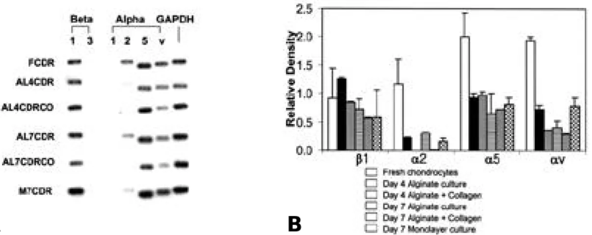
1. Integrin 표현에 대한 제 2형 교원질의 효과 관절연골로부터 새로이 분리한 연골세포는 α2 , α5, αv와 β1 integrin의 m R N A를 표현하는 것 을 관찰할 수 있었으나, α1과 β3 integrin은 표현 하지 않았다(Fig. 1A). 연골세포를 a l g i n a t e b e a d에 배양할 경우 integrin 표현의 양상은 유 지되었으나 그 정도는 변화하였다. α2, α5, αv i n t e g r i n의 표현은 새로이 분리한 관절연골세포와 비교하여 alginate bead 배양을 실시할 경우 현 저히 감소하였다. β1 integrin 표현은 a l g i n a t e b e a d에서 배양함에 따라 감소하였으나, α5의 경 우 거의 변화를 보이지 않았다. 그러나 a l g i n a t e b e a d에 제 2형 교원질을 첨가할 경우 α2 inte-g r i n의 표현이 배양 4일과 7일째에 사라지는 것을 관찰할 수 있었다. 3회 반복하여 얻은 결과를 densitometric analysis하여 평균과 표준 편차로 표시하였다(Fig. 1B). 2. 제 2형 교원질에 대한 연골세포의 부착에서 anti-integrin β1 항체( 4 B 4 )와 a n t i - a n c h o r i n CII 항체 및 RGD peptide의 영향 Anti-integrin β1 항체와 i n c u b a t i o n할 경우 제 2형 교원질에 대한 연골세포의 부착이 9 3 . 1 % 에서 억제되었으나, anti-anchorin CII 항체나 non-specific IgG에 의하여는 억제되지 않았다 (Fig. 2A). 반면에 linear RGD peptide를 처 리할 경우 연골세포의 부착은 4 6 %에서 억제되었 으며, cyclic RGD peptide로 처리할 경우 9 3 . 7 %에서 억제됨을 관찰할 수 있었으나, 음성 대조군으로 사용한 linear non-RGD peptide와Fig. 1. Results of Semiquantitative RT-PCR of integrin expression in bovine articular chondrocytes. (A)
Representa-tive reverse transcription-polymerase chain reaction analysis of RNA from bovine articular chondrocytes. With use of the primers identified in Table 1., reverse transcription-polymerase chain reaction analysis RNA from freshly isolated chondrocytes and chondrocytes cultured in different conditions was performed. Culture conditions were: freshly isolated chondrocytes(FCDR), chondrocytes cultured in monlayer for 7 days(M7CDR), chondrocytes cultured in alginate beads without collagen type II (1%) for 4 and 7 days(AL4CDR & AL7CDR), and chondrocytes cultured in alginate beads in the presence of type-II collagen for 4 and 7 days(AL4CDRCO & AL4CDRCO). (B) Densitometric analysis of integrin expression through three repetition. Results are mean±SD.
cyclic non-RGD peptide에 의하여는 억제되지 않음을 관찰 할 수 있었다(Fig. 2B). 따라서 제 2형 교원질에 대한 연골세포의 부착이 a n t i -integrin β1 항체와 RGD peptide에 의하여 특 이적으로 억제됨을 알 수 있었다. 3. 제 2형 교원질이 포함된 alginate bead에서 배양된 연골세포에서 D N A와 단백다당의 합성에 대한 antiintegrin β1 항체( 4 B 4 )와 a n t i -anchorin CII 항체의 영향 T G F -β1을 투여하지 않은 군에서는 a n t i - i n t e -grin β1 항체는 연골세포-제 2형 교원질 상호 작 용에서 별다른 효과를 보이지 않아 음성 대조군인 non-specific IgG를 처리한 군과 별다른 차이를 보이지 않았다. 그러나 T G F -β1을 투여한 군( 1 0 n g /㎖)에서는 anti-integrin β1 항체에 노출될 경우 배양 7일째에 다른 군과 비교하여 유의한 증 식의 감소를 보였다(Fig. 3). anti-anchorin CII 항체는 음성 대조군과 비교하여 유의한 차이 를 보이지 않았다. 단백다당 합성에 대한 교원질 수용체의 b l o c k-ing 효과도 유사하게 관찰되었는데, TGF-β1을 투여하지 않은 군에서는 anti-integrin β1 항체 와 anti-anchorin CII 항체는 아무런 효과를 보 이지 않았으나, TGF-β1을 투여한 군에서는 anti-integrin β1 항체는 배양 4일째에 다른 군 과 비교하여 유의한 단백다당 합성의 감소를 보였 다(Fig. 4). 4. 제 2형 교원질이 포함된 alginate bead에서 배양된 연골세포에서 D N A와 단백다당의 합성에 대한 cyclic RGD peptide의 영향 T G F -β1을 투여하지 않은 군에서는 c y c l i c RGD peptide는 세포의 증식에 별다른 영향을 미치지 못하였으나, TGF-β1을 투여한 군에서는 cyclic RGD를 노출시켰을 경우 배양 7일째에 세 포 증식의 유의한 감소를 보였다(Fig. 5). 단백 다당의 합성에 대하여도 T G F -β1을 투여하지 않
Fig. 2. Effects of collagen receptor blocking on chondrocyte attachment to type II collagen. Immunoplates(96 wells)
were coated with collagen type II(1ug/ml) and blocked for nonspecific binding with BSA(1%). Bovine chon-drocytes(100,000/well) were incubated with various kinds of antibodies(1:100 dilution) (Fig. 2A) and pep-tides(1 mM) (Fig. 2B) for 1h at 4˚C and then allowed to adhere for 1 h at 37˚C. Nonadherent cells were removed by washing, and adhesion was determined by analyzing lysosomal hexoaminidase. Adhesion is expressed as a percentage of the cell numbers of collagen-coated wells without antibodies and peptides. Attachment assays were performed in triplicate and each experiment was repeated on at least three occasions. The numbers represent the mean adhesion from three wells±SD from three experiments. (A) Blocking by monoclonal antibodies. (B) Blocking by synthetic peptide.
Fig. 3. Effects of collagen receptor blocking by
antibod-ies on DNA synthesis. Effects of collagen recep-tor blocking on DNA synthesis by bovine articu-lar chondrocytes encapsulated in alginate beads with the addition of collagen type II(1%) with T G F -β1(10ng/ml). Incorporation of [3H ] t h y m i d i n e
into chondrocytes cultured in medium supple-mented with TGF-β1 was measured as described in the Materials and Methods section. Data repre-sent mean±SD of three experiments performed in triplicate. *p<0.05 compared with cultures with no blocking by antibodies and peptides(multiple comparison test). (CON: without blocking colla-gen receptor, Beta1: anti-integrin β1 antibodies, anchorin: anti-anchorin CII antibodies, IgG: non-specific immunoglobulin G as a control)
Fig. 4. Effects of collagen receptor blocking by
antibod-ies on proteoglycan synthesis. Effects of collagen receptor blocking on proteoglycan synthesis by bovine articular chondrocytes encapsulated in alginate beads with the addition of collagen type II(1%) with TGF-β1(10ng/ml). Incorporation of [3 5S]sulfate into chondrocytes cultured in basal
medium or in medium supplemented with TGF-β 1 was measured as described in the Materials and Methods section. Data represent mean±SD of three experiments performed in triplicate. *p<0.05 compared with cultures with no block-ing by antibodies and peptides(multiple compari-son test). (CON: without blocking collagen receptor, Beta1: anti-integrin β1 antibodies, anchorin: anti-anchorin CII antibodies, IgG: non-specific immunoglobulin G as a control)
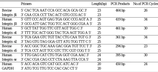
Table 1. List of Primers for Integrins
Primers Length(bp) PCR Products No.of PCR Cycles
Bovine 5’ CAC TCA AAT CCA GCC ACA GCA GC 3’ 23 464 bp 26
Integrin β1 3’ CAA CCA CCT TAC ACT GTG CCG AC 5’ 23
Human 5’ GTT CCC AGT GAG TGA GGC CCG AGT A 3’ 25 419 bp 34
Integrin β3 3’ GCG ATT GAC TGG TCC ACT GGG CGA A 5’ 25
Human 5’ GCT TAT TGG TTC GTT AGT TGG C 3’ 25 461 bp 30
Integrin α1 3’ TTT TGC ACT GGG TAC TCA AGT TGG A 5’ 25
Bovine 5’ TCA GAA GTC TGT TAC CTG CAA TGT G 3’ 25 361 bp 34
Integrin α2 3’ TAG GTG TAG GGA GTT ATG TGG TTT C 5’ 25
Bovine 5’ ACC GGC TGC AAA GAC GGA TGT TCC T 3’ 25 294 bp 23
Integrin α5 3’ TCA CCT AGT TCC GTC TTC CGT CGG T 5’ 25
Human 5’ TTG GAG CAT CTG TGA GGT CGA AAC 3’ 24 395 bp 30
Integrin αv 3’ CAC CGA CAG CCT CTA AAG TTA CCA 5’ 24
Human 5’ ACC ACA GTC CAT GCC ATC AC 3’ 20 450 bp 24
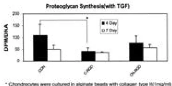
은 군에서는 단백다당의 합성에 별다른 영향을 미 치지 못하였으나, TGF-β1을 투여한 군에서는 cyclic RGD를 노출시켰을 경우 배양 4일째에 단 백다당의 합성에 유의한 감소를 관찰할 수 있었다 (Fig. 6).
고
찰
연골세포가 어떻게 세포외 기질로부터 세포 자 신의 조절 기전으로 그 신호가 전달되는 지의 기 전을 명확히 하고자 본 실험을 진행하였다. 이를 위하여는 어떠한 수용체들이 이러한 작용을 하는 지 파악하는 것이 필요하다. Integrin이 대부분 의 세포에서 세포외 기질에 부착되는데 일차적인 역할을 하는 수용체로 알려지고 있다1 3 , 3 2 ). 제 2형 교원질에 대한 연골세포의 부착 또한 i n t e g r i n에 의하여 이루어진다고 알려져 왔다2 0 ). RGD pep-t i d e는 여러 i n pep-t e g r i n에 의하여 인지되는데 이는 이러한 서열을 함유한 p e p t i d e에 의하여 제 2형 교원질에 대한 연골세포의 부착이 억제되는 현상 에서 알 수 있다. 본 실험에서는 제 2형 교원질에 대한 연골세포의 부착이 RGD peptide의 서열 뿐 아니라 cyclic conformation이 중요하게 작 용함을 알 수 있었다. Cardarelli 등7 )에 의하면 linear RGD peptide는 교원질에 대한 세포의 부착을 억제하지 못한다고 하며, cyclic RGD p e p t i d e는 세포의 부착을 억제하며, 교원질-Sepharose matrix의 추출에서 α2β1 integrin이 확인되었다고 하였다. 본 실험에서도 유사한 결과 를 보였는데, linear RGD peptide는 제 2형 교 원질에 대한 연골세포의 부착을 부분적으로 억제 하였으나, cyclic RGD peptide는 완전히 억제할 수 있었다. 이러한 현상은 2가지로 설명될 수 있 는데 첫째, 제 2형 교원질의 준비 과정에서 다른 단백질에 의하여 오염되었을 가능성이 있으며, 둘 째로 실험 도중 교원질의 c o n f o r m a t i o n에 변화Fig. 5. Effects of collagen receptor blocking by RGD
compounds on DNA synthesis. Effects of collagen receptor blocking on DNA synthesis by bovine articular chondrocytes encapsulated in alginate beads with the addition of collagen type II(1%) with TGF- β1(10ng/ml). Incorporation of [3H]thymidine into chondrocytes cultured in basal
medium or in medium supplemented with TGF-β1 was measured as described in the Materials and Methods section. Data represent mean±SD of three experiments performed in triplicate. *p<0.05 compared with cultures with no blocking by anti-bodies and peptides(multiple comparison test). (CON: without blocking, C-RGD: cyclic RGD peptide-GPenGRGDSPCA, C-NRGD: cyclic non-RGD peptide-RPmcGHRADLRCR as a control)
Fig. 6. Effects of collagen receptor blocking by RGD
compounds on proteoglycan synthesis. Effects of collagen receptor blocking by RGD compounds on proteoglycan synthesis by bovine articular chondrocytes encapsulated in alginate beads with the addition of collagen type II(1%) with TGF-β 1(10ng/ml). Incorporation of [3 5S]sulfate into
chondrocytes cultured in basal medium or in medium supplemented with TGF-β1was mea-sured as described in the Materials and Methods section. Data represent mean±SD of three exper-iments performed in triplicate. *p<0.05 compared with cultures with no blocking by antibodies and peptides (multiple comparison test). (CON: with-out blocking, C-RGD: cyclic RGD peptide-GPenGRGDSPCA, C-NRGD: cyclic non-RGD peptide-RPmcGHRADLRCR as a control)
가 온 경우이다. 다른 단백질에 의한 오염의 가능 성을 확인하기 위하여 제 2형 교원질을 p o l y-acrylamide 전기영동을 실시하여2 8 , 2 9 ) 대조군과 그 b a n d를 비교하여 다른 단백질에 의한 오염의 가능성을 배제할 수 있었다. 따라서 이러한 결과 는 제 2형 교원질의 c o n f o r m a t i o n의 변화에 의 하여 초래되었을 것으로 추론할 수 있다. Tuck-well 등에 의하면3 7 ) 교원질에 대한 연골세포의 결 합은 α2β1 integrin에 의하여 일어나지만 변성된 교원질은 α5β1-fibronectin bridge에 의하여 일 어난다고 하였다. anti-integrin β1 항체에 의하 여 제 2형 교원질에 대한 연골세포의 부착이 완전 히 억제되는 것으로 미루어 보아 이들 i n t e g r i n s u b u n i t이 존재하며, 연골세포의 부착에 능동적 으로 참여함을 알 수 있었다. T G F -β1의 자극이 없는 상태에서는 세포 부착
물질(cell adhesion molecules)들은 D N A나 단 백다당의 합성에 거의 영향을 미치지 않았으나,
T G F -β1의 자극이 있는 상태에서는 세포외 기질
과의 상호 작용을 억제함으로써 T G F -β1의 자극
에 의하여 증가된 D N A와 단백다당의 합성을 유
의하게 억제하였다. anti-integrin β1 항체와
cyclic RGD peptide로 처리한 군에서 D N A와 단백다당의 합성은 연골세포가 제 2형 교원질에 노출되지 않은 상태와 비슷한 양의 합성을 보였 다. 지금까지의 연구에 의하면 i n t e g r i n은 여러 성장인자(growth factors)와 상호작용을 하며, 세포의 부착, 분화, 성장 및 생존에서 중요한 역 할을 한다고 알려져 왔다1 , 2 1 , 2 3 ) . 연골세포와 세포외 기질간의 상호 작용은 많은 부분이 integrin β1 을 매개로 한다고 알려져 왔으며7 ) , 이러한 세포외 기질과 i n t e g r i n간의 상호작용은 특정한 세포내 의 신호 전달 물질을 활성화함이 다른 연구에서 밝혀지고 있으나 이에 대한 연골세포에서의 연구 는 아직 없는 실정이다. 또한 i n t e g r i n은 신호전 달 수용체로써의 역할도 한다고 알려지고 있는데, 세포외 기질에 대한 i n t e g r i n을 매개로 하는 세 포의 부착은 p a x i l l i n6 ) , tensin4 ) 과 같은 다수의 세포골격 단백(cytoskeletal-associated pro-teins) 및 focal adhesion kinase(FAK)와 같 은 세포내 신호 전달 단백(intracellular signal-ing proteins)들을 활성화 시킨다고 한다3 4 ). 연골 세포에서 외부 성장인자에 의하여 변화되는 이러 한 i n t e g r i n을 매개로 하는 반응들은 제 2형 교 원질과 같은 특정 연골기질 단백에 대한 i n t e-g r i n을 매개로 하는 세포의 부착을 요하게 된다. F A K의 tyrosine 인산화는 세포-세포외 기질의 상호작용, integrin β s u b u n i t의 세포질내 d o m a i n의 과표현(overexpression), 그리고 성 장인자에 의하여 유도된다고 한다1 4 ) . 여러가지 세 포내 신호전달경로(signal transduction path-w a y s )가 i n t e g r i n과 성장인자에 의하여 상호 활 성화된다는 사실이 발견되고 있으며2 5 , 2 7 , 3 8 , 3 9 ) , 이러 한 신호전달경로가 i n t e g r i n과 성장인자에 의하 여 상호 활성화될 수 있다는 이론은 i n t e g r i n과 같은 교원질 수용체가 T G F -β1에 의한 연골세포-제 2형 교원질 상호작용의 변화에서 중요한 역할 을 한다는 본실험의 결과와 일치한다. 소의 관절연골세포는 α2, α5, αv와 β1 inte-grin subunit 을 표현하였지만, α1과 β3는 표현 되지 않았다. 이러한 결과는 다른 사람들의 보고 와3 3 ) 유사하였으나, α1과 α2의 표현에 대하여는 아직 논란의 여지 있는 것으로 사료된다2 0 ) . Algi-nate bead에서 연골세포를 배양할 경우 i n t e-grin β1의 표현이 증가하는 것을 관찰할 수 있었 는데, 이러한 결과는 alginate bead 배양에서 연 골세포가 단층 배양한 연골세포보다 보다 분화된 표현형을 오래 간직하는 것으로 미루어 보아 d e d i f f e r e n t i a t i o n의 결과가 아니라고 할 수 있 다. 분화된 연골세포의 표현형과 대사의 형태는 세포외 환경을 변화시킴으로써 쉽게 변형시킬수 있다3 ). 연골세포와 제 2형 교원질간의 상호 작용 을 중재함으로 i n t e g r i n은 T G F -β1의 생리적 효 과를 변형시키는데 관여한다는 것을 알 수 있었 다. 보다 흥미로운 점은 연골세포 i n t e g r i n의 표 현형태가 세포외 기질의 존재 상태에서 세포를 배 양할 경우 영향을 받는다는 점이다. α2 integrin 은 제 2형 교원질의 존재 상태에서 그 표현이 억 제되는 것을 관찰할 수 있었다. α6와 α1 0과 같은 다른 s u b t y p e의 i n t e g r i n이 제 2형 교원질에 의 한 integrin 표현 변화에 어떤 역할 을 할 수 있 을 것으로 추론되며, 제 2형 교원질이 α2 inte-g r i n과 결합하여 α6와 α10 integrin의 표현을 유 도할 수도 있다. 그러나 본 실험에서는 소의 α6와
α10 integrin의 염기서열을 알 수 없어 관찰할 수 없었다. 또 다른 하나의 가능성은 T G F -β1이 α2 integrin의 표현을 증가시킬 가능성이다. Loeser 등1 8 )의 결과에 의하면 T G F -β1이 i n t e-g r i n의 표현을 증가시킨다고 보고하여 이의 가능 성을 뒷받침하여 주고 있다. 결과적으로 이러한 결과는 세포외 기질이 feedback mechanism을 통하여 integrin 표현의 정도와 형태를 변화 시 킬수 있다는 것을 말해준다. 또한 본 실험의 결과는 anchorin CII가 연골세 포와 제 2형 교원질간의 상호작용에 별다른 영향 을 못미친다는 것을 보여준다. 이러한 실험 결과 에 대하여는 pepsin 처리에 의한 t e l o p e p t i d e의 소실이 anchorin CII와 결합하는 부분의 소실로 이어진 결과라고 설명할 수 있다2 4 ) . 현재의 실험 p r o t o c o l은 용해 가능한 교원질을 필요로하기 때 문에 a n c h o r i n이 불용성의 교원질과 결합하여 그 생리적인 신호를 보낸다는 가능성을 배제할 수 없 다. 그런즉 anchorin CII를 제외하고 교원질에 대한 세포 표면의 수용체, 특히 i n t e g r i n이 T G F -β1에 의하여 조절되는 연골세포-제 2형 교 원질 상호작용에서 중요한 역할을 한다고 결론지 을 수 있을 것으로 사료된다.
결
론
본 연구 결과는 연골세포의 cytokine 조절과 교원질간의 상호 작용에 관한 첫번째 시도였으며, 세포외 기질에 의한 세포 조절의 개념을 연골세포 에서 처음으로 증명하였다. 더욱이 이러한 조절이 T G F -β1에 대한 연골세포의 반응을 변화시키는 작용을 한다는 것을 보여주고 있다. 이러한 연골 세포 조절에 대한 포괄적인 이해는 연골세포의 복 구 기능의 조작, 아마도 용해성 기질의 요소를 통 하여 병적인 상태의 관절연골의 내적인 치유를 촉 진하게 하는 것이 가능해질 것으로 사료된다.R E F E R E N C E S
11 ) Arner EC and Tortorella MD : Signal transduction through chondrocyte integrin receptors induces matrix metalloproteinase synthesis and synergizes with
inter-leukin-1. Arthritis Rheum, 38:1304-1314, 1995. 12 ) Aumailley M, Mann K, von der Mark H, and
Timpl R : Cell Attachment Properties of Type VI and
Arg-Gly-Asp Dependent Binding to its a2(VI) and a3(VI) Chains. Exp Cell Res, 181:463-474, 1989. 13 ) Benya PD and Shaffer JD : Dedifferentiated
chondro-cytes reexpress the differentiated collagen phenotype when cultured in agarose gels. C e l l, 30:215-224, 1982. 14 ) Bockholt SM and Burridge K : Cell spreading on
extra-cellular matrix proteins induces tyrosine phosphoryla-tion of tensin. J Biol Chem, 268:14565-14567, 1993. 15 ) Bohm BB, Wilbrink B, Kuettner KE, and
Mollen-hauer J : Structural and Functional Comparison of
Anchorin CII and Muscle Annexin V. Arch Biochem B i o p h y s, 314:64-74, 1994.
16 ) Burridge K, Turner CE, and Romer LH : Tyrosine phosphorylation of paxillin and pp125FAK accompa-nies cell adhesion to extracellular matrix: a role in cytoskeletal assembly. J Cell Biol, 119:893-903, 1992. 17 ) Cardarelli PM, Yamagata S, Taguchi I, Gorcsan F,
Chiang SL, and Lobl T : The collagen receptor alpha
2 beta 1, from MG-63 and HT1080 cells, interacts with a cyclic RGD peptide. J Biol Chem, 267:23159-23164, 1 9 9 2 .
18 ) Durr J, Goodman S, Potocnik A, von der Mark H,
and von der Mark K : Localization of beta 1-integrins
in human cartilage and their role in chondrocyte adhe-sion to collagen and fibronectin. Exp Cell Res, 207:235-244, 1993.
19 ) Enomoto M, Leboy PS, Menko AS, and Boettiger D : Beta 1 integrins mediate chondrocyte interaction with type I collagen, type II collagen, and fibronectin. E x p Cell Res, 205:276-285, 1993.
1 0 ) Eyre DR : The collagens of articular cartilage. S e m i n Arthritis Rheum, 21:2-11, 1991.
1 1 ) Guo JF, Jourdian GW, and MacCallum DK : Cul-ture and growth characteristics of chondrocytes encap-sulated in alginate beads. Connect Tissue Res, 19:277-297, 1989.
1 2 ) Hunziker EB, Herrmann W, and Schenk RK : Improved Cartilage Fixation by Ruthenium Hexaamine Trichloride (RHT)-a Prerequisite for Morphometry in
Growth Cartilage. J Ultrastruct Res, 81:1-12, 1982. 1 3 ) Hynes RO : Integrins: versatility, modulation, and
sig-naling in cell adhesion. C e l l, 69:11-25, 1992.
1 4 ) Ilic D, Furuta Y, Kanazawa S, et al : Reduced cell motility and enhanced focal adhesion contact formation in cells from FAK-deficient mice. N a t u r e, 377:539-544, 1 9 9 5 .
1 5 ) Ingber DE and Folkman J : Mechanochemical switching between growth and differenciation during fibroblast growth factor-stimulated angiogenesis in vitro: role of extracellular matrix. J Cell Biol, 109:317-330, 1989.
1 6 ) Labarca C and Paigen K : A simple, rapid, and sensi-tive DNA assay procedure. Anal Biochem, 102:344-352, 1980.
1 7 ) Landegren U : Measurement of cell numbers by means of the endogenous enzyme hexosaminidase. Applications to detection of lymphokines and cell sur-face antigens. J Immunol Methods, 67:379-388, 1984. 1 8 ) Loeser RF : Integrin-mediated attachment of articular
chondrocytes to extracellular matrix proteins. A r t h r i t i s R h e u m, 36:1103-1110, 1993.
1 9 ) Loeser RF, Carlson CS, and McGee MP : Expres-sion of beta 1 integrins by cultured articular chondro-cytes and in osteoarthritic cartilage. Exp Cell Res, 217:248-257, 1995.
2 0 ) Loeser RF, Sadiev S, Tan L, and Goldring MB : Goldring, Integrin expression by primary and immortal-ized human chondrocytes: evidence of a differential role for alpha 1 beta 1 and alpha 2 beta 1 integrins in mediat-ing chondrocyte adhesion to types II and VI collagen. Osteoarthritis & Cartilage, 8:96-105, 2000.
2 1 ) Mainiero F, Pepe A, Yeon M, Ren Y, and Giancotti
FG : The intracellular functions of alpha6beta4 integrin
are regulated by EGF. J Cell Biol, 134:241-253, 1996. 2 2 ) Mankin H : Localization of Tritated Thymidine in
Articular Cartilage of Rabbits. J Bone joint Surg AM, 44A:688-698, 1962.
2 3 ) Miyamoto S, Teramoto H, Gutkind JS, and
Yama-da KM : Integrins can collaborate with growth factors
for phosphorylation of receptor tyrosine kinases and MAP kinase activation: roles of integrin aggregation
and occupancy of receptors. J Cell Biol, 135:1633-1642, 1996.
2 4 ) Mollenhauer, J, Bee JA, Lizarbe MA, and Von der
Mark K : Role of Anchorin CII, a 31000-mol-wt
Membrane Protein, in the interaction of Chondrocytes with Type II Collagen. J Cell Biol, 98:1572-1579, 1984. 2 5 ) Pelicci G, Lanfrancone L, Grignani F, et al : A novel transforming protein(SHC) with an SH2 domain is implicated in mitogenic signal transduction. C e l l, 70: 93-104, 1992.
2 6 ) Pfaffle M, Ruggiero F, Hofmann H, et al: Biosynthe-sis, Secretion and Extracellular Localization of Anchorin CII, a Collagen-binding Protein of the Cal-pactin Family. EMBO J, 7:2335-2342, 1988.
2 7 ) Pronk GJ, McGlade J, Pelicci G, Pawson T, and
Bos JL : Insulin-induced phosphorylation of the
46-and 52-kDa Shc proteins. J Biol Chem, 268:5748-5753, 1 9 9 3 .
2 8 ) Qi WN and Scully SP : Extracellular Collagen Modu-lates the Regulation Of Chondrocytes By Transforming Growth Factor-Beta-1. J Orthop Res, 15:483-490, 1 9 9 7 .
2 9 ) Qi WN and Scully SP : Effect of type II collagen in chondrocyte response to TGF-beta 1 regulation. E x p Cell Res, 241:142-150, 1998.
3 0 ) Roskelley CD and Bissell MJ : Dynamic reciprocity revisited: a continuous, bidirectional flow of informa-tion between cells and the extracellular matrix regulates mammary epithelial cell function. Biochem Cell Biol, 73: 391-397, 1995.
3 1 ) Roskelley CD, Srebrow A, and Bissell MJ : A hierar-chy of ECM-mediated signalling regulates tissue-spe-cific gene expression. Curr Opin Cell Biol, 7:736-747, 1 9 9 5 .
3 2 ) Ruoslahti E and Yamaguchi Y : Proteoglycans as modulators of growth Factor Activities. C e l l, 64:867-869, 1991.
3 3 ) Salter DM, Hughes DE, Simpson R, and Gardner
D L : Integrin Expression by Human Articular
Chon-drocytes. Brt J Rheumatol, 31:231-234, 1992.
3 4 ) Schaller MD, Otey CA, Hildebrand JD, and
peptides mimicking beta integrin cytoplasmic domains. J Cell Biol, 130:1181-1187, 1995.
3 5 ) Sciore P, Boykiw R, and Hart DA : Semiquantitative reverse transcription-polymerase chain reaction analysis of mRNA for growth factors and growth factor recep-tors from normal and healing rabbit medial collateral ligament tissue. J Orthop Res, 16:429-437, 1998. 3 6 ) Sporn MB and Roberts AB : Transforming growth
factor-beta: recent progress and new challenges. J Cell B i o l, 119:1017-1021, 1992.
3 7 ) Tuckwell DS, Ayad S, Grant ME, Takigawa M, and
Humphries MJ : Conformation dependence of
inte-grin-type II collagen binding. Inability of collagen
pep-tides to support alpha 2 beta 1 binding, and mediation of adhesion to denatured collagen by a novel alpha 5 beta 1-fibronectin bridge. J Cell Sci, 107:993-1005, 1994. 3 8 ) Vainikka S, Joukov V, Wennstrom S, Bergman M,
Pelicci PG, and Alitalo K : Signal transduction by
fibroblast growth factor receptor-4(FGFR-4). Compari-son with FGFR-1. J Biol Chem, 269:18320-18326, 1 9 9 4 .
3 9 ) Wary KK, Mainiero F, Isakoff SJ, Marcantonio
EE, and Giancotti FG : The adaptor protein Shc
cou-ples a class of integrins to the control of cell cycle pro-gression. C e l l, 87:733-743, 1996.