THE JOURNAL OF THE ACOUSTICAL SOCIETY OF KOREA Vol.29, No.3E 2010. 9. pp. 140~145
I. Introduction
Today, dual energy X-ray absorptiometry (DEXA) providing two-dimensional (2-D) representation of bone mineral density (BMD) of the lumbar spine and the proximal femur is the gold standard method for the assessment of osteoporotic fracture risk. The problem remains that bone properties such as microarchitecture or tissue elasticity that are not captured by DEXA also contribute to bone strength independently of BMD [1]. Since their inception in 1984 [2], skeletal quantitative ultrasound (QUS) technologies have played a growing role in the diagnosis of osteoporosis [3]. This is due to the wide availability of QUS devices that provide a fracture risk assessment as an alternative to X-ray counterparts [4]. Most of the QUS devices using the transverse transmission technique measure the speed
of sound (SOS) and the broadband ultrasound attenuation (BUA) at easily accessible peripheral sites (typically the calcaneus) by using a transmitter and a receiver placed along the mediolateral axis on opposite sides of the skeletal site to be tested.
Although BMD is the most important determinant of bone strength and accounts for up to 80% of its variance, additional parameters reflecting geometrical and material properties of bones are required to predict fracture risk more accurately [5]. Changes in the human cortical bone thickness with aging and osteoporosis have been shown to be a risk factor for fracture [6]. In this regard, the axial transmission technique that was first developed in the 1950s to monitor fracture healing of cortical bone has attracted the attention of many researchers [7-12]. This method estimates the ultrasonic velocity from the transit time of the first arriving signal and the propagation distance along the axial direction of long bones (typically the tibia) by using a transmitter
Corresponding author: Kang Il Lee ([email protected]) Department of Physics, Kangwon National University, Chuncheon 200-701
Dependencies of Ultrasonic Velocities on the Wall
Thickness in Polyvinyl Chloride Cortical Bone Mimics
Kang Il Lee*
*Department of Physics, Kangwon National University, Chuncheon 200-701
(Received September 9, 2010; accepted September 30, 2010)
Abstract
In the present study, tubular polyvinyl chloride (PVC) cortical bone mimics that simulate the cortical shell of long bones were used to validate the axial transmission technique for assessing the cortical thickness by measuring the ultrasonic velocities along the cortical shell of long bones. The ultrasonic velocities in the 9 PVC cortical bone mimics with wall thicknesses from 4.0 to 16.1 mm and inner diameters from 40 to 300 mm were measured as a function of the thickness by using a pair of custom-made transducers with a diameter of 12.7 mm and a center frequency of 200 kHz. In order to clarify the measured behavior, they were also compared with the predictions from a theory of guided waves in thin plates. This phantom study using the PVC cortical bone mimics provides useful insight into the dependencies of ultrasonic velocities on the cortical thickness in human long bones. Keywords: Osteoporosis, Cortical bone, Cortical thickness, Guided ultrasonic wave, Phase velocity
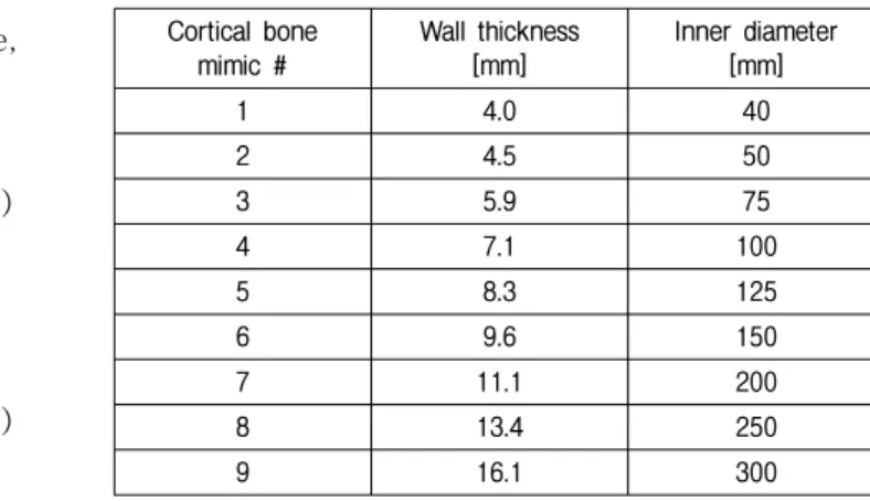
Table 1. Wall thickness and inner diameter of the 9 PVC cortical bone mimics.
Cortical bone mimic # Wall thickness [mm] Inner diameter [mm] 1 4.0 40 2 4.5 50 3 5.9 75 4 7.1 100 5 8.3 125 6 9.6 150 7 11.1 200 8 13.4 250 9 16.1 300
and a receiver placed along the bone axis on the cortical shell. The ultrasonic velocity obtained this way is claimed to reflect both structural (such as cortical thickness and cross-sectional area) and material (such as mineralization and Young’s modulus) properties of bones.
In the present study, tubular polyvinyl chloride (PVC) cortical bone mimics that simulate the cortical shell of long bones were used to validate the axial transmission technique for assessing the cortical thickness by measuring the ultrasonic velocities along the cortical shell of long bones. The ultrasonic velocities in the 9 PVC cortical bone mimics with wall thicknesses from 4.0 to 16.1 mm and inner diameters from 40 to 300 mm were measured as a function of the thickness by using a pair of custom-made transducers with a diameter of 12.7 mm and a center frequency of 200 kHz. In order to clarify the measured behavior, they were also compared with the predictions from a theory of guided waves in thin plates [13], which has been widely used in the field of nondestructive testing for the assessment of plates, pipes, and more complex structures.
II. Methods and Materials
2.1. Theoretical methods
Lamb waves are 2-D elastic waves resulting from the combination of longitudinal and shear waves propagating in a solid plate with free boundaries in a vacuum [13]. The wavenumbers of Lamb waves have to satisfy the Rayleigh-Lamb equations defined for the two types of propagation modes: symmetrical (S) and antisymmetrical (A). These equations are, respectively, given as
(
k2+s2)
cosh( )
qd sinh( )
sd −4k2qssinh( )
qd cosh( )
sd =0(1) and
(
k2+s2)
sinh( )
qd cosh( )
sd −4k2qscosh( )
qd sinh( )
sd =0(2) with 2 2 2 l k k q = − and 2 2 2 t k k s = − . In these equations, d
is the plate thickness and k=ωcp is the Lamb
wavenumber, where ω is the angular frequency and
p
c is the phase velocity. kl=ω cl and ks=ωcs are the
wavenumbers of the longitudinal and shear waves, respectively, where cl and cs are the longitudinal and
shear velocities of the plate, respectively. The Lamb wave dispersion curves that define the variation in the phase velocity cp as a function of the frequency-
thickness product are determined by solving Eqs. (1) and (2). The group velocity cg dispersion curves can
be obtained by using ω ω ω d dc c c dk d c p p p g − = = 1 . (3)
2.2. Experimental measurements
A total of 9 PVC cylindrical tubes with wall thicknesses from 4.0 to 16.1 mm and inner diameters from 40 to 300 mm were used as cortical bone mimics, as listed in Table 1. The PVC tubes that simulate the cortical shell of long bones such as the tibia were selected because they are readily available [14]. Table 2 shows the density and the bulk wave velocities of PVC and human cortical bone taken from the literature [12, 15]. Figure 1 is the schematic diagram of the experimental arrangement for measuring the ultrasonic velocities in the PVC cortical bone mimics
Table 2. Density and bulk wave velocities of PVC and human cortical bone. Material Density [kg/m3] Longitudinal velocity [m/s] Shear velocity [m/s] PVC 1300 2330 1070 Human cortical bone 1850 4000 1800
Fig. 1. Schematic diagram of the experimental arrangement for measuring the ultrasonic velocities in the PVC cortical bone mimics by using the axial transmission technique.
Fig. 2. Lamb wave dispersion curves as a function of the frequency- thickness (fd) product for a PVC plate: (a) phase velocity and (b) group velocity.
by using the axial transmission technique with a pair of custom-made transducers with a diameter of 12.7 mm and a center frequency of 200 kHz. The operation frequency of 200 kHz was selected by taking into account the acceptable resolution and signal level over propagation distances. Both transducers (a transmitter and a receiver) were positioned perpendicularly to the outer surface of the bone mimic. The transducer face was small enough to contact with the curved surface of the bone mimic. To facilitate the transmission of ultrasound into and out of the bone mimic, a low- viscosity couplant (Sonotech Echogel) was applied between the transducer and the bone mimic. The one-cycle tone burst wave with 200 kHz wave was generated by a function generator (Agilent 33250A) and amplified by a power amplifier (AR 75A250A). Received signals were averaged over 100 waveforms by a digital oscilloscope (LeCroy WS44Xs). As shown in Figure 1, the transducers had an initial separation of 20 mm that was measured from the center of both transducers and the receiver was manually moved
away from the fixed transmitter along the long axis of the bone mimic. The transducer separation was increased from 20 to 100 mm in 5-mm steps such that the responses were recorded at 16 discrete distances during the scan. This procedure was repeated five times with each bone mimic in order to obtain the mean values of the ultrasonic velocities.
III. Results and Discussion
Figure 2 shows the Lamb wave dispersion curves as a function of the frequency-thickness (fd) product
for a PVC plate. These dispersion curves were calculated by using a longitudinal velocity of 2330 m/s and a shear velocity of 1070 m/s for PVC in Table 2. The symmetrical (S) and antisymmetrical (A) Lamb modes are identified by a discrete order number that goes from 0 to the infinite value. From
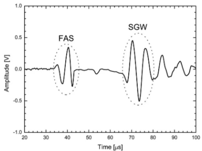
Fig. 3. Time-domain signal of the response at the transducer separation of 60 mm in the PVC cortical bone mimic with a wall thickness of 4.0 mm and an inner diameter of 40 mm.
Fig. 4. Measured velocities of the FAS and the SGW as a function of the wall thickness in the 9 PVC cortical bone mimics and theoretical velocities of the two fundamental (S0 and A0) Lamb modes as a function of the plate thickness in the PVC plate.
these dispersion curves, it can be seen that two or more modes exist at any given fd. In practice, it is
difficult to generate a pure single mode, particularly, above the cut-off fd of the first order antisymmetrical (A1) mode. The modes are also generally dispersive, which means that the shape of a propagating wave changes with the distance along the propagation path. This makes signal interpretation difficult and leads to signal-to-noise problems because the peak amplitude in the signal envelope rapidly decreases with the distance if the dispersion is strong. Therefore, the dispersion characteristics of a plate should be understood thoroughly for the appropriate application
of guided ultrasonic waves. In spite of these difficulties, the use of guided waves is very attractive because they propagate throughout the cortical thickness of long bones, which means that the entire thickness of the bones can be interrogated.
Figure 3 shows the time-domain signal of the response at the transducer separation of 60 mm in the PVC cortical bone mimic with a wall thickness of 4.0 mm and an inner diameter of 40 mm. As seen in Figure 3, two distinct waves that are, respectively, referred to as the first arriving signal (FAS) and the slow guided wave (SGW) were consistently observed from the responses measured in the 9 PVC cortical bone mimics. Their velocities were determined as a function of the wall thickness by the slope of the graph of the transducer separation versus the time of flight of the first peak of each wave.
Figure 4 shows the measured velocities of the FAS and the SGW as a function of the wall thickness in the 9 PVC cortical bone mimics. The theoretical velocities of the two fundamental (S0 and A0) Lamb modes as a function of the plate thickness in the PVC plate are also plotted in Figure 4. As seen in Figure 4, the FAS velocity in the 3 PVC cortical bone mimics with thicknesses of 11.1, 13.4, and 16.1 mm is found to be closely consistent with the longitudinal velocity of 2330 m/s for PVC. On the other hand, the FAS velocity in the 6 bone mimics thinner than 11.1 mm decreases rapidly with the decreasing thickness toward the S0 Lamb mode velocity in the PVC plate. It is also found in Figure 4 that the SGW velocity in the 9 PVC cortical bone mimics increases slightly with the increasing thickness for the thickness range ≥ 4.0 mm and is observed to be essentially consistent with the A0 Lamb mode velocity predicted in the PVC plate.
In Figure 4, for the PVC cortical bone mimics with the thickness/wavelength ratio > 1 (i.e., when the thickness is comparable with, or greater than, the wavelength of 11.6 mm @ 200 kHz in PVC), the FAS propagates at the longitudinal velocity of 2330 m/s for PVC, as expected for a lateral wave (or P-head wave) propagating along the surface [16]. On the other
hand, for the bone mimics with the thickness/wavelength ratio < 1, the FAS velocity tends to decrease rapidly with the decreasing thickness toward the S0 Lamb mode velocity in the PVC plate, resulting from the interference between the lateral wave and Lamb modes. These results suggest that the FAS is guided by the wall thickness only at wavelengths greater than the thickness and underpin the fact that the cortical thickness has a significant effect on the FAS velocity when the thickness is comparable with, or less than, the wavelength in cortical bone [17]. Therefore, the FAS velocity is expected to be more sensitive to the cortical thickness for long wavelengths (i.e., for low frequencies) compared to the thickness. The longitudinal velocity of 4000 m/s for cortical bone is higher than that of 2330 m/s for PVC (Table 2). This implies that the wavelength in cortical bone is longer than that in PVC. Therefore, the thickness effect on the FAS velocity in cortical bone would be more pronounced than that observed in the PVC cortical bone mimics.
As shown in Figure 4, the SGW velocity measured in the PVC cortical bone mimics tends to increase slightly with the increasing thickness for the thickness range ≥ 4.0 mm and shows excellent agreement with the prediction for the A0 Lamb mode in the PVC plate. In particular, the A0 Lamb mode velocity is highly dependent on the thickness when the thickness/ wavelength ratio ≪ 1, even though the measured data are only available for a limited thickness range ≥ 4.0 mm. For 200 kHz, the wavelength in the tibial cortical bone with the longitudinal velocity of 4000 m/s equals to 20 mm and thus fulfills the above condition in the human tibia with the cortical thickness of 2-8 mm. Indeed, the SGW velocity appeared to be a good discriminator for osteoporosis and an indicator for bone growth in a pilot in vivo study [18]. However, the problem remains that special signal processing techniques must be implemented for consistency of mode identification and velocity estimation because the SGW arrives after the FAS and typically interferes with other contributions, particularly, at short transmitter-receiver distances
[19, 20]. It should be also noticed that the overlying soft tissue of cortical bone may affect coupling of different modes and this can affect consistency of mode identification. Therefore, a suitable in vivo
multilayer model must be used for the unambiguous identification of the recorded mode.
IV. Conclusions
In the present study, the dependencies of the FAS and the SGW velocities on the wall thickness in the PVC cortical bone mimics were experimentally measured by using the axial transmission technique. In order to clarify the measured behavior, they were also compared with the predictions from the Lamb wave theory in the PVC plate, based on the fact that the axial Lamb modes in a cylindrical tube are similar in behavior to those in a plate provided that the wall thickness is small compared to the tube diameter [11]. While real bones are anisotropic and heterogeneous, PVC employed here is isotropic and homogeneous. However, it was demonstrated that the ultrasonic velocity is influenced by the anisotropy of cortical bone but this effect does not completely change the fundamental behavior due to the cortical thickness [6]. In conclusion, it is expected that measurements of the ultrasonic velocities in cortical bone would enhance clinical potential of QUS technologies for the diagnosis of osteoporosis.
Acknowledgments
Following are results of a study on the “Human Resource Development Center for Economic Region Leading Industry” Project, supported by the Ministry of Education, Science & Technology (MEST) and the National Research Foundation of Korea (NRF). This study was also supported by the industrial technology innovation program of Ministry of Knowledge Economy, Republic of Korea.
References
1. P. Laugier, “Instrumentation for in vivo ultrasonic characterization of bone strength,” IEEE Trans. Ultrason. Ferroelectr. Freq. Control, vol. 55, no. 6, pp. 1179-1196, 2008.
2. C. M. Langton, S. B. Palmer, and R. W. Porter, “The measurement of broadband ultrasonic attenuation in cancellous bone,” Eng. Med., vol. 13, no. 2, pp. 89-91, 1984.
3. C. C. Gluer, “A new quality of bone ultrasound research,” IEEE Trans. Ultrason. Ferroelectr. Freq. Control, vol. 55, no. 7, pp. 1524- 1528, 2008.
4. P. Laugier, “Quantitative ultrasound of bone: looking ahead,” Joint Bone Spine, vol. 73, no. 2, pp. 125-128, 2006.
5. P. Ammann and R. Rizzoli, “Bone strength and its determinants,”
Osteoporosis Int., vol. 14, suppl. 3, pp. S13-S18, 2003.
6. P. Moilanen, “Ultrasonic guided waves in bone,” IEEE Trans. Ultrason. Ferroelectr. Freq. Control, vol. 55, no. 6, pp. 1277-1286, 2008.
7. I. M. Siegel, G. T. Anast, and T. Fields, “The determination of fracture healing by measurement of sound velocity across the fracture site,” Surg. Gynecol. Obstet., vol. 107, no. 3, pp. 327-332, 1958.
8. A. J. Foldes, A. Rimon, D. D. Keinan, and M. M. Popovtzer, “Quantitative ultrasound of the tibia: a novel approach for assessment of bone status,” Bone, vol. 17, no. 4, pp. 363-367, 1995. 9. K. Raum, I. Leguerney, F. Chandelier, E. Bossy, M. Talmant, A. Saied,
F. Peyrin, and P. Laugier, “Bone microstructure and elastic tissue properties are reflected in QUS axial transmission measurements,”
Ultrasound Med. Biol., vol. 31, no. 9, pp. 1225-1235, 2005. 10. P. H. F. Nicholson, P. Moilanen, T. Karkkainen, J. Timonen, and
S. Cheng, “Guided ultrasonic waves in long bones: modelling, experiment and in vivo application,” Physiol. Meas., vol. 23, no. 4, pp. 755-768, 2002.
11. K. I. Lee and S. W. Yoon, “Feasibility of bone assessment with leaky Lamb waves in bone phantoms and a bovine tibia,” J. Acoust. Soc. Am., vol. 115, no. 6, pp. 3210-3217, 2004.
12. S. P. Dodd, J. L. Cunningham, A. W. Miles, S. Gheduzzi, and V. F. Humphrey, “Ultrasonic propagation in cortical bone mimics,”
Phys. Med. Biol., vol. 51, no. 18, pp. 4635-4647, 2006. 13. H. Lamb, “On waves in an elastic plate,” Proc. R. Soc. Lond. A,
vol. 93, no. 648, pp. 114-128, 1917.
14. P. Moilanen, V. Kilappa, P. H. F. Nicholson, J. Timonen, and S. Cheng, “Thickness sensitivity of ultrasound velocity in long bone phantoms,” Ultrasound Med. Biol., vol. 30, no. 11, pp. 1517-1521, 2004. 15. G. W. Kaye and T. H. Laby, Tables of Physical and Chemical
Constants and Some Mathematical Functions, Longman, London, 1995.
16. E. Camu, M. Talmant, G. Berger, and P. Laugier, “Analysis of the axial transmission technique for the assessment of skeletal status,”
J. Acoust. Soc. Am., vol. 108, no. 6, pp. 3058-3065, 2000. 17. P. Moilanen, P. H. F. Nicholson, V. Kilappa, S. Cheng, and J.
Timonen, “Assessment of the cortical bone thickness using ultrasonic guided waves: modelling and in vitro study,” Ultrasound Med. Biol., vol. 33, no. 2, pp. 254-262, 2007.
18. P. Moilanen, P. H. F. Nicholson, T. Karkkainen, Q. Wang, J. Timonen, and S. Cheng, “Assessment of the tibia using ultrasonic guided waves in pubertal girls,” Osteoporosis Int., vol. 14, no. 12, pp. 1020-1027, 2003.
19. D. Alleyne and P. Cawley, “A two-dimensional Fourier transform method for the measurement of propagating multimode signals,” J. Acoust. Soc. Am., vol. 89, no. 3, pp. 1159-1168, 1991.
20. W. H. Prosser, M. D. Seale, and B. T. Smith, “Time-frequency analysis of the dispersion of Lamb modes,” J. Acoust. Soc. Am., vol. 105, no. 5, pp. 2669-2676, 1999.
【Profile】
∙ Kang Il Lee
![Table 2. Density and bulk wave velocities of PVC and human cortical bone. Material Density [kg/m 3 ] Longitudinal velocity [m/s] Shear velocity[m/s] PVC 1300 2330 1070 Human cortical bone 1850 4000 1800](https://thumb-ap.123doks.com/thumbv2/123dokinfo/4909258.40565/3.892.494.796.132.604/density-velocities-cortical-material-longitudinal-velocity-velocity-cortical.webp)