저작자표시-비영리-변경금지 2.0 대한민국 이용자는 아래의 조건을 따르는 경우에 한하여 자유롭게 l 이 저작물을 복제, 배포, 전송, 전시, 공연 및 방송할 수 있습니다. 다음과 같은 조건을 따라야 합니다: l 귀하는, 이 저작물의 재이용이나 배포의 경우, 이 저작물에 적용된 이용허락조건 을 명확하게 나타내어야 합니다. l 저작권자로부터 별도의 허가를 받으면 이러한 조건들은 적용되지 않습니다. 저작권법에 따른 이용자의 권리는 위의 내용에 의하여 영향을 받지 않습니다. 이것은 이용허락규약(Legal Code)을 이해하기 쉽게 요약한 것입니다. Disclaimer 저작자표시. 귀하는 원저작자를 표시하여야 합니다. 비영리. 귀하는 이 저작물을 영리 목적으로 이용할 수 없습니다. 변경금지. 귀하는 이 저작물을 개작, 변형 또는 가공할 수 없습니다.
博士學位論文
한국의 양식 넙치에서 발생하는
여윔병의 원인 분석
濟州大學校 大學院
海洋生命科學科
金 承 鍲
2016년 2월
한국의 양식 넙치에서 발생하는
여윔병의 원인 분석
指導敎授 鄭 埈 範
金
承 鍲
이 論文을 理學 博士學位 論文으로 提出함
2016 年 2 月
金承鍲
의 理學
博
士 學位論文을 認准함
濟州大學校 大學院
2016 年 2 月
THESIS
FOR THE DEGREE OF DOCTOR OF PHILOSOPHY
Analysis of the cause of emaciation disease
in cultured olive flounder, Paralichthys
olivaceus in Korea
Seung Min Kim
(Supervised by professor Joon Bum Jeong)
Department of Life Science
GRADUATE SCHOOL
JEJU NATIONAL UNIVERSITY
ii
목 차
목 차··· i
Summary··· V LIST OF FIGURES··· ix
LIST OF TABLES··· xii
제 1장. 제주도 여윔증상 넙치로부터 분리한 점액포자충의 특성 분석··· 1 1.1. 서 론··· 1 1.2. 재료 및 방법··· 3 1.2.1. 바이러스성 및 세균성 질병검사··· 3 1.2.2. 여윔증 진단을 위한 primer set 제작··· 5 1.2.3. 실험어··· 9 1.2.4. DNA 분리··· 11 1.2.5. PCR 및 DNA sequencing··· 11 1.2.6.병리조직학적 분석··· 11 1.3. 결 과··· 12 1.3.1. 외부 및 내부증상··· 12 1.3.2. PCR 진단법 개발 및 적용 ··· 13 1.3.3.양식장별 감염률··· 15 1.3.4.개체별 감염률··· 15 1.3.5. 크기별 감염률··· 19
1.3.6. Fish weight (index) ··· 20
1.3.7.병리조직학적 검사··· 21
1.3.8.염기서열 분석··· 23
iii 제 2장. 2014-2015년도 한국산 양식 넙치에 대한 여윔증 감염 조사··· 28 2.1. 서 론··· 28 2.2. 재료 및 방법··· 30 2.2.1. 2014년도 양식넙치 여윔증 모니터링··· 30 2.2.2. 2015년도 양식넙치 여윔증 모니터링··· 32 2.2.3. DNA 분리··· 33 2.2.4. PCR··· 33 2.2.5. Cohabitation test··· 34 2.2.6. 병리조직학적 검사··· 36 2.3. 결 과··· 37 2.3.1. 2014년도 양식장 시기별 감염률··· 37 2.3.2. 2014년도 크기별 감염률··· 39 2.3.3. 2015년도 양식장 시기별 감염률··· 39 2.3.4. 검출된 넙치의 병리조직학적 검사··· 42 2.3.5. Cohabitation test ··· 44 2.4. 고 찰··· 48 제 3장. 여윔증 넙치, Paralichthys olivaceus의 비특이적 면역반응 및 혈액학적 분석···· 50 3.1. 서 론··· 50 3.2. 재료 및 방법··· 52 3.2.1. 실험어··· 52 3.2.2. DNA 분리··· 52 3.2.3. PCR··· 52 3.2.4.혈액분석 및 비특이적 면역 반응··· 53
iv 3.2.5.통계학적 분석··· 54 3.3. 결 과··· 55 3.3.1. PCR··· 55 3.3.2.혈액학적 검사··· 57 3.3.3. 비특이적인 면역 분석··· 60 3.4. 고 찰··· 62 제 4장. 여윔증의 원인규명을 위한 계통분석학적 동정··· 64 4.1. 서 론··· 64 4.2. 재료 및 방법··· 66 4.2.1. 실험어··· 66 4.2.2.현미경 검사··· 66 4.2.3. Primers 제작··· 66 4.2.4. DNA 분리··· 68 4.2.5. PCR 및 sequencing··· 68 4.2.6.계통학적 분석··· 68 4.3. 결과 및 고찰··· 71 4.3.1.계통학적 분석··· 71 4.3.2.포자 관찰··· 77 제 5장. 감염넙치의 각 내부장기에 대한 Parvicapsula sp.의 양적 분석··· 79 5.1. 서 론··· 79 5.2. 재료 및 방법··· 81 5.2.1. 실험어··· 81 5.2.2. DNA 분리··· 81
v 5.2.3. PCR··· 81 5.2.4. Real-time PCR··· 83 5.2.5. 병리조직학적 검사··· 83 5.3. 결 과··· 84 5.3.1. PCR··· 84 5.3.2. Real-time PCR의 검량선··· 86 5.3.3. Real-time PCR을 이용한 장기별 감염 농도··· 88 5.3.4. 병리 조직학적 검사··· 91 5.4. 고 찰··· 93 종합요약··· 95 참고문헌··· 99 감사의 글··· 107
vi
Summary
The olive flounder industry, a representative species of domestic aquaculture industry, has shown steady and continuous growth since artificial seedling production technique was developed in National Fisheries Research and Development Institute (NFRDI) early 1980’s. As it is a species with aquaculture history longer than 30 years, the study of its diseases has a wide variety compared with that of other species. But in Korea, researches on diseases of cultured fishes have been limited and focused only on several specific diseases (Kim et al., 2006; Cho et al., 2008; Kim et al., 2010), and there are few researches on diseases of which the causes are not known.
Recently about 20cm long olive flounders in farms on Jeju Island had been increasingly thinner and perished within 1~3 weeks. Unlike Japan, where the causes of emaciation disease were found, there’s no report on its causes in Korea.
In this study, to identify emaciation infection in cultured olive flounders in Korea, a primer set useful for aquaculturing olive flounder was developed and a test method was established and applied to the fields. In addition, the features of emaciation were identified using PCR, microscopy, histopathological examinations, molecular phylogenetic systematic, real-time PCR etc. and experiments were performed to determine the causes of emaciation.
To analyze the causes of emaciation of olive flounders with emaciation symptoms in Korea, a primer set identical with MM18Sf/MM18Sr primer set was developed. The MM18Sf/MM18Sr primer set had been made from small subunit ribosomal DNA gene (SSU rDNA) of E. leei reported as the cause of emaciated turbot in Japan. In the PCR test, for which DNA was taken from intestinal tissue of emaciated olive flounders in Korea, all of them showed negative reactions. We made the EM-F/EM-R primer set from the base sequences of the Myxidium sp., which is registered in GenBank (NCBI, USA). The primer set was applied to emaciated olive flounders in Korea, and the response was positive. Therefore we came to use it for examining emaciated olive flounders in Korea.
To observe the infected area in the emaciated olive flounders, PCR was performed on extracted kidneys, intestines, spleens, brains, livers and gills. Whereas the causes were found mostly in intestines in Japanese report on emaciation, in this study PCR was positive not only in intestines but also in kidneys. In case of significantly emaciated fishes, it was positive in all internal organs except gills. Detection frequency was in order of kidneys, intestines and spleens.
To identify the infection situation of emaciation disease, 216 olive flounders out of 24 farms were investigated in the period of 2010 to 2013. Most of the infected fishes showed significant emaciation in abdominal area. The weight of emaciated fish was 30~40% lower than that of uninfected fish and 33 (50%) out of 66 farms were positive in infection test. Yearly infection rates were so high with 8 out of 21 farms (38%) in 2010, 12 out of 25 farms (48 %) in 2011, 2 out of 4 farms (50%) in 2012 and 11
vii
out of 16 farms (63.1%) in 2013 that they can cause serious damage to farms.
The tissue sections of organs were observed with optical microscope, and many spores of circular or oval shape were detected in kidneys and intestines of emaciated olive flounders. In other organs except for kidneys and intestines, some spores were observed, but it was difficult to examine because the number of detected spores was small compared with that in kidneys and intestines or no specific histological abnormalities were observed. Therefore, histopathologic emaciation examination should be performed mainly in kidneys and intestines.
According to gene sequencing analysis of PCR products, the gene homology between pathogens detected in emaciated olive flounders was more than 99%. According to gene comparison analysis using the GenBank database, it showed partial agreement with the gene sequences of Myxosporean sp. and was found to be a new species not registered in GenBank.
To investigate the outbreak trend of emaciation infection in wider range, total 900 fish were systematically examined in April, May, Sept., Nov., Dec., 2014 and the infection rate was checked. In 2015, the same examination was conducted in March, May, July and October with the different fish number. As a result, the infection rate was 18.3%~71.6% in 2014 and 16.3%~90.3% in 2015, higher than in 2014. In addition, according to the infection trend analysis depending on the sample size, the infection occurred in any size and the size of 11~30cm showed the highest infection rate. In periodic infection rate, Sept. and Dec. in 2014 and March, July and October in 2015 showed relatively higher infection rate. The reason for the high infection rate was not identified yet, but it is assumed that it might be related with marine environment of Jeju Island region. It might also be related with the fact that olive flounder seedlings are brought in from other regions to Jeju Island.
To identify whether emaciation disease could be transmitted within a same fishes or among the other fishes (red sea bream, black sea bream), two groups were built after PCR infection test a donor group showing positive reactions and a recipient group showing negative reactions. The test result showed that infection occurred within olive flounders, the same fishes, but was not observed in red sea bream and black sea bream, the other species. This result, in relation to species specificity, is estimated that emaciated disease causes infection only to olive flounder of the same fishes not to those of different fishes because factors such as host environment and living conditions do not fit for fish of different fishes. However, up to now, more investigation is required and in the future, a more detailed study is required to find out infection occurs to fish of different species. Moreover, according to the result of the histological examination of the renal tissue after cohabitation, the same spores as those where emaciation is generally observed were observed for the same group (recipient group) detected in PCR and for red sea bream and black sea bream of different fishes, the PCR result and spores were not observed.
viii
To establish diagnosis index of emaciation disease, hematologic analysis was performed and non-specific immune response to emaciated and non-emaciated fish was observed. The result of the hematological analysis shows that hematocrit and GPT (farm-B) displayed higher experimental values than control values, and that GPT, GLU, total glucose, total cholesterol, and total protein exhibited lower experimental values than control values. As a result of measuring Lysozyme, NBT and MPO which are non-specific immune responses, lower experimental values were observed in three experiments. It is assumed that the stress by emaciation cause and the damage of internal organs destroy the primary defense system.
According to phylogenetic systematics analysis, all of 4 isolates belong to Parvicapsula petuniae and gene homology with the 4 isolates identified in Korea was 99.7%~99.76 %. In addition, according to the result of the gene comparative analysis using GenBank database, P. petuniae and high homology of 92% were observed for 18S rRNA and emaciation occurring domestically has been identified as
Parvicapsula sp.
For morphological identification, according to the result of the microscopic inspection of the fish species suffering from emaciation, the spores were classified into two forms. One from form was where two spores gathered in the same or different direction and spores with lengths of 16~18㎛ And widths of 4~5㎛ Were observed. Also, spores with two polar capsules gathered on one side were observed. The other form was a form of small spores with lengths of 5~8㎛ and widths of 7~9㎛ And a form with one polar capsule and lacking part of spore was observed. In this result, spores morphologically similar to P. anisocaudata and P. pseudobranchicola n. sp. occurring in a olive flounder farm has been observed and spores typically observed only in Parvicapsula sp. have been confirmed. Therefore, emaciation occurring domestically has been identified as Parvicapsula sp. in the same way shown by phylogenetic analysis result.
Real-time PCR we analyzed the number of pathogens in tissues of Parvicapsula sp. and the order of infection. Through Histopathologic examination we checked its correlation with the results of real-time PCR. Infection concentration of Parvicapsula sp. in tissues was investigated. The highest infection concentration was observed in kidney with 2.6 × 10³ ~1.7 × 10⁷copies/mg, followed by intestines, spleens, brains and livers. With the result we could identify more infected cases that were not detected on PCR. It is assumed that minimum infection concentration for PCR examination of
Parvicapsula sp. is 103copy/mg.
To check the result of real-time PCR of Parvicapsula sp. we performed correlation analysis through Histopathologic examination. A number of spores were observed in all of the organs, and the presence or absence of infecting spores corresponds with the results of real-time PCR. As Histopathologic examination is accurate but unfavorable in immediacy, it is difficult to respond to infection outbreaking immediately. Real-time PCR is assumed to be more useful for the Parvicapsula sp.
ix
x
LIST OF FIGURES
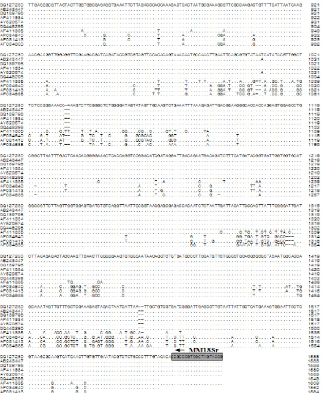
Fig. 1. The small subunit ribosomal DNA (SSU rDNA) gene of Enteromyxum leei which was reported to be a causative agent of the olive flounder (Paralichthys olivaceus) emaciation disease in Japan.···6 6
Fig. 2. (A) Total DNA separated from the gut of Paralichthys olivaceus having emaciation disease and PCR results obtained from a reference primer set made by using SSU rDNA gene of Enteromyxum leei. 1-8; A: Total DNA seperated from the gut of P. olivaceus having emaciation disease, 9-15; B: Total DNA separated from the gut of P. olivaceus having emaciation disease, (B) 1-3; DNA of E.leei provided by Japan, M; 100 bp DNA ladder.···8 8
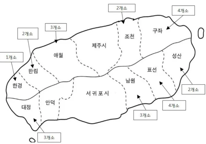
Fig. 3. Location of the Jeju olive flounder sampling station.··· ··· ···110 0
Fig. 4. The external (A) and internal (B) signs of the emaciated olive flounder (P. olivaceus).···112 2
Fig. 5. PCR amplification from nucleic acids of intermal organs from the emaciated olive flounder (P. olivaceus)
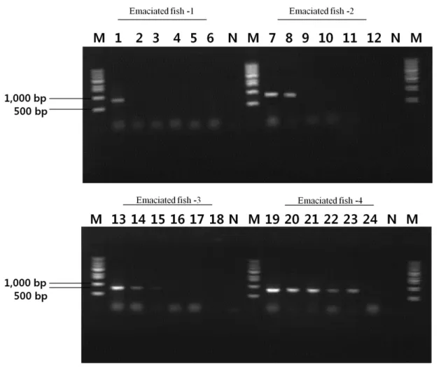
in the level stages of infection. Lanes 1, 7, 13 and 19, Kidney; lane 2, 8, 14 and 20, Intestine; lanes 3, 9, 15 and 21, Spleen; lanes 4, 10, 16 and 22, Liver; lanes 5, 11, 17 and 23, Brain; lanes 6, 12, 18 and 24, Gill; M, 1kb DNA ladder.···114 4 Fig. 6. Gel electrophoresis of the products amplified with the primer set EM-F/R from the extracted sample of
the kidney of affected olive flounder cultured in emaciated disease. Lane1, Farm-A (June, 2010); lane 2, Farm-B (October, 2011); lane 3, Farm-C (October, 2012); lane 4, Farm-D (November, 2011); lane 5, Farm-E (October, 2012); lane 6, Farm-F (November, 2012); lane 7, Farm-G (July, 2013); lane 8, Farm-N (December, 2010); lane 9, Farm-O (December, 2010); M, 1kb DNA ladder; N, negative control; P, positive control.···116 6
Fig. 7. Monitoring of the index content in the emaciated olive flounder (P. olivaceus). ◯: Uninfected olive flounder, ●: infected olive flounder.···220 0
Fig. 8. Histological findings of kidney (A, ×200 and B, ×400) and intestine (C, ×200 and D, ×400) from the emaciated olive flounder (P. olivaceus). Note microsporean spores both in emaciated disease the glomerulus (arrows) and in the gut epithelium (arrows). H & E stain. Bar = 20μm.···222 2
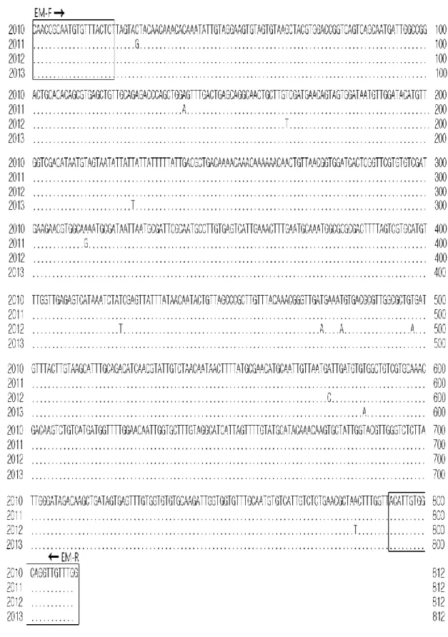
Fig. 9. Comparison analysis of the DNA nucleotide sequences of pathogens detected form the emaciated olive flounder (P. olivaceus). Primers used in PCR are in boxes. ···224 4
Fig.10. Location of the Jeju olive flounder sampling station.···330 0
Fig.11. Experimental transmission of emaciation disease to olive flounder (P. olivaceus), red sea bream (P.
xi
Fig.12. Gel electrophoresis of the products amplified with the primer set EM-F/R from the extracted sample of the kidney of affected olive flounder cultured in emaciated disease. Lanes 1, 3, 4, 5, 6, 7, 8, 10, 11, 12, 13 and 14, positive samples; Lanes 2 and 9, negative smaples; M, 1kb DNA ladder; N, negative control. ·
···338 8 Fig.13. Gel electrophoresis of the products amplified with the primer set EM-F/R from the extracted sample of the kidney of affected olive flounder cultured in emaciated disease. Lanes 3, 4, 5, 6, 8, 9, 10, 11 and 13, positive samples; Lanes 1, 2, 7 and 13, negative smaples; M, 1kb DNA ladder; N, negative control. ·
···441 1 Fig.14. Histological features of kidneys (A and B ×100) from the emaciated olive flounder (P. olivaceus). Note microsporean spores in emaciated disease the gloerulus (arrows). H & E stain. Bar = 20μm. ·
···443 3 Fig.15. Cumulative mortality (%) of normal olive flounder (P. olivaceus) after cohabitated with the emaciated olive flounder.···445 5 Fig.16. Histological findings of kidney (A, x200 and B, x400) from the recipient olive flounder (P. olivaceus)
cohabitated with the emaciated olive flounder. Note microsporean spores in emaciated disease the gloerulus (arrows). H & E stain. Bar = 20μm.···445 5 Fig.17. Cumulative mortality (%) of normal red sea bream (P. major) after cohabitated with the emaciated olive flounder (P.olivaceus).···446 6 Fig.18. Cumulative mortality (%) of normal black sea bream (A. schlegelii) after cohabitated with the emaciated olive flounder (P. olivaceus).···446 6 Fig.19. Results of PCR using EM-F/R primers on extracted kidney DNA of olive flounder. Lane1 to lane3, control; lane 4 to lane 6, Farm-A; lane 7 to lane 9, Farm-B, lane 10 to lane 12, Farm-C; M, 1kb DNA ladder; N, negative control.···556 6 Fig.20. Analysis of hematocrit (A), GPT (B), GOT (C) in the serum of olive flounder (P. olivaceus). *
Significant difference between control group and emaciation group based on the t-test (p < 0.05). ·
···558 8 Fig.21. Analysis of glucose (D), Total cholesterol (E), Total protein (F) in the serum of olive flounder, (P.
olivaceus). * Significant difference between control group and emaciation group based on the t-test ( P <
0.05).···559 9 Fig.22. Analysis of lysozyme activity (A), myeloperoxidase activity (B), nitroblue tetrazolium (C) in the serum of olive flounder, (P. olivaceus).*Significant difference between control group and emaciation group based on the t-test ( P < 0.05).···661 1 Fig.23. PCR analysis of LSU and SSU rDNA sequences. Lanes 1 and 5 PP1F-W3R; lanes 2 and 6 PPNF-W3R; lanes 3 and 7 EMF-EMR; lanes 4 and 8 W3F-W3NR; M, 1kb DNA ladder; N, negative control. ·
xii
Fig.24. Phylogenetic tree of showing the genetic relatic relationships 49 myxosporeans based on the nucleotide sequences of the SSU, internal transcribed spacer (ITS) region (ITS1, 5.8S rRNA, ITS2), and LSU gene. Phylogenetic analysis indicated that most of Korea emaciation belong to the parvicapsula sp. ·
···773 3 Fig.25. Comparison analysis of the DNA nucleotide sequences of pathogens detected from the emaciated olive flounder (P. olivaceus). Primers used in PCR are in boxes.···774 4 Fig.26. A hypothetical sequence of developmental stages of emaciation. A: first stage of shizont visible with microscopy (×1000, bar=10μm); B~C: Sporont (×1000, bar=10μm), E~G: Sporonts (×400, bar=10μm); H: Pansporoblast (×400, bar=10μm); I~K: Spore in frontal view (×400, bar=10μm); L: Atypical smaller spore (×400, bar=10μm); N~O: triactinospores (×1000, bar=20μm).···778 8 Fig.27. PCR amplification from nucleic acids of intermal organs from the emaciated olive flounder (P.
olivaceus) in the level stages of infection. Lanes 1, 6 and 11, Kidney; lane 2, 7 and 12 Intestine; lanes 3, 8 and 13, Spleen; lanes 4, 9 and 14, Brain; lanes 5, 10 and 15, Liver; M, 1kb DNA ladder; N, negative control.···885 5 Fig.28. Standard curve derived from 10-fold (101, 102, 103, 104, 105) serially diluted plasmid DNA containing a partial 28S rDNA.···887 7 Fig.29. Micrographs of the kidney (A: EM-A ×100, E: EM-B ×200 and I: EM-C ×100), intestine (B: EM-A ×200, F: EM-B ×200 and J: EM-C ×200), spleen (C: EM-A ×200,G: EM-B ×200 and K: EM-C ×200) and liver (D: EM-A ×200, H: EM-B ×200 and L: EM-C ×200) from olive flounder (P. olivaceus) with control and Parvicapsula sp. Note the serous tissue destruction and many spore in the internal organs (arrow). H&E stain Bar= 20μm.···992 2
xiii
LIST OF TABLES
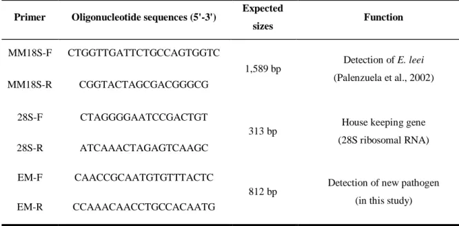
Table 1. Culture mediums used in this study···4 4 Table 2. Oligonucleotide primers and conditions used in PCR amplification···4 4 Table 3. PCR primers used for the detection of new pathogen ···8 8
Table 4. Prevalence of emaciation disease in cultured olive flounder (P. olivaceus) of Jeju from 2010 to 2013 ···117 7
Table 5. Prevalence of causative agent of the emaciated disease according to the size of olive flounder (P.
olivaceus)···119 9 Table 6. Sample information employed for the investigation of emaciation infection in rearing olive flounder (P.
olivaceus) of Korea in 2014···331 1 Table 7. Sample information employed for the investigation of emaciation infection in rearing olive flounder (P.
olivaceus) of Korea in 2015···332 2 Table 8. Monitoring of emaciation disease for olive flounder (P. olivaceus) farms in 2014···338 8 Table 9. Monitoring of emaciation disease for olive flounder (P. olivaceus) farms in 2015···441 1 Table 10. Results of experiments testing for transmission of emaciated disease to olive flounder (P. olivaceus),
red sea bream (P. major) and black sea bream (A. schlegelii) by close association with infected olive flounder···447 7 Table 11. Sequences of the oligonucleotide primers used for PCR amplification and sequencing ···667 7
Table 12. Myxosporean species, host, site of infection, country and GenBank accession numbers used for
phylogenetic analyses···6699 Table 13. List of primers used for the detection of Parvicapsula sp. from olive flounder (P. olivaceus) by PCR and r eal-time PCR···882 2 Table 14. PCR, histological examination and real-time PCR using kidney, intestine, spleen, brain and liver of olive flounder (P. olivaceus) in Korea. P, positive; N, negative···889 9 Table 15. PCR, histological examination and real-time PCR using kidney, intestine, spleen, brain and liver of olive flounder (P. olivaceus) in Korea. P, positive; N, negative···990 0
- 1 -
제 1장 제주도 넙치양식장에서 발생하는 원인불명 여윔증에 대한
분자생물학적 방법 및 조직병리학적 진단
1.1 서론
여윔증은 1990년 일본의 자주복(Takifugu rubripes) 양식장에서 처음 발견되었으나, 최근 에는 해산 양식 어류인 참돔(Pagrus major), 돌돔(Oplegnathus fasciatus), 넙치(Paralichthys
olivaceus) 등에서도 여윔 증상을 보이는 질병이 확인되어, 양식어민들에게 많은 피해가
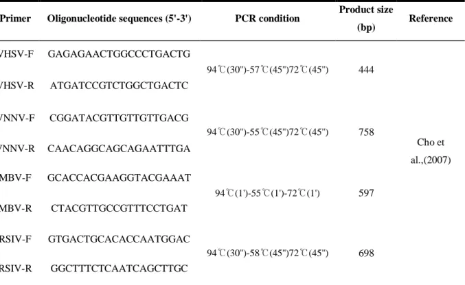
발생하고 있다. 이 질병에 감염된 어류는 안구함몰, 두부돌출 및 어체중 감소 등의 증상 을 나타내다가 폐사에 이르게 된다고 보고하였다(Tun et al., 2000). Tun et al. (2002)의 연구 에 따르면, 이러한 여윔 증상을 나타내는 어류의 장에서 3종의 점액포자충이 발견되었으 며, Enteromyxum fugu, Enteromyxum leei 및 Leptotheca fugu인 것으로 보고하였다. 자주복 여 윔증의 원인은 명확하게 밝혀지지 않았지만, 앞서 동정된 점액포자충들이 한 종 또는 두 종 이상이 감염되었을 때 동일한 증상이 나타나는 것으로 확인되었다(Ogawa and Yokoyama, 2001). Yasuda et al. (2005)은 조직병리학적 분석을 통해 장 상피세포에서 다량의 포자를 관찰하였고, 상피의 탈락 및 파괴를 초래한다고 조사하였으며, 3종의 점액포자충 에 대한 primers를 제작한 후 PCR 방법을 사용하여 원인체를 진단하였다.
제주 양식산업의 대표적 품종인 넙치는 전국 양식넙치 생산량의 50%이상을 차지하고 국내 전체 넙치 수출량의 95%를 점유하고 있는 실정이다. 그러나 제주 양식 넙치의 질 병에 의한 연간 피해액이 700억원대에 이르며 해마다 급증하고 있다. 대표적인 폐사 원 인으로는 바이러스성 질병인 viral hemorrhagic septicemia (VHS) 와 Scuticociliatida에 의한 기생충성 질병이 높은 비율을 차지하고 있으며, 세균성 질병인 연쇄구균병과 에드워드병 외에도, 원인불명으로 인한 폐사가 심각한 수준인 것으로 밝혀졌다. 국내의 여윔증은 2007년부터 제주도 넙치 양식장에서 약 20cm급 크기 전후에서 발생 하고 있으며, 1~3주 동안 발병하여 폐사를 야기시키고 있다. 일본의 자주복과는 달리, 국 내 넙치의 여윔증 발병에 대한 원인은 아직 보고되어 있지 않은 상황이며, 원인불명 질 병에 의한 피해를 최소화하기 위해서는 신속한 진단법을 마련하여 질병이 확산되는 것을 미연에 방지하는 것은 매우 중요한 점이라 할 수 있다. 따라서 본 연구에서는 제주지역에서 여윔증상을 보이는 넙치를 대상으로, 여윔증의 원 인을 밝혀내기 위해 분자생물학적 분석 및 조직병리학적 분석을 실시하였으며, 또한 여 윔증의 감염현황을 파악하기 위해 2010~2013년까지 모니터링을 실시하여 감염률을 조사
- 2 -
- 3 -
1.2 재료 및 방법
1.2.1 바이러스성 및 세균성 질병 검사
국내의 넙치 양식장에서 발생하는 여윔증의 원인체를 밝혀내기 위해, 여윔증상을 보이 는 넙치를 대상으로 바이러스성 및 세균성 질병에 대한 일반적인 검사방법에 따라 진단 하였다(Table 1 and 2). 그 결과 바이러스성 질병의 경우, 여윔 증상을 보이는 넙치에서 한 건의 발생률도 관찰되지 않아(data not shown), 여윔증의 원인체가 바이러스성 질병이 아닌 것으로 조사되었으며, 세균성 질병의 경우 Vibrio sp. Streptococcus sp. Edwardsiella sp.가 검 출되었으나 여윔 증상을 보이는 넙치에서 공통적으로는 관찰되지 않았다(data not shown). 하지만, 검출된 세균에 의해 여윔 증상을 보일 가능성이 있어, 검출된 세균들을 사용하여 인위감염 실험을 실시한 결과, 분리된 세균들이 여윔증 발병에 직접적인 영향이 없는 것 으로 조사되었다.- 4 -
Table 1. Culture mediums used in this study
Fish Target organs Medium Incubation temperature (℃)
Olive flounder Kidney, Spleen, Liver TSA, TCBS, SS, BAP 25
Table 2. Oligonucleotide primers and conditions used in PCR amplification
Primer Oligonucleotide sequences (5'-3') PCR condition Product size
(bp) Reference VHSV-F GAGAGAACTGGCCCTGACTG 94℃(30'')-57℃(45'')72℃(45'') 444 Cho et al.,(2007) VHSV-R ATGATCCGTCTGGCTGACTC VNNV-F CGGATACGTTGTTGTTGACG 94℃(30'')-55℃(45'')72℃(45'') 758 VNNV-R CAACAGGCAGCAGAATTTGA MBV-F GCACCACGAAGGTACGAAAT 94℃(1')-55℃(1')-72℃(1') 597 MBV-R CTACGTTGCCGTTTCCTGAT RSIV-F GTGACTGCACACCAATGGAC 94℃(30'')-58℃(45'')72℃(45'') 698 RSIV-R GGCTTTCTCAATCAGCTTGC 1)
VHSV, viral hemorrhagic septicaemia virus; VNNV, viral nervous necrosis virus; MBV; marine birnavirus; RSIV, red sea bream iridovirus.
- 5 -
1.2.2 여윔증 진단을 위한 primer set 제작
일본의 터봇 여윔증의 원인체로 보고된 E. leei의 small subunit ribosomal DNA gene (SSU rDNA)으로부터 제작된 MM18Sf/MM18Sr primer set (1,589 bp)를 동일하게 제작하였고(Table 3 and Fig. 1) 국내의 여윔증상 넙치의 장 조직으로부터 DNA를 분리하여 PCR 실험을 실 시한 결과, 모두 PCR 음성반응을 나타내었다(Fig. 2). 국내에서 발생하는 여윔증을 진단하 기 위해 GenBank (NCBI, USA)에 등록된 점액포자충(Myxidium sp.)의 염기서열로부터 degenerated primers를 제작하였고, 여윔증상 넙치로부터 DNA를 분리하여 PCR을 실시한 결과 양성 반응을 확인하였으며, DNA sequencing을 통한 염기서열 분석 후 국내 넙치의 여윔증 진단을 위한 새로운 EM-F/EM-R primer set를 제작하였다(Table 3).
- 7 -
Fig. 1. The small subunit ribosomal DNA (SSU rDNA) gene of Enteromyxum leei which was reported to be a causative agent of the olive flounder (Paralichthys olivaceus) emaciation disease in Japan.
- 8 -
Fig. 2. (A) Total DNA separated from the intestine of Paralichthys olivaceus having emaciation disease and PCR results obtained from a reference primer set made by using SSU rDNA gene of
Enteromyxum leei. 1-8; A: Total DNA seperated from the gut of P. olivaceus having emaciation
disease, 9-15; B: Total DNA separated from the gut of P. olivaceus having emaciation disease, (B) 1-3; DNA of E. leei provided by Japan, M; 100 bp DNA ladder.
Table 3. PCR primers used for the detection of new pathogen Primer Oligonucleotide sequences (5'-3') Expected
sizes Function MM18S-F CTGGTTGATTCTGCCAGTGGTC 1,589 bp Detection of E. leei (Palenzuela et al., 2002) MM18S-R CGGTACTAGCGACGGGCG 28S-F CTAGGGGAATCCGACTGT
313 bp House keeping gene (28S ribosomal RNA) 28S-R ATCAAACTAGAGTCAAGC
EM-F CAACCGCAATGTGTTTACTC
812 bp Detection of new pathogen (in this study) EM-R CCAAACAACCTGCCACAATG
- 9 -
1.2.3 실험어
제주도 양식 넙치를 대상으로 여윔증 원인체의 감염 현황을 조사하기 위해, 2010년부터 2013년까지 4년 동안 양식 넙치를 대상으로 2010년에 21회(6, 7, 8, 11, 12월), 2011년 25회(3, 7, 8, 10, 11월), 2012년 4회(7, 10, 11월), 2013년 16회(1, 3, 7, 8, 11, 12월)에 걸쳐 동.하절기 및 중복된 양식장을 포함하여 24개소 양식장에서 66회의 샘플링을 실시하여 총 216마리를 대상으로 실험을 수행하였고(Fig. 3), 확보된 샘플은 육안상 여윔증상을 보이며 폐사가 발 생하여 의뢰를 맡긴 양식장과 무작위로 넙치를 제공받은 양식장을 대상으로 감염조사를 실시하였다. 실험 전 수질측정기 YSI Model 650XL (YSI, USA)을 사용하여 수온측정을 실 시하였고, 실험실로 운반된 어류의 외부 및 내부증상을 확인하고, 어체의 전장 및 무게를 측정하였다.- 10 -
- 11 -
1.2.4 DNA 분리
여윔증상을 보이는 넙치로부터 간, 비장, 신장, 장, 뇌 및 아가미 등의 장기들을 적출하
였고, DNA를 분리하기 위하여 DNeasyⓇ
Blood & Tissue Kit (Qiagen Hilden, Germany)를 사용 하였다. 먼저 각 조직의 10mg에 ATL buffer 180μL와 proteinase K 20μL를 첨가하여 56℃에 서 조직이 녹을 때까지 반응시켰다. 반응 후, AL buffer 200μL를 넣어 섞은 다음 ethanol 200μL를 더하여 spin column에 옮겨 6,000 × g에서 1분간 원심분리하였다. Column을 새로 운 tube로 옮긴 후 AW1 buffer 500μL와 AW2 buffer 500μL를 이용하여 세척과정을 거친 후, AE buffer 50μL를 첨가하여 최종적으로 DNA를 분리하였다. 분리된 DNA는 실험 전까지 -80℃에 보관하였다.
1.2.5 PCR 및 DNA sequencing
PCR은 microtube에 1μM의 각 primer, 2.5 mM의 각 dNTP, 10 × G-Taq Buffer, 2.5 U G-Taq DNA polymerase (Gene Pro Themal Cycler Cosmo, Korea) 및 template DNA로서 추출된 핵산을 첨가한 후 distilled water로 PCR 혼합물의 최종 volume이 20μL가 되게 하였다. PCR 조건 은 95℃에서 3분간 pre-denaturation 시킨 후, 95℃에서 30초 denaturation, 55℃에서 30초 annealing, 72℃에서 30초 extension의 반응을 1 cycle로 하여, 35 cycles를 반응시켰다. 그리 고, 72℃에서 7분간 post-extension시켰다. PCR 후 증폭 산물은 1 × TAE buffer (40 Mm Tris-acetate, 1 mM EDTA)를 전기영동을 위한 완충액으로 하여, 0.5μg/μL ethidium bromide가 첨가 된 1% agarose gel 상에서 전기 영동한 후, UV 검출기에서 band를 관찰하였다. PCR 증폭
산물은 gel purification kit (Bioneer, Korea)를 이용하여 회수한 후, ToPo TA cloning®
kit (Invitrogen, USA)로 cloning하여 염기서열 분석을 의뢰하였다(Solgent, Korea).
1.2.6 병리조직학적 분석
병리조직학적 분석을 위해 여윔 증상을 보이는 넙치로부터 간, 비장, 신장, 장, 뇌 및 아가미 조직을 적출하여 Bouin's solution에 24시간 동안 고정한 후 70% EtOH를 사용하여 탈수하였다. 이후 파라핀 침투를 시키고(Leica EG 1150HC, Germany) 포매기(Leica Jung 820, Germany)를 사용하여 포매를 실시하였다. 이 후 마이크로톰으로 4~5μm 두께의 절편을 잘라 유리 슬라이드에 부착시켜 건조시켰다. 제작된 조직표본은 haematoxylin과 eosin (H&E)으로 염색을 실시한 후 광학현미경(Zeiss LT60, Germany)으로 검경하였다.
- 12 -
1.3 결 과
1.3.1 외부 및 내부증상
여윔증에 감염된 넙치의 대부분은 복부 부위가 심각하게 여위는 것으로 관찰되었고, 일부 체색흑화 및 해부 시 간 출혈 등의 증상이 관찰되었으나, 일본의 넙치에서 보고된 안구함몰이나 두부돌출과 같은 증상은 보이지 않았다(Fig. 4A& B).- 13 -
1.3.2 PCR 진단법 개발 및 적용
제주도 넙치 양식장에서 여윔 증상을 보이는 넙치를 대상으로 새롭게 제작한 specific EM primer set를 이용하여 PCR을 실시한 결과, 일본의 여윔증에 대한 연구에서는 원인체 가 주로 장에서 검출되는 것으로 보고하였으나, 본 연구에서는 넙치의 장뿐만 아니라 신 장에서도 양성의 결과를 나타낸다는 것을 확인하였다(Fig. 5). 여윔증에 감염된 다양한 넙 치를 대상으로 PCR을 수행한 결과, 여윔증 원인체의 검출 정도가 신장, 장, 비장의 순서 로 높은 것을 확인하였으며, 병원체의 주요 감염 표적장기는 신장 및 장 부위인 것으로 추정되었다. 또한, 심각한 경우에는 아가미를 제외한 모든 검사대상 조직에서 PCR 양성 의 결과를 확인할 수 있었다(Fig. 5).- 14 -
Fig. 5. PCR amplification from nucleic acids of intermal organs from the emaciated olive flounder (P.
olivaceus) in the level stages of infection. Lanes 1, 7, 13 and 19, Kidney; lane 2, 8, 14 and 20,
Intestine; lanes 3, 9, 15 and 21, Spleen; lanes 4, 10, 16 and 22, Liver; lanes 5, 11, 17 and 23, Brain; lanes 6, 12, 18 and 24, Gill; M, 1kb DNA ladder; N, negative control.
- 15 -
1.3.3 양식장별 감염률
2010년 6월부터 2013년 12월까지 총 66회에 걸쳐 216마리의 넙치를 대상으로 여윔증 원인체의 감염현황을 조사한 결과, 66회에 걸친 조사기간 동안의 대상 양식장 중에서 50% (33회)에서 여윔증 양성임을 확인하였다(Table 4). 그 중, 양식장별 감염률은 2010년도 21개소 중 8개소(38%), 2011년도 25개소 중 12 개소(48%), 2012년도 4개소 중 2개소(50%), 2013년도 16개소 중 11개소(60%)에서 양성임을 확인하였다(Fig. 6, Table. 4).1.3.4 개체별 감염률
총 넙치 216마리 중 93마리(43%)에서 여윔증 감염 양성임을 확인하였다. 그 중, 개체별 감염률은 2010년도 60마리 중 17마리(28.3%), 2011년도 75마리 중 32마리(42.6%), 2012년도 24마리 중 8마리(33.3%), 2013년도 57마리 중 36마리(63.1%)의 감염률을 관찰하였다(Table 4).- 16 -
Fig. 6. Gel electrophoresis of the products amplified with the primer set EM-F/R from the extracted sample of the kidney of affected olive flounder cultured in emaciated disease. Lane1, Farm-A (June, 2010); lane 2, Farm-B (October, 2011); lane 3, Farm-C (October, 2012); lane 4, Farm-D (November, 2011); lane 5, Farm-E (October, 2012); lane 6, Farm-F (November, 2012); lane 7, Farm-G (July, 2013); lane 8, Farm-N (December, 2010); lane 9, Farm-O (December, 2010); M, 1kb DNA ladder; N, negative control; P, positive control.
- 17 -
Table 4. Prevalence of emaciation disease in cultured olive flounder (P. olivaceus) of Jeju from 2010 to 2013 Year Month Water temp. (℃) Farm & no. of fish Infection rate (%) (positive no. / total no.)
Fish Farm
PCR test Total Histological examination PCR test Total 2010 June 18.5 A(6)*, B(3) 33.3% (3/9) 28.3% (17/60) 44.4% (4/9) 50% (1/2) 38% (8/21) July 19.5 C(3), D(3), E(3), F(3), G(3) 0% (0/15) 0% (0/15) 0% (0/5) August 20 E(3)*, H(3)* 66.6% (4/6) 66.6% (4/6) 100% (2/2) November 18.1 I(6)* 83.3% (5/6) NT 100% (1/1) December 15 A(1)*,D(1), J(1), K(3), L(3)*M(3)*, N(2), O(3), P(3), Q(1)*, R(3) 20.8% (5/24) NT 36.3% (4/11) 2011 May 15 S(6)* 100% (6/6) 42.6% (32/75) NT 100% (1/1) 48% (12/25) July 19.5 A(3)*,M(3),T(3) ,U(3)*,V(3)*,W(3), X(3) 28.5% (6/21) 33.3% (7/21) 28.5% (3/7) August 21.5 A(3), B(3)*,M(3), T(3)*, V(3)*,W(3), X(3), 33.3% (7/21) NT 28.5% (3/7) October 20 A(3), B(3)*,C(1)*, M(3)*,V(3), X(3), W(3),Y(2) 33.3% (7/21) 42.8% (9/21) 37.5% (3/8)
- 18 - November 18.5 T(3)*, D(3)* 100% (6/6) NT 100% (2/2) 2012 July 20 D(9), F(3) 0% (0/12) 33.3% (8/24) NT 0% (0/2) 50% (2/4) October 21 E(6)* 33.3% (2/6) 33.3% (2/6) 100% (1/1) November 18 F(6)* 100% (6/6) 100% (6/6) 100% (1/1) 2013 January 14.8 J(6) 0% (0/6) 63.1% (36/57) NT 0% (0/1) 60% (11/16) May 16 Q(4)* 100% (4/4) 100% (4/4) 100% (1/1) July 19.7 G(4)*, Q(4)* 87.5% (7/8) 100% (8/8) 100% (2/2) August 22 E(5)*, K(5)* 80% (8/10) NT 100% (2/2) September 21 S(2)* 100% (2/2) 100% (2/2) 100% (1/1) November 18.8 K(3)*, J(4)*,U(4)* 63.6% (7/11) NT 100% (3/3) December 15.6 C(1), J(3)*, Q(3), R(3), V(3)*, Y(3) 50% (8/16) 62.5% (10/16) 33.3% (2/6) Total 43% (93/216) 50% (33/66) * : Positive NT: not tested
- 19 -
1.3.5 크기별 감염률
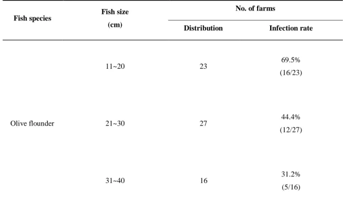
2010~2013년도까지 총 66번의 샘플링 중 11~20cm가 23개소, 21~30cm 27개소, 31~40cm 16개소로 분류되었으며, 크기별 감염 현황 결과, 11~20cm 23개소 중 16개소(69.5%), 21~30cm 27개소 중 12개소(44.4%), 31~40cm 16개소 중 5개소(31.2%)의 검출률을 나타내었 다(Table 5).Table 5. Prevalence of causative agent of the emaciated disease according to the size of olive flounder (P. olivaceus).
Fish species Fish size (cm)
No. of farms
Distribution Infection rate
Olive flounder 11~20 23 69.5% (16/23) 21~30 27 44.4% (12/27) 31~40 16 31.2% (5/16)
- 20 -
1.3.6 Fish weight (index)
PCR에서 양성반응과 음성반응을 나타낸 넙치를 대상으로 체장에 따른 체중을 관찰한 결과, 여윔증에 감염된 넙치가 여윔증이 발병하지 않는 넙치보다 30~40% 낮은 체중이 관찰되었다(Fig. 7).
Fig. 7. Monitoring of the index content in the emaciated olive flounder (P. olivaceus). ◯: Uninfected olive flounder,
●
: infected olive flounder.- 21 -
1.3.7 병리조직학적 검사
각 장기의 조직 절편을 광학현미경으로 관찰한 결과, 여윔증에 감염된 넙치의 신장과 장 부위에서 원형 또는 난원형의 포자가 다수 관찰되었고, 장 부위보다 신장 부위에서 더 많은 수의 포자가 관찰되었으며, 기부골절, 사구체, 핵 등의 파괴와 변형체 (plasmodium)가 일부 관찰되었다(Fig. 8A and B). 감염된 장 상피 부위에서는 포자가 관찰 되었지만 변형체는 관찰되지 않았으며, 심한 경우 조직의 탈락 및 파괴가 나타났다(Fig. 8C and D). 신장 및 장을 제외한 다른 장기에서는 일부 포자가 관찰되었지만, 포자의 수 가 신장, 및 장에 비해 적은 수가 관찰되었거나 특이적인 조직학적 이상소견이 관찰되지 않았다(data not shown).
- 22 -
Fig. 8. Histological findings of kidney (A, ×200 and B, ×400) and intestine (C, ×200 and D, ×400) from the emaciated olive flounder (P. olivaceus). Note microsporean spores both in emaciated disease the glomerulus (arrows) and in the gut epithelium (arrows). H & E stain. Bar = 20μm.
- 23 -
1.3.8 염기서열 분석
검출된 PCR products에 대한 유전자 염기서열을 분석한 결과, 다른 시기에 제주도의 다 양한 양식장의 여윔증상 넙치로부터 검출된 병원체들 간의 유전자 상동성은 서로 99% 이상인 것으로 나타났고(Fig. 9), 제주도의 넙치양식장에서 발생하는 여윔증의 원인체는 모두 동일한 기생충 종인 것으로 확인되었다. GenBank database를 활용한 유전자 비교 분 석을 실시한 결과, 일부 염기서열인 150 bp가 Myxobolus sp. (Accession No. JN616264.1) 및Cystodiscus axonis (Accession No. JN977605.1)와 부분적으로 86% 및 88%의 유사성을 각각
- 24 -
Fig. 9. Comparison analysis of the DNA nucleotide sequences of pathogens detected form the emaciated olive flounder (P. olivaceus). Primers used in PCR are in boxes.
- 25 -
1.4 고 찰
본 연구에서는 일본의 여윔증에 대한 연구방법 및 결과들을 국내의 여윔증 연구를 위 하여 적용해 보았고, 우리나라의 여윔증 원인체는 일본에서 보고된 여윔증 원인체와 형 태학적으로는 유사하지만, 분자유전학으로는 전혀 다른 종이라는 것을 밝혀내었다. 먼저 분자생물학적 진단방법을 적용하고자 일본 여윔증의 원인체인 E. leei의 primer set (Palenzuela et al., 2002)를 제작한 후 국내 여윔증상 넙치의 장을 이용하여 PCR을 실시하 였지만 음성 반응을 나타내었다. 이러한 결과는 Choi et al. (2012)의 보고와 일치하였으며, 국내에서 발병하는 넙치의 여윔증 원인체는 일본에서 보고된 primer set를 사용한 PCR에 서는 검출되지 않는다는 것을 확인하였다. 본 연구에서는 국내 양식넙치의 여윔증 진단 을 위한 primer set (EM-F/EM-R)를 처음으로 개발하였고(Table 3), 여윔증상을 나타내는 넙 치를 대상으로 한 PCR 실험에서 명확한 band가 나타나는 것을 확인하였으며(Fig. 5 and 6), 향후 국내의 넙치에서 여윔증의 감염 진단이 필요한 경우, 이와 같은 방법이 유용하게 활용될 것으로 여겨진다. 여윔증에 대한 모니터링을 실시한 결과, 여윔증에 감염된 넙치의 대부분은 복부 부위 에 심각한 여윔이 관찰되었으며(Fig. 4A), 여윔증에 감염된 넙치가 여윔증이 발병하지 않 는 넙치보다 30~40% 낮은 체중이 관찰되었고(Fig. 7), 간혹 육안상 복부부위가 여위지 않 은 넙치에서도 감염이 발생하여 여윔증은 감염 후 여위는 것으로 판단된다. 양식장별 감염률은 2010년도 21개소 중 8개소(38%), 2011년도 25개소 중 12개소(48%), 2012년도 4개소 중 2개소(50%), 2013년도 16개소 중 11개소(60%)에서 양성임을 확인하였 다(Table 2). 그 중 양식장에서 여윔증 발생이 의심되어 분석을 의뢰해온 양식장의 경우에 는 71.4% (25개소/35개소)의 높은 검출율을 보였고, 무작위로 여윔증 모니터링을 실시한 양식장의 경우에는 25.8% (8개소/31개소)의 낮은 검출률을 나타내었다(data not shown). 또 한, 검출된 양식장은 이후의 감염 발생률이 높은 것으로 확인되었고, 향후에는 발병경로, 기회감염성, 숙주의 감수성 등 질병에 대한 전반적인 연구가 뒤따라야 할 것이다. 여윔증 원인체에 의한 연도별 감염률은 2010년도 60마리 중 17마리(28.3%), 2011년도 75마리 중 32마리(42.6%), 2012년도 24마리 중 8마리(33.3%), 2013년도 57마리 중 36마리 (63.1%) 등으로 나타났고, 조사 기간 중 수온에 따른 감염률은 여름철부터 겨울철까지 다 양하게 나타나는 것으로 조사되었다(Table 4). 제주도의 양식장은 대부분 연중 17℃ 내외 의 지하해수를 혼합 사용하여 여름철에도 사육수온을 23℃ 내외, 겨울철에는 14℃ 이상 을 유지하고 있어(Oh et al., 1998), 여윔증이 연중 발생하는 원인으로서 제주도의 시기적인 수온변동이 크지 않은 것도 연관성이 있을 것으로 추정된다.
- 26 - 현재까지 어류에서 보고되는 여윔증 원인체에 대한 생활사가 밝혀진 바는 없으며, 감 염되는 어류의 크기 및 감염경로 등에 대해서도 조사가 부족한 실정이다. 본 연구에서 넙치의 크기별 감염현황을 조사한 결과, 11~20cm가 23개소 중 16개소(69.5%), 21~30cm가 27개소 중 12개소(44.4%), 31~40cm가 16개소 중 5개소(31.2%)에서 검출률을 나타내었다 (Table 5).
이전 연구들에서는 sharpsnout sea bream Puntazzo puntazzo, red sea bream Pagrus major, red drum Sciaenops ocellatus이 Myxidium leei에 감염되었을 때 공통적으로 장 조직에서 포자를 관찰할 수 있었다고 보고하였다(Le Breton and Marques, 1995; Diamant, 1998; Athanassopoulou et al., 1999). 또한, Myxosporea에 감염된 스페인의 turbot, Scophthalmus maximus 에서도 포자 를 소화관이나 장에서 확인할 수 있었으며 이러한 결과는 turbot의 성장에 영향을 미쳤을 것이라 제시하였다(Beaman et al., 1999). Bartholomew et al. (1989)은 Ceratomyxa shasta 에 감염된 연어과 어류에서도 포자형태의 기생충을 장 조직에서 관찰할 수 있었다고 보고하 였는데 이와 같이 유럽의 여러 연구결과에서 점액포자충에 감염된 어류는 주로 장에서 감염이 이루어지는 것으로 보고되었다. 또한, 일본에서 발생하는 여윔증의 원인 기생충도 장과 담낭에서 확인되었다고 보고하였다(Tun et al., 2002). 이러한 결과는 우리나라 여윔증 과 조직학적으로 유사한 경향을 보여주고 있으며, 여윔증에 심하게 감염된 넙치의 경우, 전 장관의 상피, 점막고유층, 근층 등에서 포자가 관찰되는 부분과도 일치하였다. 그러나, 감염부위에 있어서 유럽 및 일본에서는 장 조직을 주요한 감염 조직으로 보고하였지만, 본 연구결과에서 국내 넙치의 여윔증 원인체는 장 조직보다 신장 조직에서 더욱 높게 감 염되는 것으로 조사되어(Fig. 8), 외국에서의 보고와 차이가 있는 것으로 나타났다. 또한 기생충성 질병으로 잘 알려진 Ichthyobodo sp., Cryptocaryon sp., Myxobolus sp., Miamiensis sp.,
Myxidium sp. 등과 조직학적으로 비교했을 때, 대부분 다른 형태가 관찰되어 새로운 기생
충 종인 것으로 추정되며, 향후에는 여윔증에 대한 형태학적 동정에 대한 연구가 이루어 져야 할 것이다.
PCR에서 검출된 샘플을 대상으로 염기서열을 분석한 결과, 국내 넙치의 여윔증 원인 체들 간의 DNA nucleotide sequences의 상동성은 99% 이상인 것으로 나타났다(Fig. 9). 그리 고, GenBank에 등록된 Myxobolus sp. 및 Cystodiscus axonis 의 일부 염기서열과 80% 정도의
상동성이 있는 것으로 확인되어 아직 GenBank database에 등록되지 않은 새로운 기생충
종인 것으로 판명되었고, 향후에는 이 병원체에 대한 DNA walking과 같은 분자생물학적 분석을 통한 더욱 세부적인 연구가 뒤따라야 할 것이다.
- 27 - 일본에서 보고된 결과와 비교했을 때, 장 조직에서 형태학적으로 유사한 포자가 관찰된 것은 동일하지만, 아직 GenBank에 등록되지 않은 새로운 기생충 종인 것으로 조사되었다. 그리고, 국내의 넙치 양식장에서 많은 문제를 야기시키는 여윔증에 대한 진단법을 처음 개발하여 제시하였고, 제주도 내에서 양식되는 유사한 크기의 넙치에서 발생할 수 있는 여윔증을 추정할 수 있는 기초 자료로서 활용될 수 있을 것으로 사료되며, 여윔증과 같 이 양식장에서 새로운 질병 유입에 대한 위험성을 낮추기 위해서는 지속적인 질병 예찰 및 감시가 필요할 것으로 판단되며, 향후에는 여윔증 원인 기생충의 동정에 대한 연구가 이루어져야 할 것이다.
- 28 -
제 2장 2014~2015년도 제주도 양식 넙치에 대한 여윔증 감염 조
사
2.1 서론
1990년대부터 각종 해수어의 양식이 활발하게 이루어져 각국에서 종묘 수입이 성해지 자 방역체계가 미비한 상태에서 수입종묘와 같이 질병도 함께 도입되어 우리나라에 없었 던 기생충이 유행하기 시작하였으며(Chun, 2006), 질병의 발병 양상 또한, 양식 초창기에 는 고수온기에 기생충 및 세균에 의한 단독 감염이 주를 이루었으나 최근에는 수온과 상 관없이 연중 다양한 병원체가 혼합감염의 형태로 질병을 일으키고 있어 수산생물의 대량 폐사를 유발시키기도 한다(Kim et al.,2006). 최근 들어 외래 질병의 유입 가능성이 증가하 면서 질병을 전반적으로 모니터링하거나 질병과의 상관관계를 규명하고자 하는 연구가 점차 증가하고 있는 추세이다(Cho et al., 2009; Jung et al., 2012; Song et al., 2013). 그러나 현 재까지 국내 양식 어류에 대한 질병 조사는 대부분 특정 질병을 중심으로 조사되어 왔으 며(Kim et al., 2006; Cho et al., 2008; Kim et al., 2010), 원인불명의 질병에 대해서는 연구가 미 미한 실정이다.최근 제주도 내 양식장의 약 20cm 전후 크기의 넙치에서 여위어가는 원인불명의 질병 이 발생하여, 최초 발병증상을 보인 이후 1~3주 동안 폐사가 되는 사례가 증가하고 있으 며, 이 질병에 감염된 넙치는 체색흑화, 어체중 감소 및 간 출혈 등의 증상을 나타낸다 (Kim et al., 2015). Kim et al. (2015)의 연구에 따르면, 주변 국가인 일본의 터봇 여윔증의 원 인체로 보고된 E. leei의 primer set (1,589 bp)를 제작하여(Palenzuela et al., 2002) 국내 여윔증 넙치를 대상으로 PCR실험을 실시한 결과, 음성반응이 관찰되어 동일한 원인체가 아닌 것으로 확인되었다. 또한, Kim et al. (2015)은 GenBank (NCBI, USA)에 등록된 점액포자충 (Myxidium sp.)의 염기서열로부터 degenerated primers를 제작하여, 여윔증상 넙치로부터 DNA를 분리한 후 PCR을 실시한 결과 양성반응이 나타났고, DNA sequencing을 통해 염기 서열을 분석한 결과 일부 염기서열이 점액포자충과 일치하였으나 아직 GenBank에 등록 되지 않은 새로운 기생충 종인 것으로 보고하였다(Kim et al., 2015). 하지만, 국내에서 발 생하는 여윔증 원인체의 발병 동향에 대한 연구는 아직 보고되어 있지 않은 실정이며, 양식 어류에 대한 질병 모니터링은 시기별 질병 관리에 유용한 자료를 제공하여, 자국 내 특정 질병에 대한 기초 자료로서 매우 중요한 역할을 하며(OIE, 2008), 양식생물에서 발생하는 질병에 대하여 효과적인 방제 대책을 수립하기 위해서는 양식장별 질병 발생
- 29 -
현황에 대한 정확한 모니터링을 수집하는 것이 선행되어야 한다.
따라서, 본 연구에서는 2014~2015년도에 제주 지역 60개소 넙치 양식장을 대상으로 여 윔증 감염현황을 파악하기 위하여, Kim et al. (2015)이 보고한 primer set를 사용한 PCR법을 통해 시기별, 어체 크기별 발생 현황에 대한 기초자료로 제공하고자 감염률을 조사하였 으며, 또한 cohabitation test를 통하여 동일한 어종 및 타 어종으로의 전이가 발생하는지를 확인하고자 하였다.
- 30 -
2.2 재료 및 방법
2.2.1 2014년도 양식넙치 여윔증 모니터링
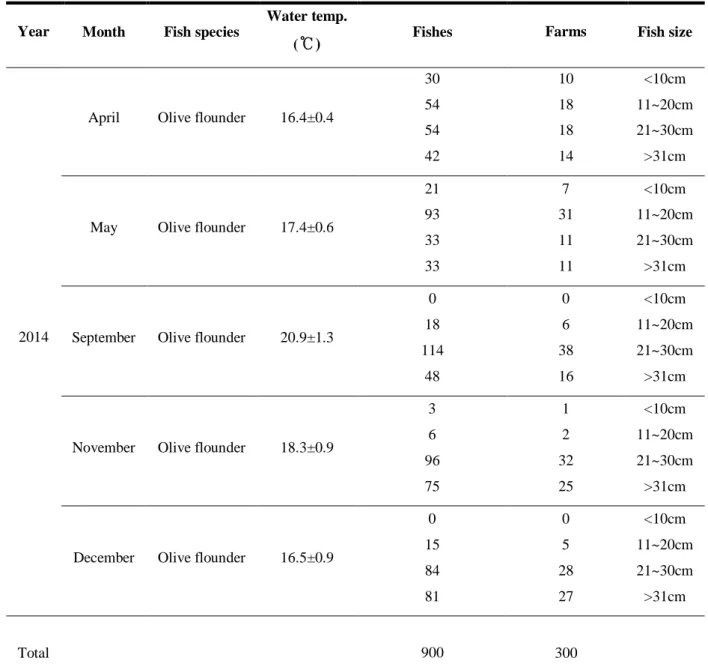
여윔증 감염현황 조사를 위해, 2014년도에 4, 5, 9, 11, 12월에 모니터링을 수행하였으며, 1 년간 총 5회 실시하였다. 조사 지역 양식장은 Fig. 10과 같이 제주도의 제주시와 서귀포 시 해역의 넙치를 양성하는 60개소 양식장을 대상으로 질병여부와 상관없이 각각의 양식 장에서 넙치를 무작위로 3마리씩(5~600g 이내)을 수거하여 1회에 180마리씩 총 900마리 를 조사하였고, 수온측정은 수질측정기 YSI Model 650XL (YSI, USA)를 사용하여 수온을 측정하였다(Table. 6).- 31 -
Table 6. Sample information employed for the investigation of emaciation infection in rearing olive flounder (P. olivaceus) of Korea in 2014
Year Month Fish species
Water temp.
(℃) Fishes Farms Fish size
2014
April Olive flounder 16.4±0.4
30 54 54 42 10 18 18 14 <10cm 11~20cm 21~30cm >31cm
May Olive flounder 17.4±0.6
21 93 33 33 7 31 11 11 <10cm 11~20cm 21~30cm >31cm
September Olive flounder 20.9±1.3
0 18 114 48 0 6 38 16 <10cm 11~20cm 21~30cm >31cm
November Olive flounder 18.3±0.9
3 6 96 75 1 2 32 25 <10cm 11~20cm 21~30cm >31cm
December Olive flounder 16.5±0.9
0 15 84 81 0 5 28 27 <10cm 11~20cm 21~30cm >31cm Total 900 300
- 32 -
2.2.2 2015년도 양식넙치 여윔증 모니터링
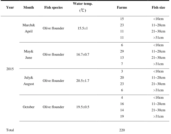
2015년도에는 3~4월 60개소, 5~6월 55개소, 7~8월 52개소, 10월 53개소 양식장을 대상으 로 1개의 양식장 당 3마리씩 신장 조직을 1.5mL microtube에 모아 양식장별 감염 조사를 2014년도와 동일하게 실시하였다(Table 7).
Table 7. Sample information employed for the investigation of emaciation infection in rearing olive flounder (P. olivaceus) of Korea in 2015
Year Month Fish species
Water temp.
(℃) Farms Fish size
2015
March&
April Olive flounder 15.5±1
15 23 11 11 <10cm 11~20cm 21~30cm >31cm May&
June Olive flounder 16.7±0.7
6 29 13 7 <10cm 11~20cm 21~30cm >31cm July&
August Olive flounder 20.5±1.7
3 20 23 6 <10cm 11~20cm 21~30cm >31cm
October Olive flounder 19.5±0.5
4 16 14 19 <10cm 11~20cm 21~30cm >31cm Total 220
- 33 -
2.2.3 DNA 분리
DNA를 분리하기 위하여 Kim et al. (2015)에 보고된 방법을 참고하여 신장부위를 대상
으로 DNeasyⓇ
Blood & Tissue Kit (Qiagen Hilden, Germany)를 사용하여 DNA를 분리하였다. 먼저 ATL buffer 180μL와 proteinase K 20μL를 첨가하여 56℃에서 조직이 녹을 때까지 반응 시켰다. 반응 후, AL buffer 200μL를 넣어 섞은 다음 ethanol (100%) 200μL를 더하여 spin column에 옮겨 6000 × g에서 1분간 원심분리 하였다. Column을 새로운 tube로 옮긴 후 AW1 buffer 500μL와 AW2 buffer 500μL를 이용하여 세척과정을 거친 후, AE buffer 50μL를 첨가하여 DNA을 분리하였다. 분리된 DNA는 실험 전까지 -80℃에 보관하였다.
2.2.4 PCR
PCR에 사용한 primer는 Kim et al. (2015)이 여윔증 진단을 위하여 제시하는 primers를 사 용하였으며(Table 3), PCR은 microtube에 1μM의 각 primer, 2.5 mM의 각 dNTP, 10 × G-Taq Buffer, 2.5 U G-Taq DNA polymerase (Gene Pro Themal Cycler Cosmo, Korea) 및 template DNA 로서 추출된 핵산을 첨가한 후 distilled water로 PCR 혼합물의 최종 volume이 20μL가 되 게 하였다. PCR 조건은 95℃에서 3분간 pre-denaturation 시킨 후, 95℃에서 30초 denaturation, 55℃에서 30초 annealing, 72℃에서 30초 extension의 반응을 1회로 하여, 35회 반복하여 반응시켰다. 그리고, 72℃에서 7분간 post-extension시켰다. PCR 후 증폭 산물은 1ⅹTAE buffer를 전기영동을 위한 완충액으로 하여, 0.5 μg/μL EtBr이 첨가된 1% agarose gel 상에서 전기영동한 후, UV 검출기를 이용하여, ultraviolet상에서 검출되는 산물의 크기를 관찰하였다. 증폭된 PCR 산물은 gel purification kit (Bioneer, Korea)를 이용하여 회수한 후,
Topo TA cloning® kit (Invitrogen, USA)로 cloning하여 염기서열을 확인하였다. 염기서열의 분
- 34 -
2.2.5 Cohabitation test
감염어로부터 물을 통한 감염이 가능한지를 확인하기 위하여 Ishimatsu et al., (2007)의 방법을 사용하여 cohabitation 실험을 실시하였다. 먼저 PCR에 의해 여윔증 감염이 확인 된 넙치를 donor group으로 하였고, 음성으로 판정된 넙치, 참돔, 감성돔을 recipient group 으로 지정하여 두 가지 실험을 실시하였다. 첫 번째 실험에서는 여윔증에 걸린 넙치 (16.3±0.4 cm, 40±5.23 g) 15마리와 여윔증이 감염되지 않은 넙치(16.7±0.9 cm, 53.4±8.47 g) 15 마리를 이용하여 cohabitation 실험을 실시하였다(Fig. 11). 실험에 사용된 넙치는 100L 플 라스틱 수조에 recipient group의 꼬리 지느러미를 조금 잘라내어 구별하였다. 두 번째 실 험에서는 넙치 외 타 어종인 참돔(Pagrus major)과 감성돔(Acanthopagrus schlegelii)으로 여 윔증의 전이가 이루어지는지를 조사하고자 하였다. 참돔 및 감성돔이 잡아먹히는 것을 방지하기 위하여 100L 수조 내에 플라스틱 그물망을 사용하여 구역을 나눈 후, 여윔증 감염이 확인된 넙치 10마리와 여윔증 음성 판정이 확인된 참돔(6.5±0.5 cm, 3.5±0.7 g)과 감 성돔(17.3±1cm, 20.5±1.03 g)을 각각 10마리를 이용하여 cohabitation 실험을 실시하였다. 실 험기간 동안의 사육 수온은 19±1℃를 유지시켜 주었으며 1일 1회씩 상업용 사료를 공급 한 후, 사육수를 환수시켰다. 매일 폐사 정도를 확인하였고, 폐사어는 조직병리학적 관찰 및 PCR 방법을 사용하여 여윔증 감염 여부를 확인하였다.
- 35 -
여윔넙치 → 정상넙치(꼬리표시) 넙치
참돔 감성돔
넙치→참돔, 감성돔 사료공급, 환수, 폐사어 관찰
Fig. 11. Experimental transmission of emaciation disease to olive flounder (P. olivaceus), red sea bream (P.
- 36 -
2.2.6 병리조직학적 검사
여윔증에 감염된 넙치를 대상으로 신장부위를 적출하여 Bouin’s solution에 24시간 동안 고정한 후 70% EtOH 탈수하였다. 이후 파라핀 침투를 시키고(Leica EG 1150HC, Germany) 포매기(Leica Jung 820, Germany)를 사용하여 포매를 실시한 후 마이크로톰으로 4~5μm 두 께의 절편을 잘라 유리슬라이드에 부착시켜 건조시켰다. 제작된 조직표본은 haematoxylin 과 eosin (H&E)으로 염색을 실시한 후 광학현미경(Zeiss LT60, Germany)으로 검경하였다.
- 37 -
2.3 결 과
2.3.1 2014년도 양식장 시기별 감염률
본 연구에서는 국내 주요 넙치 양식 지역인 제주도를 대상으로 총 60개소 양식장으로 부터 4, 5, 9, 11, 12월에 양식 넙치 총 900마리에 대하여 여윔증 감염 현황을 조사하였다. 제주지역 넙치 양식장에 심각한 피해를 주고 있는 여윔증에 감염된 넙치는 개체에 따라 다소 차이가 있었으나, 전반적인 주요 외부증상은 체색흑화 및 복부 부위에 심각한 여윔 증상이 관찰되었고, 주요 내부증상은 간 출혈 및 간 위축 증상이 관찰되었다(data not shown). 제주지역 넙치 양식장에 심각한 피해를 주고 있는 여윔증에 대한 감염 현황을 조사한 결과, 60개소 양식장으로부터 채집한 180마리 중 2014년 4월 13개소의 26마리(14.4%), 5월 11개소의 12마리(6.6%), 9월 32개소의 44마리(24.4%), 11월 23개소의 36마리(20%), 12월 42개소의 78마리(43.3%)에서 PCR 양성반응을 나타내었다(Table 8 and Fig. 12). 또한, 시기별 감염률은 봄철부터 겨울철까지 다양한 감염률이 관찰되었으며(Table 8), 그 중, 수온이 떨 어지기 시작하는 9월(가을)부터 12월(겨울)에 가장 높은 감염률이 관찰되었다(Table 8).
- 38 -
Table 8. Monitoring of emaciation disease for olive flounder (P. olivaceus) farms in 2014
Year Month
Detection rate (%) by size(cm) group
>10 11~20 21~30 <31 Total Fish Farm 2014 April 10 (1/10) 22.2 (4/18) 44.4 (8/18) 0 (0/14) 14.4 (26/180) 21.6 (13/60) May 14.2 (1/7) 9.6 (3/31) 54.5 (6/11) 9 (1/11) 6.6 (12/180) 18.3 (11/60) September - 100 (6/6) 55.2 (21/38) 31.2 (5/16) 24.4 (44/180) 53.3 (32/60) November 0 (0/1) 100 (2/2) 50 (16/32) 20 (5/25) 21.1 (38/180) 38.3 (23/60) December - 100 (5/5) 75 (20/28) 62.9 (16/27) 42.2 (76/180) 71.6 (43/60) Total 11.1 (2/18) 32.2 (20/62) 56.6 (72/127) 30.1 (28/93) 21.7 (196/900) 40.6 (122/300)
Fig. 12. Gel electrophoresis of the products amplified with the primer set EM-F/R from the extracted sample of the kidney of affected olive flounder cultured in emaciated disease. Lanes 1, 3, 4, 5, 6, 7, 8, 10, 11, 12, 13 and 14, positive samples; Lanes 2 and 9, negative samples; M, 1kb DNA ladder; N, negative control.
- 39 -