Translationally Controlled Tumor Protein
induces epithelial to mesenchymal
transition and promotes cell migration,
invasion and metastasis
Seong-Yeon Bae, Hyun Jung Kim, Kong-Joo Lee & Kyunglim Lee
Graduate School of Pharmaceutical Sciences, College of Pharmacy, Ewha Womans University, Seoul 120-750, Korea.
Translationally controlled tumor protein (TCTP), is a highly conserved protein involved in fundamental processes, such as cell proliferation and growth, tumorigenesis, apoptosis, pluripotency, and cell cycle regulation. TCTP also inhibits Na,K-ATPase whose subunits have been suggested as a marker of epithelial-to-mesenchymal transition (EMT), a crucial step during tumor invasiveness, metastasis and fibrosis. We hypothesized that, TCTP might also serve as an EMT inducer. This study attempts to verify this hypothesis. We found that overexpression of TCTP in a porcine renal proximal tubule cell line, LLC-PK1, induced EMT-like phenotypes with the expected morphological changes and appearance of EMT related markers. Conversely, depletion of TCTP reversed the induction of these EMT phenotypes. TCTP overexpression also enhanced cell migration via activation of mTORC2/Akt/GSK3b/b-catenin, and invasiveness by activating MMP-9. Moreover, TCTP depletion in melanoma cells significantly reduced pulmonary metastasis by inhibiting the development of mesenchymal-like phenotypes. Overall, these findings support our hypothesis that TCTP is a positive regulator of EMT and suggest that modulation of TCTP expression is a potential approach to inhibit the invasiveness and migration of cancer cells and the attendant pathologic processes including metastasis.
T
ranslationally controlled tumor protein (TCTP) is a highly conserved multifunctional protein present in all eukaryotes, across animal and plant kingdoms. TCTP regulates numerous fundamental processes by inter-acting with many cellular proteins. Since its discovery in ascites tumor cells, TCTP has been implicated in tumorigenesis and cancer progression. Several studies revealed that TCTP acts as a cell survival protein modu-lating apoptosis. TCTP also regulates cell cycle, interacting with microtubules1. Depletion of TCTP by shRNA in colon cancer cell lines significantly reduced cell migration, invasion and hepatic metastasis2. However, the mechanisms by which TCTP contributes to cancer metastasis are not fully understood.Following our finding that TCTP interacts with the third cytoplasmic domain of Na,K-ATPase a subunit and inhibits the pump activity3, inhibition of Na,K-ATPase has been implicated in pathologic states including hypertension, cataract, and tumorigenesis4–6. Moreover, Na,K-ATPase subunits have been suggested as markers of epithelial to mesenchymal transition (EMT)7. Na,K-ATPase expression was found reduced during TGF-b1 mediated EMT. These findings together, suggest a possible association of TCTP and EMT.
EMT is a pivotal biological process that allows a well-polarized epithelial cell, which is immotile which normally interacts with basement membrane, to undergo multiple biochemical changes to mesenchymal cell phenotypes, including enhanced migratory capacity, invasiveness, elevated resistance to apoptosis and greatly increased production of ECM components8,9. During EMT, epithelial cells change from cobble stone-like mor-phology and acquire scattered, fibroblast-like mormor-phology that characterizes mesenchymal cells, along with altered cell adhesion molecules, enhanced migratory capacity, invasiveness, elevated resistance to apoptosis, greatly increased production of extracellular matrix (ECM) components8,9and invasiveness derived via extra-cellular matrix degradation. EMT harmonizes with the reverse process, known as mesenchymal-epithelial trans-ition (MET) has been shown to play important roles in developmental process and tissue repair10,11. Aberrant regulation of EMT results in pathological processes such as fibrosis, tumor invasiveness, and metastasis, the process by which cancer cells leave the primary tumor environment and migrate to distant sites12,13.
OPEN
SUBJECT AREAS: METASTASES EPITHELIAL-MESENCHYMAL TRANSITION Received 4 August 2014 Accepted 29 December 2014 Published 27 January 2015 Correspondence and requests for materials should be addressed to K.L. ([email protected]. kr)The reported reduction in Na,K-ATPase expression during TGF-b1 mediated EMT process suggested to us a possible relationship between TCTP which inhibits Na,K-ATPase and EMT and led us to hypothesize that TCTP induces EMT and contributes to meta-stasis by promoting EMT process. In this study we describe our attempts to test this hypothesis by focusing on the roles of and interrelationship between TCTP, and EMT in metastasis.
Results
Ectopic overexpression of TCTP promotes EMT and enhances cell migration.Several studies showed that TCTP levels increase in colon cancer14, prostate cancer15and hepatocellular carcinoma (HCC)16. In addition, a strong correlation between the expression levels of TCTP and degree of metastasis was observed in ovarian cancer17, colon
cancer cell2, and human glioma18. It has been well established that TCTP acts as an anti-apoptotic protein and contributes to malignancy19. Although TCTP is clearly associated with cancer progression and metastasis, the exact role of TCTP on cancer metastasis is unclear. We tested our hypothesis that TCTP increases metastasis by inducing EMT, employing LLC-PK1- renal proximal tubular epithelial cells transiently altered by adenoviral vector to overexpress TCTP. Phase contrast microscopic studies indicated that the TCTP-overexpressing cells lost cell-cell contacts and acquired dispersed appearance, which are hallmarks of cellular/ morphologic changes during EMT (Figure 1a)20. Immunoblotting studies demonstrated alterations in the epithelial and mesenchymal markers in these cells. We also observed reduction in the epithelial marker; E-cadherin, and increases in the mesenchymal markers,
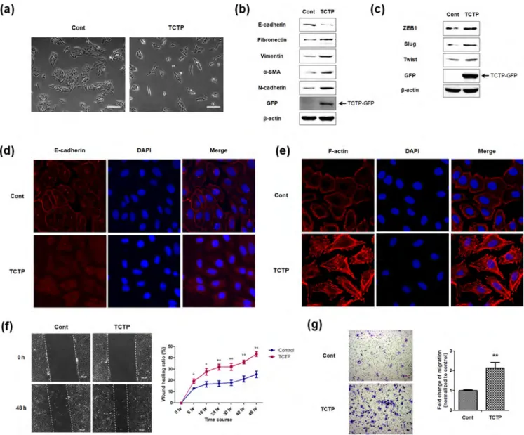
Figure 1|Ectopic overexpression of TCTP promotes EMT and enhances cell migration. (a) Phase-contrast microscopic images of LLC-PK1 cells infected with control Ad-GFP virus (Cont) or Ad-TCTP-GFP virus (TCTP). Images were taken with 3100 magnification. scale bar 200mm (b) Expression levels of epithelial marker as well as mesenchymal markers were examined by immunoblotting of control and TCTP overexpressed cells. (c) Expression levels of transcription repressors of E-cadherin were examined by immunoblotting. b-actin was used as a loading control. (d, e) Fluorescence microscopic staining of E-cadherin (d) and F-actin (e) were performed with control and TCTP overexpressed cell (red). Nuclear DNA was stained with DAPI (blue). (f) Wound healing assays were performed with control and TCTP overexpressed cells for 48 hours. Representative images were taken at 0 h and 48 h after wounding, as indicated. Images were taken with 3100 magnification using. Quantification was carried out by measuring the distance migrated compared with the controls. Values are means 6 S. E. M. of three experiments. *P , 0.05, **P , 0.01 (g) Transwell migration assays were performed with LLC-PK1 cells infected with adeno-GFP virus or adeno-TCTP-GFP virus over 18 h. Experiments were done in triplicate and representative images were taken with 3100 magnification using. Quantification was carried by counting the number of migratory cells that had infiltrated the filter. Values are means 6 S. E. M. **P , 0.01.
fibronectin, vimentin, a-smooth muscle actin (a-SMA) and N-cadherin, hallmarks of EMT induced by ectopic expression of TCTP (Figure 1b). Because of the demonstrated role of transcriptional repressors in the loss of E-cadherin21, we also examined the ex-pression levels of E-cadherin transcription repressors such as ZEB1, slug and twist, by immunoblotting, and found that these repressors were elevated by TCTP overexpression (Figure 1c). Furthermore, we also confirmed that TCTP induces the expression of mesenchymal markers in cancer cell lines, A549 and HeLa cells (Figure S1). Next, we immunostained E-cadherin and mapped the changes in the localization of cadherin caused by TCTP overexpression. E-cadherin was found localized in areas of cell-cell contact in the control cells, in contrast TCTP overexpressing cells which showed reduced membrane localization of E-cadherin (Figure 1d).
We also examined whether TCTP overexpression induces cyto-skeletal reorganization of actin cytoskeleton, which has been shown to lead to morphologic and motility changes22. Control cells exhib-ited cortical actin staining, and actin filament bundles below the plasma membrane, whereas TCTP overexpressing cells showed F-actin stress fibers (Figure 1e). Because acquisition of EMT phenotype alters migratory ability of epithelial cells, we examined the effect of TCTP overexpression on cell migration using both wound-healing and transwell migration assays. In wound healing assay, cells were allowed to starve enough to minimize the effect of TCTP on cell proliferation and complicate the interpretation of migration assays. We found that the wound healing capacities of TCTP overexpressing cells increased for 48 hours after wounding (Figure 1f). Transwell migration assay revealed that the migration capacity of the TCTP
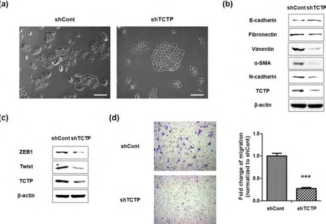
overexpressing cells increased over two (2.14) fold (Figure 1g). These results indicate that ectopic expression of TCTP induces all the EMT phenotypes examined, and thereby promotes cell migration. Silencing TCTP reverses EMT phenotypes and suppresses cell migration.Next, we examined whether down-regulation of TCTP can reverse EMT phenotypes and suppress migration of LLC-PK1 cells. For this, we depleted TCTP using shRNA transfection. As shown in Figure 2, the TCTP-depleted cells exhibited: 1. Cobblestone-like cell clusters (Figure 2a); 2. Elevated expression of E-cadherin, as well as reduced expression of mesenchymal markers in immunoblotting experiment (Figure 2b); 3. Decreased expression of ZEB1 and Twist, transcription repressors of E-cadherin (Figure 2c). These results were also confirmed in both A549 and HeLa cells (Figure S2); and 4. Significantly slower serum-induced directional migration, compared with control cells in transwell migration assays (Figure 2d). These findings clearly show that depletion of TCTP inhibits the development of mesenchymal-like phenotypes and confirm TCTP’s role in positively regulating EMT.
TCTP enhances cell invasion through MMP-9 activation.EMT is implicated in the remodeling of the cytoskeleton and the formation of structures which promote cell invasion23. Therefore, we examined whether TCTP overexpression increases the cell’s invasive ability. Performing matrigel-coated transwell invasion assay, we observed over two fold (2.33-fold) augmentation in invasive capacities of TCTP overexpressing cells (Figure 3a). Since the invasive ability following EMT, correlated with increased activity in the matrix
Figure 2|Knockdown of TCTP reverses EMT phenotypes and suppresses cell migration. (a) Phase-contrast microscopic images of LLC-PK1 cells transfected with control shRNA (shCont) or TCTP shRNA (shTCTP). Images were taken with 3100 magnification. scale bar 200 mm (b) EMT phenotypic markers including E-cadherin and mesenchymal markers were examined by immunoblotting in both LLC-PK1 shCont and shTCTP cells. b-actin was used as a loading control. (c) Expression levels of transcription repressors of E-cadherin in shCont and shTCTP cells were examined by immunoblotting. b-actin was used as a loading control. (d) Transwell migration assays were performed with shCont and shTCTP cells. Experiments were done in triplicate and representative images were taken with 3100 magnification using. Quantification was carried by counting the number of migratory cells that had infiltrated the filter. Values are means 6 S. E. M. ***P , 0.001.
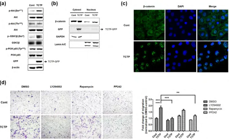
metalloproteinases (MMPs), that degrade components of the basement membrane24, we investigated whether TCTP activates MMPs. The expression levels of MMPs in both TCTP overexpressing and TCTP-depleted LLC-PK1 cell lysates were compared with that in control cells, using gelatinase specific antibodies to MMP-2 and MMP-9. We found that the expression level of MMP-9 increased in TCTP overexpressing cells, while that of MMP-2 was unchanged (Figure 3b). Conversely, the expression level of MMP-9 decreased following TCTP depletion (Figure 3c). Furthermore, by immunoblotting, we detected secreted MMP-9 and not MMP-2, in the conditioned media of these cells (Figure 3d). Also, gelatin zymographic study demonstrated that TCTP activates MMP-9, concomitantly with the expression of MMP-9 (Figure 3e). Taken together, these results indicate that TCTP facilitates cell invasion through activation of MMP-9. TCTP promotes cell migration via mTORC2/Akt/GSK3b/b-catenin pathway.EMT is regulated by ERK, Ras/MAPK, PI3K/Akt, Smads, RhoB and b-catenin pathways among others8. In our previous studies, we examined the activation of Akt by overexpression of TCTP6,25, focusing on Akt signaling and found that phosphorylation of Akt at Ser473residue significantly increased in TCTP overexpressing cells, with no change in the phosphorylation of Akt at Thr308 (Figure 4a). Full activation of Akt is known to be achieved by PDK-1-dependent phosphorylation at Thr308 and mTORC2-dependent phosphorylation at Ser473 26,27. We found, as expected, that PI3K
which is upstream of PDK-1, is not activated by TCTP overexpression. Since GSK3b is well-known as downstream target of Akt28, we examined whether overexpression of TCTP alters the activity of GSK3b. As shown in Figure 4a, overexpression of TCTP increased the phosphorylation of GSK3b (Ser9) and deactivates it while total GSK3b levels remained unchanged, indicating the absence of GSK3b activation. Phosphorylation and functional inactivation of GSK3b stabilize cytoplasmic b-catenin thereby inducing nuclear translocation of b-catenin. Nuclear accumulation of b-catenin is another characteristic change during EMT29. Based on these results, we hypothesized that TCTP activates b-catenin signaling pathways by inducing nuclear translocation of b-catenin. To test this hypothesis, we performed subcellular fractionation and evaluated nuclear translocation of b-catenin following TCTP overexpression. Figure 4b shows nuclear accumulation of b-catenin in TCTP overexpressing cells. Immunofluorescence microscopy also showed translocation of b-catenin from membrane to nucleus following overexpression of TCTP (Figure 4c). Moreover, phospho-Akt (Ser473) is known to be a key downstream molecule in mTOR complex 2 (mTORC2). Therefore, we investigated whether mTORC2 is involved in TCTP-mediated cell migration using several signaling molecule inhibitors: PI3K/Akt/mTOR kinase inhibitor, LY294002; mTORC1 inhibitor, rapamycin; and mTOR kinase inhibitor, PP242, which acts on both mTORC1 and mTORC2. Treatment with LY294002 and PP242 suppressed the migration of TCTP overexpressing LLC-PK1
Figure 3|TCTP enhances cell invasion through MMP-9 activation. (a) Transwell invasion assay was performed with LLC-PK1 cells infected with adeno-GFP virus or adeno-TCTP-adeno-GFP virus over 24 hr. Experiments were done in triplicate and representative images were taken with 3100 magnification using. Quantification was carried by counting the number of migratory cells that had infiltrated the filter. Values are means 6 S. E. M. **P,0.01. (b, c) The expression levels of MMP-2 and MMP-9 in cell lysates were examined by immunoblotting. (d) Secreted MMP-9 was detected in conditioned media of control and TCTP overexpressed cells by immunoblotting. (e) Conditioned media were collected at indicated time course from adenoviral infection. Gelatinolytic activity of pro and active MMP-9 were visible as a clear area on the gel.
cells (Figure 4d). In contrast, inhibition of mTORC1 by rapamycin did not affect TCTP induced effects on cell migration. Thus, activation of mTORC2 seems to be involved in TCTP induced cell migration. Overall, these findings suggest that overexpression of TCTP enhances cell motility through mTORC2/Akt/GSK3b/b-catenin pathways and that TCTP plays a role in EMT through these signaling pathways.
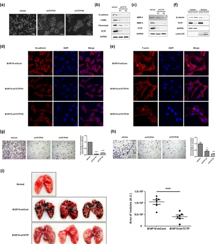
Downregulating TCTP inhibits mesenchymal-like changes and suppresses pulmonary metastasis of mouse melanoma cells. Having established that TCTP is a positive regulator of EMT process and knockdown of TCTP inhibits EMT transition in vitro, using LLC-PK1 cells, and that EMT is a critical regulator of cancer metastasis23, we investigated whether downregulation of TCTP represses development of mesenchymal-like features and decreases the metastatic potential of cancer cells in vivo. To this end, we infected stable mouse melanoma B16F10 cells with TCTP specific shRNA expressing lentivirus or control and obtained two distinct clones in which TCTP was significantly downregulated. We examined morphological changes in the stable cell lines by phase contrast microscopy. We found that lack of TCTP led to close cell-cell contacts, which indicates diminished mesenchymal changes. Also, diminished spike-like structures were observed in TCTP downregulated B16F10 cells (Figure 5a). More-over, mesenchymal markers such as N-cadherin, a-SMA, and fibronectin were significantly reduced in TCTP downregulated B16F10 cells (Figure 5b). When we examined MMP expressions in stable cell lysates, MMP-9 expression significantly diminished in
TCTP downregulated stable cells whereas MMP-2 levels did not change, consistent with results depicted in Figure 3 (Figure 5c). Immunostaining showed reduced N-cadherin in TCTP downre-gulated B16F10 cells (Figure 5d). In addition, significant changes were observed in the morphology of TCTP downregulated B16F10 cells compared to control cells, following F-actin staining with phalloidin. We noticed that B16F10-shCont cells exhibited elongated fibroblast-like morphology whereas B16F10-shTCTP cells showed flattened cell shapes (Figure 5e). As shown in Figure 5f, knockdown of TCTP decreased the nuclear accumulation of b-catenin. Since cancer metastasis is a complex multi-step process involving both migration and invasion8,23, we measured the migration and invasion capacities of B16F10-shCont and B16F10-shTCTP #1 cells by trans-well migration (Figure 5g) and invasion assays (Figure 5h), respec-tively. Both the migration and invasion of TCTP downregulated B16F10 cells were significantly reduced compared to those of control B16F10 cells. To understand the role of TCTP in metastatic potential, we also performed in vivo pulmonary metastasis assay. We injected both B16F10-shCont and B16F10-shTCTP #1 cells intravenously into the tail veins of male BALB C/nude mice and monitored metastasis in lungs. We observed significant metastasis after 11 days of intravenous injection, of B16F10-shCont cells. The numbers and areas of metastatic colonies on the surface of lungs were significantly less in mice injected with B16F10-shTCTP cells, than in those injected with B16F10-shCont cells (Figure 5i). These findings clearly demonstrate that downregula-tion of TCTP inhibits pulmonary metastasis of melanoma cells by reducing the invasiveness and migration of melanoma cells.
Figure 4|TCTP promotes cell migration through mTORC2/Akt/GSK3b/b-catenin pathway. (a) Phosphorylation states of Akt (Ser473) and GSK3b
(Ser9) in control and TCTP overexpressed cells were examined by immunoblotting. (b) The nuclear translocation of b-catenin was evaluated by
immunoblotting after subcellular fractionation. The purity of each fraction was monitored by the respective markers, GAPDH for the cytosolic fraction, and lamin A/C for the nuclear fraction. (c) Fluorescent microscopic visualization of b-catenin (green) in control and TCTP overexpressed cells. Nuclear DNA was stained with DAPI (blue). (d) Both control and TCTP overexpressed LLC-PK1 cells treated with 10 mM of LY294002, 100 nM of rapamycin, and 5 mM of PP242 for 18 h were subjected to transwell migration assay. Experiments were done in triplicate and representative images were taken with 3100 magnification using. Quantification was carried by counting the number of migratory cells that had infiltrated the filter. Values are means 6 S. E. M. **P , 0.01, ***P,0.001.
Figure 5|Silencing TCTP promotes MET process and suppresses pulmonary metastasis of mouse melanoma cell line. (a) Phase-contrast microscopic images of B16F10 cells infected with lentiviral control shRNA (shCont) or shRNA vector targeting TCTP (shTCTP #1 and #2). Images were taken with 3100 magnification. scale bar 200 mm (b) Expression levels of mesenchymal markers were determined by immunoblotting. GAPDH was used as a loading control. (c) The expression levels of MMP-2 and MMP-9 in cell lysates were examined by immunoblotting. (d, e) Fluorescence microscopic staining of N-cadherin (d) and F-actin (e) were performed with B16F10-shCont, shTCTP #1 and #2 (red). Nuclear DNA was stained with DAPI (blue). (f) The nuclear translocation of b-catenin was evaluated by immunoblotting after subcellular fractionation. GAPDH was used for the cytosolic fraction, and lamin A/C was used for the nuclear fraction. (g, h) B16F10-shCont, B16F10-shTCTP #1 and #2 cells were subjected to the transwell migration (g) and transwell invasion (h) assay. Experiments were done in triplicate and representative images were taken with 3100 magnification using. Quantification was carried by counting the number of migratory cells that had infiltrated the filter. Values are means 6 S. E. M. *P,0.05, **P,0.01, ***P,0.001. (i) Effect of TCTP depletion on the pulmonary metastasis was determined. Both B16F10-shCont and shTCTP #1 cells were injected intravenously into the tail vein of male BALB C/nude mice with 1 3 106cells (n56/group). After 11days, the lungs were extirpated and areas of nodules were calculated. Three
representatives per each set are shown. Normal lung indicates control injected with PBS.
Discussion
The present study is the first demonstration that TCTP is a positive regulator of EMT. We showed that TCTP overexpression in well polarized epithelial LLC-PK1 cells enhanced cell motility and inva-sivity via EMT process, evidenced by the expression of EMT-related markers and morphological changes. This study also demonstrated that TCTP-induced phosphorylation at Akt at Ser473by activation of mTORC2, and nuclear translocation of b-catenin by inhibiting GSK3b activity. We also showed that downregulation of TCTP inhi-bits EMT transition in both renal epithelial cells and melanoma cells and suppresses pulmonary metastasis of melanoma.
Reorganization of cytoskeleton and concomitant alteration of cell morphology are essential for cancer cell motility and invasivity and are thus regarded as typical phenomena during EMT process23. Several observations clearly indicate that TCTP is associated with cytoskeletal modification. In xenopus XL2 cells, TCTP was shown to colocalize with F-actin at the leading edge of lamellipodia-like struc-ture and regulate cell shape in a cytoskeleton-dependent manner30. It was also demonstrated that cofilin, an actin binding protein, pro-motes metastasis31and that TCTP influences F-actin cytoskeleton modification by competing with cofilin at the cofilin-binding site of actin32. In this context, we stained F-actin cytoskeleton with phalloi-din and observed that overexpression of TCTP in LLC-PK1 cells led to rearrangement of actin cytoskeleton and formation of stress fibers (Figure 1e). Stress fibers, induced by RhoA, connected to focal adhe-sions, have been shown to play a critical role in cell motility regu-lation33,34. Kloc et al observed the positive correlation with expression level of TCTP and RhoA in ovarian cancer cell line17. Thus, RhoA may be a downstream agent responsible for TCTP induced stress fiber formation. Further studies need to be performed to elucidate the detailed mechanism for TCTP-induced stress fiber formation.
The mammalian target of rapamycin (mTOR) which plays pivotal roles in cell growth, proliferation, survival and migration35,36, exists in two distinct multiprotein complexes, rapamycin senstive mTOR complex 1 and rapamycin insensitive mTOR complex 2 (mTORC2). mTORC1 plays critical roles in protein synthesis and cell growth by phosphorylation of translational regulators such as ribosomal S6 kinase1 (S6K1) and 4E-BP137. Whether TCTP regulates mTORC1 signaling in cell growth, proliferation and protein synthesis is being debated38–40. Studies in Drosophila revealed that dTCTP directly interacts with dRheb GTPase, a positive regulator of mTORC1, and acts as its guanine nucleotide exchange factor (GEF)41. mTORC2 regulates actin cytoskeleton rearrangement thereby promoting cell migration36,42. We observed previously6,25 as well as in the current study, elevation of Akt phosphorylation at Ser473residue in TCTP overexpressing cells (Figure 4a). Ser473is a key downstream residue in mTORC2. Our data indicate that TCTP affects mTORC2 signaling, which this study, using pharmacological inhibitors, showed, is involved in TCTP induced cell migration (Figure 4d). Thus, TCTP seems to affect and also use both mTORC1 and mTORC2 pathways; it regulates cell cycle and metabolisms by activating mTORC1 and alters cytoskeletal organization and motility through mTORC2.
Loss of E-cadherin is a hallmark of EMT process and is strongly associated with poor clinical outcome43,44in cancer. We found that ectopic expression of TCTP reduced E-cadherin expression and redirected its localization from membrane to cytosol (Figure 1b, d). One principal mechanism which reduces E-cadherin expression involves transcriptional repressors. Therefore, we examined whether TCTP is associated with E-cadherin transcriptional repressors. We found that TCTP elevated ZEB1, Slug, and twist, transcriptional repressors of E-cadherin (Figure 1c). The exact mechanism of how TCTP upregulates these transcriptional repressors has not been elu-cidated yet. Several studies demonstrated that b-catenin/TCF4 binds directly to the ZEB1 and Slug promoter and activates their transcrip-tions45,46We observed nuclear accumulation of b-catenin in TCTP overexpressing LLC-PK1 cells using subcellular fractionation and
immunofluorescence techniques (Figure 4b, c), and diminished nuc-lear b-catenin in TCTP downregulated melanoma cells (Figure 5f). Based on these findings, TCTP might regulate the expression of E-cadherin transcriptional repressors through activation of b-catenin signaling. However, additional studies using b-catenin specific shRNA/siRNA are needed to clarify the role of b-catenin in TCTP-induced EMT process. Also, understanding of TCTP’s role in the mTORC2/Akt/GSK3b axis is needed to establish the specific TCTP signaling in invasion and metastasis.
Tumor invasion and metastasis are multistep processes involving proteolysis of extracellular matrix (ECM) and destruction of base-ment membranes as initial events in the metastatic cascade47. Matrix metalloproteinases (MMPs), are a family of zinc-dependent endo-peptidases that plays an important role in proteolysis of ECM48, MMP-2 and MMP-9 playing especially important roles in tumor invasion and metastasis49,50. Our study shows that ectopic expression of TCTP in LLC-PK1 cells significantly enhanced cell invasion (Figure 3a). Also, the expression of MMP-9 increased in TCTP over-expressing cells (Figure 3b) and the activity of secreted MMP-9 was concomitantly elevated (Figure 3d, e). In a previous study of MMP expression levels in TCTP overexpressing MCF10A cells, we found that only MMP-3 and MMP-13 were upregulated while MMP-2 and MMP-9 levels did not change6. The difference in the cell line used may explain the observed variation.
The role of TCTP in metastasis is less well characterized than in tumorigenesis. A previous study showed that knocking down of TCTP in colon cancer cells inhibited liver metastasis2. We confirmed this effect of TCTP knockdown in the metastasis of melanoma. TCTP downregulated B16F10 cells also exhibited significantly sup-pressed pulmonary metastasis (Figure 5i). In addition, we showed that TCTP downregulated B16F10 cells markedly reduced mesench-ymal markers such as N-cadherin, a-SMA, and fibronectin (Figure 5b). This study is the first to report that knocking down TCTP, and the resultant inhibition of mesenchymal-like changes is a rational approach to inhibiting cancer cell migration and invasion, involved in metastasis. Furthermore, metastasis, which is a multi-step process that involves proliferation, migration, and invasion, the proliferative effect by TCTP might play a role in metastasis, at least in part. A unified view of TCTP’s regulation in signaling pathways (e. g. Src, EGFR, mTORC2/Akt etc.) and in proliferation and invasion is necessary to propose TCTP as a target for cancer metastasis. A fuller understanding of TCTP’s regulatory role in proliferation and inva-sion is needed before one can propose TCTP as a target for cancer metastasis.
Methods
Cells and Cell culture.Porcine renal proximal tubule LLC-PK1 cells were purchased from the American Type Culture Collection (ATCC) (Manassas, VA), and were maintained in Medium 199 supplemented with 5% fetal bovine serum at 37uC and 5% CO2. Mouse melanoma B16F10 cells were grown in Dulbecco’s modified Eagle’s
medium supplemented with 10% fetal bovine serum at 37uC and 5% CO2.
Sources of antibodies and other reagents.Antibodies against E-cadherin and N-cadherin were obtained from BD Bioscience (NJ, USA); ZEB1/TCF8, 2, MMP-9, phospho-Akt (Ser473), phospho-Akt (Thr308), Akt, phospho-GSK3b (Ser9), GSK3b,
phospho-PI3K, PI3K p85a, b-catenin, b-actin and lamin A/C antibodies were from Cell Signaling Technology (MA, USA); fibronectin and a-smooth muscle actin (SMA) antibodies were from Sigma-Aldrich Biotechnology (LP, USA); vimentin, green fluorescent protein (GFP), Twist, and Slug antibodies were from Santa Cruz Biotechnology (CA, USA); and TCTP antibodies were obtained from LabFrontier (Seoul, Korea). 2-(4-Morpholinyl)-8-phenyl-4H-1-benzopyran-4-one (LY294002), rapamycin, and PP242 were obtained from Sigma-Aldrich Biotechnology (LP, USA). Overexpression of TCTP by adenoviral infection and transient transfections with TCTP shRNA.Cells were infected to overexpress TCTP as described previously with minor modification6. Adenovirus vectors expressing the transgene TCTP, containing
green fluorescent protein (Ad-TCTP-GFP) or empty vectors (Ad-Vector) were used to infect the LLC-PK1 cells. When multiplicity of infection (MOI) was 10, LLC-PK1 cells were co-cultured with Ad-TCTP-GFP or Ad-GFP at 37uC, in a humidified atmosphere with 5% CO2, for 2 h. Construct for intracellular synthesis of TCTP
siRNA vector (pSUPER-shTCTP) was generated as previously described51.
Transfection of TCTP shRNA was performed by using WelFect-EXTMPLUS
transfection reagent (WelGENE, Korea). The level of suppression of TCTP gene was determined by immunoblotting.
Generation of stable cell lines by lentiviral transduction.Lentiviruses were generated as described52with minor modification. Briefly, TCTP shRNA cloned
PLKO.1, pVSV-G, and D8.9 were cotransfected in HEK293FT cells with Lipofectamine 2000 (Invitrogen, CA, USA). Viral particles were harvested at 48 and 72 hr, and infected into cells in the presence of 10 mg/ml polybrene. Infected cells were selected with puromycin (0.8 mg/ml).
Immunoblotting.Immunoblotting were performed as described previously with minor modification53. Briefly, cell extracts were prepared in modified RIPA buffer
[50 mM Tris–Cl (pH 7.4), 1% Triton-X 100, 0.25% sodium deoxycholate, 150 mM sodium chloride (NaCl), 1 mM ethylenediaminetetraacetic acid (EDTA), 2 mM sodium orthovanadate (Na3VO4) and 1 mM sodium fluoride (NaF)] with protease
inhibitor cocktails (Roche Molecular Biochemicals) and phosphatase inhibitor cocktails (Sigma-Aldrich Biotechnology, LP, USA). Proteins were separated by 10% sodium dodecyl sulfate-polyacrylamide gel electrophoresis (SDS-PAGE) and transferred to nitrocellulose membrane.
Preparation of conditioned medium.Proteins were precipitated from equal-volume aliquots of supernatant using 15% ice-cold trichloroacetic acid (TCA). The precipitates were washed twice with 100% acetone, air-dried, dissolved in sample buffer. Samples were detected using immunoblotting method.
Gelatin Zymography.Gelatin zymography was performed as described previously with minor modification54. Briefly, cells were infected and incubated in the serum free
media for indicated time. Cell supernatants were collected and centrifuged at 12,000 rpm for 10 min at 4uC to remove cellular debris. Conditioned medium was then concentrated with Amicon Ultra-4 filter (30 K) (Millipore, USA) at 7,500 g for 30 min at 4uC. Samples were electrophoresed under nonreducing conditions onto 10% SDS-PAGE gels polymerized with 1 mg/ml gelatin. The gels were washed with 2.5% Triton X-100, and incubated at 37uC for 24 h in incubation buffer containing 50 mM Tris-HCl, pH 7.4, 10 mM CaCl2, 50 mM NaCl, and 0.05% Brij 35. The gel
was stained with Coomassie blue stain (0.5% Coomassie blue R-250, 40% methanol, and 10% acetic acid), followed by destaining (40% methanol, 10% acetic acid). The image was taken with the LAS-3000 (Fujifilm, Japan).
Wound-healing assay.Wound healing assay was performed as described previously with minor modification55. Briefly, LLC-PK1 cells were grown to confluence in a
6-well plate and were infected with Ad-GFP or Ad-TCTP-GFP virus. After 24 hours, cells were wounded using 1 ml sterile pipette tip. The wounded area was photographed at each time courses for 48 hours. The wound healing capacities were calculated by measuring the distance of the migrating edge. Mean values were obtained from at least 3 separate experiments.
Transwell migration/invasion assay.Migration assays were performed using uncoated cell culture inserts with 8 mm pores (Corning Life Sciences, NY, USA) according to manufacturer’s instructions. Briefly, LLC-PK1 cells were infected with Ad-GFP or Ad-TCTP-GFP virus; or were transfected with pSUPER or pSUPER-shTCTP. After 24 hours, Cells were harvested with Trypsin/EDTA and resuspended in serum-free medium at a density of 5.0 3 105cells/ml. B16F10 stable cells were
resuspended in serum-free medium at a density of 1.0 3 106cells/ml. 100 m, of cells
were seeded into the upper chamber. Lower chambers were filled with 650 m, medium containing 10% FBS as a chemoattractant. After 18 hours cells were fixed and permeablized with 100% methanol and stained with 0.5% crystal violet/20% methanol. The non-migrating cells on the upper surface of the filter were removed by cotton swab. The number of migratory cells was measured by counting at 3 100 magnifications using a microscope. Invasion assays were performed using the BD BioCoatTMBD MatrigelTMInvasion Chamber (BD Bioscience, NJ, USA) according to
the manufacturer’s instructions. Briefly, infected LLC-PK1 cells were resuspended in serum-free medium at a density of 1.0 3 106cells/ml and B16F10 stable cells were
resuspended in serum-free medium at a density of 2.0 3 105cells/ml. 500 m, of cells
were seeded into the upper chamber and lower chambers were filled with 750 m, medium containing 10% FBS as a chemoattractant. After 24 hours, the transwell invasion assay was performed with the same method as transwell migration assay. Immunofluorescence confocal microscopy.Confocal microscopy was performed as described previously with minor modification6. Briefly, cells were grown on glass
cover slips to 70% confluence and were infected with 10 MOI Ad GFP or TCTP-GFP virus. Cells were incubated for 24 hours and then fixed with 4% paraformaldehyde in PBS for 15 minutes at room temperature. For staining E-cadherin, cells were fixed with 100% methanol for 15 minutes at 220uC. After washing with PBS, the cells were permeabilized in 0.2% Triton X-100 in PBS and blocked in 1% bovine serum albumin. Cells were than stained with mouse monoclonal anti-E-cadherin antibody (15200), anti-N-cadherin antibody (15200) and rabbit polyclonal anti-b-catenin (15200) and then probed with Alexa Fluor 488 or 594 conjugated goat anti-mouse IgG (Invitrogen, CA, USA). For F-actin staining, Cells were stained with rhodamine conjugated phalloidin (Molecular Probes, OR, USA). Counterstain with DAPI was performed by mounting in ProLong Gold antifade reagent (Invitrogen, CA, USA). Cells were photographed at 3 400 magnifications with a fluorescence confocal microscope
(LSM510, Zeiss, Germany). Cell morphology was examined under an inverted microscope (Zeiss, Germany) at 3 400 magnification.
Preparation of Nuclear fraction.Nuclear protein was extracted using NE-PER nuclear and cytoplasmic extraction reagents (Pierce, Rockford, IL) following the manufacturer’s supplied protocol
Experimental metastasis assay.Experimental metastasis assay was performed as described previously56with mild modification. Briefly, B16F10 cells were trypsinized
and suspended in PBS. Cells (1 3 106in 100 m,) were injected intravenously into the
tail vein of BALB/c nude mice (Jung-Ang Lab, Animal, Inc., Seoul, Korea). After 11 days of intravenous injection, the lung was extirpated and the areas of black spherical B16F10 colonies were analyzed using ImageJ software (NIH, USA). All animal studies were conducted in accordance with IACUC guidelines and were approved by IACUC committee at Ewha Womans University (Approval ID: 2013-01-114).
Statistical analysis.Data are presented as means and their standard errors. Data were analyzed using GraphPad Prisms 5 software (GraphPad Software Inc., CA, USA). Statistical significance was determined using Student’s t-test.
1. Gachet, Y. et al. The growth-related, translationally controlled protein P23 has properties of a tubulin binding protein and associates transiently with microtubules during the cell cycle. J. Cell Sci. 112, 1257–1271 (1999). 2. Ma, Q. et al. The role of translationally controlled tumor protein in tumor growth
and metastasis of colon adenocarcinoma cells. J. Proteome Res. 9, 40–49, doi:10.1021/pr9001367 (2010).
3. Jung, J. et al. Translationally controlled tumor protein interacts with the third cytoplasmic domain of Na,K-ATPase alpha subunit and inhibits the pump activity in HeLa cells. J. Biol. Chem. 279, 49868–49875, doi:10.1074/ jbc.M400895200 (2004).
4. Kim, M. J. et al. Transgenic overexpression of translationally controlled tumor protein induces systemic hypertension via repression of Na1,K1-ATPase. J. Mol. Cell Cardiol. 44, 151–159, doi:10.1016/j.yjmcc.2007.09.017 (2008).
5. Kim, M. J. et al. Over-expression of translationally controlled tumor protein in lens epithelial cells seems to be associated with cataract development. Transgenic Res. 18, 953–960, doi:10.1007/s11248-009-9283-y (2009).
6. Jung, J., Kim, H. Y., Kim, M., Sohn, K. & Lee, K. Translationally controlled tumor protein induces human breast epithelial cell transformation through the activation of Src. Oncogene 30, 2264–2274, doi:10.1038/onc.2010.604 (2011). 7. Rajasekaran, S. et al. Na,K-ATPase subunits as markers for
epithelial-mesenchymal transition in cancer and fibrosis. Mol. Cancer Ther. 9, 1515–1524, doi:10.1158/1535-7163.mct-09-0832 (2010).
8. Kalluri, R. & Weinberg, R. The basics of epithelial-mesenchymal transition. The J. Clin. Invest. 119, 1420–1428, doi:10.1172/jci39104 (2009).
9. Thiery, J. & Sleeman, J. Complex networks orchestrate epithelial-mesenchymal transitions. Nat. Rev. Mol. Cell Biol. 7, 131–142, doi:10.1038/nrm1835 (2006). 10. Locascio, A. & Nieto, M. A. Cell movements during vertebrate development:
integrated tissue behaviour versus individual cell migration. Curr. Opin. Genet. Dev. 11, 464–469 (2001).
11. Lee, J., Dedhar, S., Kalluri, R. & Thompson, E. The epithelial-mesenchymal transition: new insights in signaling, development, and disease. J. Cell Biol. 172, 973–981, doi:10.1083/jcb.200601018 (2006).
12. Chaffer, C. L. & Weinberg, R. A. A perspective on cancer cell metastasis. Science 331, 1559–1564, doi:10.1126/science.1203543 (2011).
13. Liang, X. EMT: new signals from the invasive front. Oral Oncol. 47, 686–687, doi:10.1016/j.oraloncology.2011.04.016 (2011).
14. Chung, S., Kim, M., Choi, W., Chung, J. & Lee, K. Expression of translationally controlled tumor protein mRNA in human colon cancer. Cancer Lett. 156, 185–190 (2000).
15. Arcuri, F. et al. Translationally controlled tumor protein (TCTP) in the human prostate and prostate cancer cells: expression, distribution, and calcium binding activity. Prostate 60, 130–140, doi:10.1002/pros.20054 (2004).
16. Chan, T. et al. Translationally controlled tumor protein induces mitotic defects and chromosome missegregation in hepatocellular carcinoma development. Hepatology 55, 491–505, doi:10.1002/hep.24709 (2012).
17. Malgorzata, K. et al. Inverse relationship between TCTP/RhoA and p53//cyclin A/ actin expression in ovarian cancer cells. Folia Histochem. Cyto. 50, 358–367 doi:10.5603/19745 (2012).
18. Gu, X. et al. TCTP promotes glioma cell proliferation in vitro and in vivo via enhanced b-catenin/TCF-4 transcription. Neuro-oncology 16, 217–227, doi:10.1093/neuonc/not194 (2014).
19. Graidist, P. et al. Fortilin binds Ca21 and blocks Ca21-dependent apoptosis in vivo. Biochem. J. 408, 181–191, doi:10.1042/BJ20070679 (2007).
20. Acloque, H., Adams, M., Fishwick, K., Bronner-Fraser, M. & Nieto, M. Epithelial-mesenchymal transitions: the importance of changing cell state in development and disease. J. Clin. Invest. 119, 1438–1449, doi:10.1172/jci38019 (2009). 21. Tiwari, N., Gheldof, A., Tatari, M. & Christofori, G. EMT as the ultimate survival
mechanism of cancer cells. Semin. Cancer Biol. 22, 194–207, doi:10.1016/ j.semcancer.2012.02.013 (2012).
22. Gulhati, P. et al. mTORC1 and mTORC2 regulate EMT, motility, and metastasis of colorectal cancer via RhoA and Rac1 signaling pathways. Cancer Res. 71, 3246–3256, doi:10.1158/0008-5472.CAN-10-4058 (2011).
23. Yilmaz, M. & Christofori, G. EMT, the cytoskeleton, and cancer cell invasion. Cancer metast. Rev. 28, 15–33, doi:10.1007/s10555-008-9169-0 (2009). 24. Chambers, A. F. & Matrisian, L. M. Changing views of the role of matrix
metalloproteinases in metastasis. J. Natl. Cancer Inst. 89, 1260–1270 (1997). 25. Kim, M., Jung, J. & Lee, K. Roles of ERK, PI3 kinase, and PLC-gamma pathways
induced by overexpression of translationally controlled tumor protein in HeLa cells. Arch. Biochem. Biophys. 485, 82–87 (2009).
26. Alessi, D. R. et al. Characterization of a 3-phosphoinositide-dependent protein kinase which phosphorylates and activates protein kinase Balpha. Curr. Biol. 7, 261–269 (1997).
27. Sarbassov, D. D., Guertin, D. A., Ali, S. M. & Sabatini, D. M. Phosphorylation and regulation of Akt/PKB by the rictor-mTOR complex. Science 307, 1098–1101, doi:10.1126/science.1106148 (2005).
28. Manning, B. D. & Cantley, L. C. AKT/PKB signaling: navigating downstream. Cell 129, 1261–1274, doi:10.1016/j.cell.2007.06.009 (2007).
29. Mu¨ller, T., Bain, G., Wang, X. & Papkoff, J. Regulation of epithelial cell migration and tumor formation by beta-catenin signaling. Exp. Cell Res. 280, 119–133, doi:10.1006/excr.2002.5630 (2002).
30. Bazile, F. et al. Complex relationship between TCTP, microtubules and actin microfilaments regulates cell shape in normal and cancer cells. Carcinogenesis 30, 555–565, doi:10.1093/carcin/bgp022 (2009).
31. Wang, W. et al. The activity status of cofilin is directly related to invasion, intravasation, and metastasis of mammary tumors. J. Cell Biol. 173, 395–404, doi:10.1083/jcb.200510115 (2006).
32. Tsarova, K., Yarmola, E. G. & Bubb, M. R. Identification of a cofilin-like actin-binding site on translationally controlled tumor protein (TCTP). FEBS Lett. 584, 4756–4760, doi:10.1016/j.febslet.2010.10.054 (2010).
33. Ridley, A. J. Rho GTPases and actin dynamics in membrane protrusions and vesicle trafficking. Trends Cell Biol. 16, 522–529, doi:10.1016/j.tcb.2006.08.006 (2006).
34. Cheng, I. K. et al. GEF-H1 over-expression in hepatocellular carcinoma promotes cell motility via activation of RhoA signalling. J. Pathol. 228, 575–585, doi:10.1002/path.4084 (2012).
35. Kim, D. H. et al. mTOR interacts with raptor to form a nutrient-sensitive complex that signals to the cell growth machinery. Cell 110, 163–175 (2002).
36. Sarbassov, D. et al. Rictor, a novel binding partner of mTOR, defines a rapamycin-insensitive and raptor-independent pathway that regulates the cytoskeleton. Curr. Biol. 14, 1296–1302, doi:10.1016/j.cub.2004.06.054 (2004).
37. Hay, N. & Sonenberg, N. Upstream and downstream of mTOR. Gene. Dev. 18, 1926–1945, doi:10.1101/gad.1212704 (2004).
38. Wang, X. et al. Re-evaluating the roles of proposed modulators of mammalian target of rapamycin complex 1 (mTORC1) signaling. J. Biol. Chem. 283, 30482–30492, doi:10.1074/jbc.M803348200 (2008).
39. Dong, X., Yang, B., Li, Y., Zhong, C. & Ding, J. Molecular basis of the acceleration of the GDP-GTP exchange of human ras homolog enriched in brain by human translationally controlled tumor protein. J. Biol. Chem. 284, 23754–23764, doi:10.1074/jbc.M109.012823 (2009).
40. Kim, D. K. et al. Translationally controlled tumour protein is associated with podocyte hypertrophy in a mouse model of type 1 diabetes. Diabetologia 55, 1205–1217, doi:10.1007/s00125-012-2467-7 (2012).
41. Hsu, Y.-C., Chern, J., Cai, Y., Liu, M. & Choi, K.-W. Drosophila TCTP is essential for growth and proliferation through regulation of dRheb GTPase. Nature 445, 785–788, doi:10.1038/nature05528 (2007).
42. Jacinto, E. et al. Mammalian TOR complex 2 controls the actin cytoskeleton and is rapamycin insensitive. Nat. Cell Biol. 6, 1122–1128, doi: 10.1038/ncb1183 (2004). 43. Cavallaro, U. & Christofori, G. Cell adhesion and signalling by cadherins and
Ig-CAMs in cancer. Nat. Rev. Cancer 4, 118–132, doi:10.1038/nrc1276 (2004). 44. Perl, A., Wilgenbus, P., Dahl, U., Semb, H. & Christofori, G. A causal role for
E-cadherin in the transition from adenoma to carcinoma. Nature 392, 190–193, doi:10.1038/32433 (1998).
45. Conacci-Sorrell, M. et al. Autoregulation of E-cadherin expression by cadherin-cadherin interactions: the roles of beta-catenin signaling, Slug, and MAPK. J.Cell Biol. 163, 847–857, doi:10.1083/jcb.200308162 (2003).
46. Sanchez-Tillo, E. et al. beta-catenin/TCF4 complex induces the epithelial-to-mesenchymal transition (EMT)-activator ZEB1 to regulate tumor invasiveness. Proc. Natl. Acad. Sci. U S A 108, 19204–19209, doi:10.1073/pnas.1108977108 (2011).
47. Stetler-Stevenson, W. G. Type IV collagenases in tumor invasion and metastasis. Cancer Metast. Rev. 9, 289–303 (1990).
48. Vihinen, P. & Kahari, V. M. Matrix metalloproteinases in cancer: prognostic markers and therapeutic targets. Int. J. Cancer 99, 157–166, doi:10.1002/ijc.10329 (2002).
49. Hadler-Olsen, E., Winberg, J. O. & Uhlin-Hansen, L. Matrix metalloproteinases in cancer: their value as diagnostic and prognostic markers and therapeutic targets. Tumor Biol. 34, 2041–2051, doi:10.1007/s13277-013-0842-8 (2013).
50. Lamar, J. M., Pumiglia, K. M. & DiPersio, C. M. An immortalization-dependent switch in integrin function up-regulates MMP-9 to enhance tumor cell invasion. Cancer Res. 68, 7371–7379, doi:10.1158/0008-5472.CAN-08-1080 (2008). 51. Yoon, T., Kim, M. & Lee, K. Inhibition of Na,K-ATPase-suppressive activity of
translationally controlled tumor protein by sorting nexin 6. FEBS Lett. 580, 3558–3564, doi:10.1016/j.febslet.2006.05.042 (2006).
52. Kim, E. et al. Selective activation of Akt1 by mammalian target of rapamycin complex 2 regulates cancer cell migration, invasion, and metastasis. Oncogene 30, 2954–2963, doi:10.1038/onc.2011.22 (2011).
53. Sheverdin, V., Bae, S. Y., Shin, D. H. & Lee, K. Expression and localization of translationally controlled tumor protein in rat urinary organs. Microsc. Res. Tech. 75, 1576–1581, doi:10.1002/jemt.22103 (2012).
54. Kasai, H., Allen, J. T., Mason, R. M., Kamimura, T. & Zhang, Z. TGF-beta1 induces human alveolar epithelial to mesenchymal cell transition (EMT). Respir. Res. 6, 56, doi:10.1186/1465-9921-6-56 (2005).
55. Meng, X. et al. Knockdown of BAG3 induces epithelial-mesenchymal transition in thyroid cancer cells through ZEB1 activation. Cell Death Dis. 5, e1092, doi:10.1038/cddis.2014.32 (2014).
56. Kim, H. et al. Ubiquitin C-terminal hydrolase-L1 is a key regulator of tumor cell invasion and metastasis. Oncogene 28, 117–127, doi:10.1038/onc.2008.364 (2009).
Acknowledgments
This study was supported by a grant of the Korea Health Technology R&D Project, Ministry of Health & Welfare [A111417(HI11C1371)] and National Research Foundation of Korea (NRF) grant funded by the Ministry of Science, ICT & Future Planning (MSIP) [2012R1A1A2042142] [2012M3A9A8053272] to K. Lee, NRF Grant funded by the Korean Government (NRF-2011-Global Ph.D. Fellowship Program) to S. Y. Bae and RP-Grant 2013 of Ewha Womans University to H. J. Kim.
Author contributions
S.Y.B. designed and performed all experiments and wrote manuscript. H.J.K. and K.J.L. conducted experimental metastasis assay as well as data analysis. K.L. participated in the conception and design of the study and the writing of the manuscript.
Additional information
Supplementary informationaccompanies this paper at http://www.nature.com/ scientificreports
Competing financial interests:The authors declare no competing financial interests. How to cite this article:Bae, S.Y., Kim, H.J., Lee, K.J. & Lee, K. Translationally Controlled Tumor Protein induces epithelial to mesenchymal transition and promotes cell migration, invasion and metastasis. Sci. Rep. 5, 8061; DOI:10.1038/srep08061 (2015).
This work is licensed under a Creative Commons Attribution-NonCommercial-ShareAlike 4.0 International License. The images or other third party material in this article are included in the article’s Creative Commons license, unless indicated otherwise in the credit line; if the material is not included under the Creative Commons license, users will need to obtain permission from the license holder in order to reproduce the material. To view a copy of this license, visit http:// creativecommons.org/licenses/by-nc-sa/4.0/

![OPEN BID INVITATION[the Provision of Security Service]](data:image/gif;base64,R0lGODlhAQABAIAAAP///wAAACH5BAEAAAAALAAAAAABAAEAAAICRAEAOw==)