TNF-related Activation-induced Cytokine (TRANCE) Induces
Angiogenesis through the Activation of Src and Phospholipase C
(PLC) in Human Endothelial Cells*
Received for publication, October 1, 2001, and in revised form, December 7, 2001 Published, JBC Papers in Press, December 10, 2001, DOI 10.1074/jbc.M109434200
Young-Mi Kim‡§, Young-Myoung Kim¶, You Mie Lee储, Hae-Sun Kim储, Jong Dai Kim§,
Yongwon Choi**, Kyu-Won Kim储‡‡, Soo-Young Lee§§¶¶, and Young-Guen Kwon‡储储
From the ‡Department of Biochemistry, College of Natural Sciences, the §Department of Food Science and Technology, College of Agriculture and Life Sciences, and the¶Department of Molecular and Cellular Biochemistry, School of Medicine, Kangwon National University, Chunchon, Kangwon-Do 200-701, Korea, the储Laboratory of Biochemistry, College of Pharmacy, Seoul National University, Seoul 151-742, Korea, the **Department of Pathology and Laboratory Medicine, Abramson Family Cancer Research Institute, University of Pennsylvania, Philadelphia, Pennsylvania 19104, and the §§Division of Molecular Life Science and Center for Cell Signaling Research, Ewha Womans University, Seoul 120-750, Korea
Angiogenesis is an essential step for many physiolog-ical and pathologphysiolog-ical processes. Tumor necrosis factor (TNF) superfamily cytokines are increasingly recog-nized as key modulators of angiogenesis. In this study, we tested whether TNF-related activation-induced cyto-kine (TRANCE), a new member of the TNF superfamily, possesses angiogenic activity in vitro and in vivo. TRANCE stimulated DNA synthesis, chemotactic motil-ity, and capillary-like tube formation in primary cul-tured human umbilical vein endothelial cells (HUVECs). Both Matrigel plug assay in mice and chick chorioallan-toic membrane assay revealed that TRANCE potently induced neovascularization in vivo. TRANCE had no ef-fect on vascular endothelial growth factor (VEGF) ex-pression in HUVECs and TRANCE-induced angiogenic activity was not suppressed by VEGF-neutralizing anti-body, implying that TRANCE-induced angiogenesis may be the result of its direct action on endothelial cells. TRANCE evoked a time- and dose-dependent activation of the mitogen-activated protein kinases ERK1/2 and
focal adhesion kinase p125FAK in HUVECs, which are
closely linked to angiogenesis. These signaling events were blocked by the Src inhibitor PP1 or the phospho-lipase C (PLC) inhibitor U73122. Furthermore, these
in-hibitors and the Ca2ⴙchelator BAPTA-AM suppressed
TRANCE-induced HUVEC migration. These results indi-cate that the angiogenic activity of TRANCE is mediated
through the Src-PLC-Ca2ⴙ signaling cascade upon
re-ceptor engagement in endothelial cells, suggesting the role of TRANCE in neovessel formation under physio-logical and pathophysio-logical conditions.
Angiogenesis, the formation of new blood vessels from pre-existing endothelium, is a fundamental step in a variety of
physiological and pathological conditions including wound healing, embryonic development, chronic inflammation, and tumor growth (1–3). The angiogenic process is tightly con-trolled by a wide variety of positive or negative regulators, which are composed of growth factors, cytokines, lipid metab-olites, and cryptic fragments of hemostatic proteins (4), and many of these factors are initially characterized in other bio-logical activities. Positive regulators of angiogenesis are clas-sified into two groups: direct inducers, such as vascular endo-thelial growth factor (VEGF)1 and bFGF that can induce
proliferation, migration, and morphogenesis of endothelial cells and indirect inducers that act on endothelial cells via produc-tion of direct angiogenic factors from accessory cells such as immune cells and tumor cells (4).
Tumor necrosis factor (TNF)-related activation-induced cy-tokine (TRANCE), also called ODF, RANKL, and OPGL, is a novel member of the TNF family ligands that regulates im-mune responses and bone remodeling (5– 8). TRANCE ex-pressed on activated T cells promotes the survival of dendritic cells and modulates T helper cell responses to viral infections (9, 10). TRANCE is also highly expressed on osteoblasts and plays a key role in osteoclast differentiation from hematopoietic precursors and calcium metabolism (11, 12). Recently, it has been also reported that, in a T cell-dependent model of rat adjuvant arthritis, TRANCE regulates bone loss and cartilage destruction (13). These effects of TRANCE are exerted by its binding to the transmembrane receptor RANK (receptor acti-vator of NF-B). RANK is detected on mature dendritic cells, chondrocytes, osteoclast precursors, and mature osteoclasts (9, 14) and induces activation of extracellular signal-regulated kinase (ERK) and c-Jun N-terminal kinase (JNK), AKT, and NF-B in dendritic cells and osteoclasts (15–17).
The vasculature is crucial for the processes of bone formation by paving the way for a variety of cells essential for bone morphogenesis, including osteoblasts, to migrate into cartilage
* This work was supported by Molecular Medical Science Research Grant M1-0106-00-0015 (to Y. G. K.) and M1-0106-00-0025 (to S. Y. L.), and a Vascular System Research Center grant. The costs of publication of this article were defrayed in part by the payment of page charges. This article must therefore be hereby marked “advertisement” in ac-cordance with 18 U.S.C. Section 1734 solely to indicate this fact.
‡‡ Recipient of the National Research Laboratory Fund from the Ministry of Science and Technology.
¶¶To whom correspondence may be addressed. Tel.: 82-2-3277-3770; Fax: 82-2-3277-3760; E-mail: [email protected].
储储To whom correspondence may be addressed. Tel.: 82-33-250-8517; Fax: 82-33-242-0459; E-mail: [email protected].
1The abbreviations used are: VEGF, vascular endothelial growth
factor; bFGF, basic fibroblast growth factor; TNF, tumor necrosis factor; TRANCE, TNF-related activation-induced cytokine; RANK, receptor activator of NF-B; NF, nuclear factor; ERK, extracellular signal-reg-ulated kinase; JNK, c-Jun N-terminal kinase; PMA, phorbol 12-myris-tate 13-ace12-myris-tate; HUVEC(s), human umbilical vein endothelial cell(s); FBS, fetal bovine serum; RT, reverse transcriptase; CAM(s), cho-rioallantoic membrane(s); IPT,total inositol phosphate; IP3, inositol
1,4,5-trisphosphate; FAK, focal adhesion kinase; PLC, phospholipase C; NO, nitric oxide; PKC, protein kinase C.
© 2002 by The American Society for Biochemistry and Molecular Biology, Inc. Printed in U.S.A.
This paper is available on line at http://www.jbc.org
6799
at Ewha Medical Library on October 26, 2016
http://www.jbc.org/
was purified from insect cells as described previously (15). bFGF and VEGF were from Upstate Biotechnology (Lake Placid, NY). U73122, U73343, PP1, GF109203X, and U0126 were from BIOMOL. Phorbol 12-myristate 13-acetate (PMA) were from Alexis (Laufelfingen, Switzerland). M199, heparin, and Trizol Reagent were purchased from Invitrogen (Grand Island, NY). [3H]Thymidine (25 Ci/mmol) was from
Amersham Biosciences, Inc. Matrigel was from Collaborative Biomed-ical Products (Bedford, MA). Transwell plate was from Corning Costar (Cambridge, MA). Antibodies for phospho-specific ERK (Thr-202/ Tyr-204) and ERK were obtained from New England Biolabs (Beverly, MA). Antibody for p125FAK was from Upstate Biotechnology.
Anti-phosphotyrosine antibody was from Transduction Laboratories (Lexington, KY). Antibody for VEGF was from R&D Systems (Minneapolis, MN). All other reagents were purchased from Sigma unless indicated otherwise.
Cell Culture—Human umbilical vein endothelial cells (HUVECs) were
isolated from human umbilical cord veins by collagenase treatment as described previously (20) and used in passages 2–7. The cells were grown in M199 medium supplemented with 20% fetal bovine serum (FBS), 100 units/ml penicillin, 100g/ml streptomycin, 3 ng/ml bFGF, and 5 units/ml heparin at 37 °C within humidified 5% CO2/95% air.
Reverse Transcriptase (RT)-PCR Analysis—Total RNA was obtained
from HUVECs using the Trizol Reagent kit. Fiveg of total RNA was converted to cDNA by treatment with 200 units of reverse transcriptase and 500 ng of oligo(dT) primer in 50 mMTris-HCl (pH 8.3), 75 mMKCl, 3 mMMgCl2, 10 mMdithiothreitol and 1 mMdNTPs at 42 °C for 1 h. The
reaction was stopped by heating at 70 °C for 15 min. 3l of the cDNA mixture was used for enzymatic amplification. Polymerase chain reac-tion was performed in 50 mMKCl, 10 mMTris-HCl (pH 8.3), 1.5 mM MgCl2, 0.2 mMdNTPs, 2.5 units of Taq DNA polymerase, and 0.1M
each of primers for RANK or VEGF. The reaction mixture was heated at 94 °C for 1 min, annealed at 55 °C for 2 min, and extended at 72 °C for 3 min for 30 repetitive cycles. The primers used were 5 ⬘-CTCCAG-CGGGCAGGTGATGAACTT-3⬘ (sense) and 5⬘-TCAAGCCTTGGCCCC-GCCTTGCTC-3⬘ (antisense) for the RANK or 5⬘-GGCCTCCGAAACC-ATGAACTTTCTGCT-3⬘ (sense) and 5⬘-CCTCCTGCCCGGCTCACCG-C-3⬘ (antisense) for the VEGF.-actin was used as an internal control.
In Vivo Matrigel Plug Assay—Matrigel plug assay was performed as
previously described (21). Briefly, C57/BL6 mice were injected subcu-taneously with 0.5 ml of Matrigel containing the indicated amount of TRANCE with heparin (40 units/ml). The injected Matrigel rapidly formed a single, solid gel plug. After 5 days, mice were killed, and the Matrigel plugs were recovered, fixed with 3.7% formaldehyde/phos-phate-buffered saline, and embedded in paraffin. The plugs were then sectioned and examined with Trichrome-Masson stain. The neovessel area was quantified using TINA 2.0 software (Fuji Inc.).
Chorioallantoic Membrane (CAM) Assay—Angiogenic assay with
chick CAM was performed as previously described (22). Fertilized chick embryos were preincubated for 9 days at 38 °C with 70% humidity. A hole was drilled over the air sac at the end of the eggs, and an avascular zone was identified on the CAMs. A 1⫻ 1-cm window in the shell was made to expose the CAM. Thermanox discs were sterilized and loaded with TRANCE (3g/ml) and PMA (0.12 g/ml). After air-drying under the laminar flow, the discs were applied to the CAM surface of 9-day-old chick embryos. The windows were sealed with clear tape, and the eggs were incubated for 3 more days. Intrapos (Green-Cross) was injected under the upper CAM to increase contrast between vessels and back-ground. Capillary formation was inspected using a light microscope.
[3H]Thymidine Incorporation Assay—[3H]Thymidine incorporation
assay was performed as previously described (21). Briefly, HUVECs were plated at a density of 2⫻ 104cells/well in a 24-well plate. Cells were
incubated in growth media and allowed to attach for 24 h. Cells were washed twice with M199 and incubated for 6 h with M199 containing 1% FBS. Cells were stimulated by the addition of indicated concentration of TRANCE or 10 ng/ml VEGF for 30 h and followed by the addition of 1 Ci/ml [3H]thymidine for 6 h. High molecular mass3H-labeled
radioac-tivity was precipitated using 5% trichloroacetic acid at 4 °C for 30 min.
temperature before seeding. One hundredl of the cell suspension was loaded into each of the upper wells. The chamber was incubated at 37 °C for 4 h. Cells were fixed and stained with hematoxylin and eosin. Nonmigrating cells on the upper surface of the filter were removed by wiping with a cotton swab, and chemotaxis was quantified by counting the cells that migrated to the lower side of the filter with optical microscope (⫻200). Ten fields were counted for each assay.
Tube Formation Assay—Tube formation assay was performed as
previously described (21). Briefly, 250l of growth factor-reduced Ma-trigel (10 mg protein/ml) was pipetted into a 16-mm diameter tissue culture well and polymerized for 30 min at 37 °C. HUVECs incubated in M199 containing 1% FBS for 6 h were harvested after trypsin treat-ment, resuspended in M199, plated onto the layer of Matrigel at a density of 1⫻ 105or 4⫻ 105cells/well, and followed by the addition of
3 g/ml TRANCE. Matrigel cultures were incubated at 37 °C. After 20 h, the cultures were photographed (⫻40). The area covered by the tube network was determined using an optical imaging technique in which pictures of the tubes were scanned in Adobe Photoshop and quantitated using Image-Pro Plus (Media Cybernetics).
Measurement of Total Inositol Phosphate (IPT) in HUVECs—
HUVECs were plated at a density of 2⫻ 105cells/well in six-well plates
and allowed to recover for 24 h. The medium was then aspirated, and cells were incubated for 12 h in myo-inositol-free M199 media supple-mented with [3H]myo-inositol (1Ci/ml, 25 mCi/mmol) (DuPont
Bio-technology Systems) and 0.5% FBS. The cells were washed with phos-phate-buffered saline and incubated in M199 media supplemented with 20 mMLiCl for 30 min, followed by additional incubation in the absence or presence of 0.5 g/ml TRANCE for 20 min. The incubation was terminated by adding perchloric acid to a final concentration of 5% (w/v). Cells were scraped into Eppendorf tubes and subjected to centrif-ugation. The supernatant was equilibrated with 2MKOH/1 mMEDTA and was applied to SAX column. Bound inositol phosphates were eluted by the application of linear gradient (0.1Mammonium phosphate) at a flow rate of 2 ml/min. The amount of radioactivity in the resulting fraction, corresponding to liberated [3H]inositol triphosphate (IP
3), was
measured by a liquid scintillation counter.
Immunoprecipitation—Confluent HUVECs were incubated for 6 h in
M199 containing 1% FBS before addition of TRANCE. After stimula-tion, cells were lysed in 1 ml of lysis buffer containing 20 mMTris/HCl, pH 8.0, 2 mMEDTA, 137 mMNaCl, 1 mMNa3VO4, 1 mM
phenylmeth-ylsulfonyl fluoride, 10% glycerol, and 1% Triton X-100. Lysates were clarified by centrifugation at 15,000⫻ g for 10 min, and the resulting supernatants were immunoprecipitated with 1g/ml of anti-FAK an-tibody for 3 h at 4 °C, followed by the addition of protein A-agarose beads for 1 h at 4 °C. Immunoprecipitates were washed three times with lysis buffer, solubilized in SDS-PAGE sample buffer, and further analyzed by Western blotting.
Western Blotting—Cell lysates or immunoprecipitates from HUVECs
were electrophoresed on SDS-PAGE gel and transferred to polyvinyldi-fluoride membrane. The blocked membranes were then incubated with the indicated antibody, and the immunoreactive bands were visualized using chemiluminescent reagent as recommended by Amersham Bio-sciences, Inc.
Data Analysis and Statistics—The data are presented as means⫾
S.E. or as percentages of control⫾ S.E. from three different experi-ments with triplicate. Statistical comparisons between groups were performed using the Student’s t test.
RESULTS
TRANCE Induces DNA Synthesis, Migration, and Tube For-mation of HUVECs—The process of angiogenesis is complex and involves several discrete steps, including extracellular ma-trix degradation, proliferation, and migration of endothelial cells and morphological differentiation of endothelial cells to form tubes (4). To determine whether TRANCE induces angio-genesis, the ability of TRANCE as an angiogenic stimulus has
at Ewha Medical Library on October 26, 2016
http://www.jbc.org/
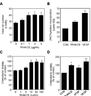
been assessed in in vitro angiogenesis models. We have first examined the effect of TRANCE on HUVEC proliferation. TRANCE increased the total number of cells at 48 h following TRANCE treatment in a dose-dependent manner (Fig. 1A). The effect of TRANCE on DNA synthesis of HUVECs was moni-tored by [3H]thymidine incorporation assay (Fig. 1B). TRANCE
increased the DNA synthesis of HUVECs by 1.5-fold at 3g/ml. The activity at 3g/ml TRANCE was lower than that of VEGF at 10 ng/ml.
We next determined the effect of TRANCE on chemotactic motility of HUVECs by employing a modified Boyden chamber assay. TRANCE stimulated the chemotactic motility of HUVECs in a does-dependent manner with near maximal ac-tivity at 50 ng/ml (Fig. 1C). The migratory acac-tivity at 50 ng/ml of TRANCE was 40% increase over the control, and the effect of TRANCE was comparable with those of the optimal concentra-tion of bFGF (25 ng/ml) and VEGF (25 ng/ml), which are the known stimuli of HUVEC migration (Fig. 1D).
The effect of TRANCE on the morphological differentiation of HUVECs was investigated using two-dimensional Matrigel. When placed on growth factor-reduced Matrigel in the absence of angiogenic factors, HUVECs formed incomplete and narrow tube-like structures (Fig. 2A). In contrast, the treatment of 3 g/ml TRANCE led to the formation of elongated and robust tube-like structures, which were organized by much larger number of cells compared with the control (Fig. 2A). By meas-uring the area covered by the tube network using an image analysis program, TRANCE stimulated tube formation by 2-fold over the control (Fig. 2B). We also observed that
TRANCE induces tube formation of human microvascular en-dothelial cell line, HMEC-1 (data not shown). These results indicate that TRANCE has a novel angiogenic activity in in vitro human endothelial cell culture system.
TRANCE Induces Angiogenesis in Vivo—To determine whether TRANCE is capable of promoting angiogenesis in vivo, experiments were first performed on the CAMs using Ther-manox discs. After 72 h of contact with the CAM, the disc containing 3g/ml TRANCE was associated with the signifi-cant induction of neovascularization (Fig. 3A). The relative quantitation of the CAMs with a disc loaded with vehicle alone, a disc with PMA as a positive control, and a disc with 3g/ml TRANCE are shown in Fig. 3B. The in vivo angiogenic activity of TRANCE was further evaluated by an established in vivo angiogenesis model, the mouse Matrigel plug assay. Matrigel with or without TRANCE (3 g/ml) was injected subcutane-ously into C57/BL6 mice. The solid gel plug was removed from the mice at 5 days after implantation for histological examina-tion. As shown in Fig. 3C, TRANCE produced more neovessels within gels than Matrigel alone. The vascular density and the number of mature vascular structures were significantly in-creased by TRANCE. When quantified by measurement of vas-cular areas in the fixed Matrigel plugs, the angiogenic activity of TRANCE was comparable with 50 ng/ml bFGF (data not shown). Taken together, these results indicate that TRANCE has a potent angiogenic activity in vivo.
TRANCE-induced Angiogenesis Does Not Require VEGF Ex-pression in Endothelial Cells—The angiogenic activity of TRANCE may be the result of its direct action on endothelial cells or through the induction of other genes involved in angio-genesis (4). VEGF is a selective mitogen and motogen for en-dothelial cells. A number of angiogenic inducers including TNF␣, transforming growth factor-, interleukin-1, prostag-landins, and endothelins have been shown to induce expression of VEGF in various cell types, which is in part responsible for their role in angiogenesis (4). We tested the possibility that the effect of TRANCE on angiogenesis is mediated through expres-sion of VEGF.
The expression of VEGF by TRANCE in HUVECs was as-sessed by RT-PCR analysis. As previously reported, TNF␣ markedly increased the VEGF transcript in HUVECs, whereas TRANCE had no effect on the levels of VEGF mRNA (Fig. 4A). To confirm the results of RT-PCR analysis, we used a VEGF-neutralizing antibody to test whether the angiogenic activities
FIG. 1. TRANCE induces proliferation and migration in
HUVECs. A, effect of TRANCE on proliferation of HUVECs. Various
concentrations of TRANCE were added to HUVECs. After 48 h, the number of cells was counted under the microscope. B, for comparison of the effects of TRANCE and VEGF on DNA synthesis, HUVECs were stimulated with 3g/ml TRANCE or 10 ng/ml VEGF and allowed to proliferate for 36 h. [3H]Thymidine was present during the last 6 h of
incubation. C, chemotactic motility of HUVECs by different doses of TRANCE. HUVECs were placed in the upper chamber, and M199 (1% FBS) with various concentrations of TRANCE placed in the lower wells of chemotaxis chamber. D, the comparison of the effects of TRANCE and other angiogenic factors on the chemotactic motility of HUVECs. M199 (1% FBS) with 50 ng/ml TRANCE, 25 ng/ml bFGF, or 25 ng/ml VEGF were placed in the lower wells. After 4 h, chemotactic motility of HUVECs was measured as described under “Materials and Methods.” The basal migration in the absence of TRANCE was 51.4⫾ 3.3 cells/ field. All data are expressed as percentage⫾ S.E. from three different experiments with triplicate. *, p⬍ 0.05; **, p ⬍ 0.01 versus control.
FIG. 2. TRANCE induces tube formation of HUVECs. A, effect of TRANCE on tube formation of HUVECs. HUVECs were plated on Matrigel-coated wells at a density of 4⫻ 105cells/well without (CON) or
with 3g/ml TRANCE. After 20 h, photographs were taken (⫻40). B, the area covered by the tube network was quantitated using Image-Pro Plus software. Data are expressed as percentage ⫾ S.E. from three different experiments with triplicate. *, p⬍ 0.01 versus control.
at Ewha Medical Library on October 26, 2016
http://www.jbc.org/
of TRANCE are mediated by VEGF. VEGF-neutralizing anti-body significantly reduced VEGF-induced HUVEC prolifera-tion, but had no effect on TRANCE-induced proliferation (Fig. 4B). We also observed that TRANCE-induced HUVEC tube formation was not affected by VEGF-neutralizing antibody (data not shown). These results indicate that TRANCE-induced angiogenesis is not caused by the induction of VEGF expres-sion, suggesting that TRANCE may act as a direct angiogenic modulator in endothelial cells. TRANCE and TNF␣ are classi-fied as the same TNF superfamily ligand based on amino acid sequence homology and have fairly similar biological actions, mainly in immune response and the development of osteoclasts (6, 23). The present study showed that TRANCE also had an angiogenic activity in addition to TNF␣ as previously
charac-terized. However, since TNF␣ has been shown to require VEGF expression for its angiogenic activity and have no direct action on endothelial cell proliferation and migration, the mechanism of the angiogenic action of TRANCE is likely to be distinct from that of TNF␣.
TRANCE Activates ERKs and p125FAK in HUVECs—
TRANCE induces intracellular signaling through the interac-tion with its receptor RANK. We confirmed the presence of RANK mRNA in HUVECs using RT-PCR analysis (Fig. 5A). Immunoblot analysis showed expression of RANK protein in HUVECs, which is similar size to that reported in mouse den-dritic cells (data not shown). To characterize the function of the TRANCE receptor in stimulating angiogenesis, we attempted to examine TRANCE-induced intracellular signals in endothe-lial cells. Recent studies have shown that activation of ERKs is closely involved in proliferation, migration, or morphogenesis of endothelial cells induced by various angiogenic factors. Therefore, we investigated whether TRANCE can stimulate ERKs in HUVECs. Subconfluent HUVECs were exposed to various concentrations of TRANCE, and the activation of ERK1/2 was analyzed by Western blot analysis using antibody directed against the phosphorylated form of ERK1/2 (p44 ERK1 and p42 ERK2). As shown in Fig. 5B, TRANCE induced ERK activation in a dose- and time-dependent manner, and maximal activation was observed after 20 min of TRANCE stimulation and declined thereafter. In contrast to the effect of TRANCE on ERK1/2, TNF␣ was not able to induce ERK acti-vation in HUVECs (data not shown), suggesting that the in-tracellular signaling property of TRANCE is also distinct from that of TNF␣ in endothelial cells.
Focal adhesion kinase p125FAK is believed to represent a
primary signaling mediator for the dynamic changes in actin cytoskeleton reorganization that are a prerequisite for the pro-motion of cell migration (24, 25). Recent work has shown that VEGF stimulates tyrosine phosphorylation of p125FAK in
HUVECs, and the resulting tyrosine phosphorylation is sug-gested to have a role in the migratory cell response to VEGF (26). Thus, we next examined the effect of TRANCE on tyrosine
FIG. 3. Identification of TRANCE-induced angiogenesis by
mouse Matrigel plug assay and the chicken chorioallantoic membrane assay. Angiogenesis was induced by Themanox discs
con-taining TRANCE (3g/ml) or PMA (0.12 g/ml) on the CAM of 9-day-old chick embryos. The angiogenic response was assessed as positive after appearance of a significantly increased vascularization under or in the vicinity of the disc. A, the effect of TRANCE on the formation of new blood vessels in the CAMs. The representative areas of the CAM were pictured using a light microscope (⫻16). Arrow indicates the formation of new blood vessels by TRANCE. B, comparison of the effect of TRANCE and PMA on the CAMs. Numerical values on the bars are expressed as the number of positive discs/total number of eggs. For the Matrigel plug assay, C57/BL6 mice were injected subcutaneously with 0.5 ml of Matrigel with or without TRANCE (3g/ml). C, the effect of TRANCE on neovessel formation in the Matrigel (⫻100). Arrow indi-cates the active neovessels containing red blood cells. D, quantitative effect of TRANCE on neovessel formation. Neovessel areas in the Ma-trigel plug were quantified using TINA 2.0 software. Four mice were used as a group, and the experiment was repeated twice.
FIG. 4. TRANCE does not induce VEGF expression. A, the effect TRANCE on VEGF expression in HUVECs. HUVECs were cultured with 3g/ml TRANCE or 50 ng/ml TNF␣. Total mRNAs were isolated, and RT-PCR was performed as described under “Material and Meth-ods.” B, the effect of VEGF-neutralizing antibody on TRANCE-induced DNA synthesis in HUVECs. HUVECs were stimulated with or without 3g/ml TRANCE or 10 ng/ml VEGF in the presence or absence of 1 g/ml VEGF-neutralizing antibody and allowed to proliferate for 36 h. [3H]Thymidine was present during the last 6 h of incubation. Data
represent the means⫾ S.E. of three different experiments with tripli-cate. *, p⬍ 0.05; **, p ⬍ 0.01 versus control.
FIG. 5. Expression of RANK and TRANCE-induced ERK and
p125FAKactivation in HUVECs. A, RT-PCR analysis of RANK mRNA
expression in HUVECs. HUVECs were cultured with or without TRANCE (5g/ml) for 8 h. Total mRNAs were isolated, and RT-PCR was performed as described under “Material and Methods.” B, ERK activation by TRANCE. HUVECs were stimulated with various concen-trations of TRANCE for 20 min (left panel) or stimulated with 5g/ml TRANCE for the indicated time periods (right panel). Phosphorylated forms of ERKs (P-ERK1 and P-ERK2) in whole cell extracts were detected with phospho-specific antibodies. The membranes were stripped and reprobed with antibody against ERK2. C, TRANCE-in-duced tyrosine phosphorylation of p125FAK in HUVECs. Cells were
stimulated with various concentrations of TRANCE for 10 min (left
panel) or stimulated with 0.1g/ml TRANCE for the indicated time
periods (right panel). Cell lysates were prepared and immunoprecipi-tated with anti-p125FAKantibody. The immune complexes were
ana-lyzed by immunoblotting with anti-phosphotyrosine antibody to assay for p125FAKtyrosine phosphorylation (P-p125FAK) or anti-p125FAK
an-tibody for p125FAKprotein levels (p125FAK).
at Ewha Medical Library on October 26, 2016
http://www.jbc.org/
phosphorylation of p125FAKin HUVECs. We observed tyrosine
phosphorylation of p125FAKat 0.05g/ml TRANCE (Fig. 5C).
The effective dose of TRANCE for p125FAK activation was
lower by one order than that for ERK activation, suggesting that p125FAKis likely to be more sensitively triggered upon the
TRANCE receptor engagement than ERKs. Kinetic analysis revealed phosphorylated p125FAKto be detectable within 10
min after addition of TRANCE and declined to a basal level at 1 h (Fig. 5C). Thus, this study demonstrates that, in addition to TRANCE-induced ERKs, NF-B, and AKT/PKB activation in other cells, TRANCE can activate p125FAKin endothelial cells.
This signaling event may provide support for a signaling cas-cade in which TRANCE induces cytoskeletal reorganizations resulting in cell spreading and migration.
TRANCE-induced ERKs and p125FAK Activation in
HUVECs Are Blocked by Inhibitors of Src Family Tyrosine Kinases and PLC—To further delineate the TRANCE-mediated signaling pathway in endothelial cells, we were prompted to explore the upstream signaling mechanisms re-lated to activation of ERK and p125FAKfrom receptor
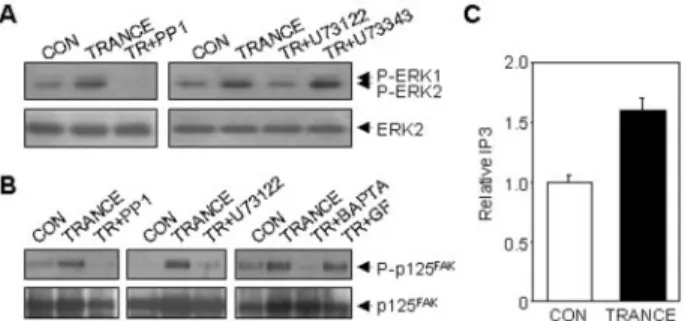
engage-ment. In dendritic cells and osteoclasts, the nonreceptor tyro-sine kinase c-Src is shown to play a critical role in AKT/PKB activation by TRANCE (16). We tested the involvement of Src family kinases on the TRANCE signaling pathway in HUVECs. Pretreatment of HUVECs with PP1, an inhibitor for Src family kinases, completely blocked both ERK and p125FAKactivation
by TRANCE (Fig. 6). These results indicate that Src family tyrosine kinases lie upstream of the ERK and p125FAK
cas-cades initiated by the TRANCE receptor in HUVECs. c-Src is previously shown to interact with RANK upon receptor engage-ment, and the resulting activation of c-Src is responsible for TRANCE-induced cell survival in dendritic cells (16). Our data indicate that activation of Src is likely to be linked to TRANCE-induced angiogenic signaling pathway.
Since evidence has been presented that a Src family tyrosine kinase lies upstream of PLC-␥1 (27, 28), we further examined the ability of PLC inhibitor to block TRANCE-induced ERK and p125FAK activation. Pretreatment of HUVECs with the
PLC inhibitor U73122 blocked TRANCE-induced ERK activa-tion, while U73343, a structurally similar derivative of U73122 but inactive for PLC, had no effect (Fig. 6A). U73122 also completely blocked TRANCE-induced p125FAK
phosphoryla-tion (Fig. 6B). PLC is known to generate IP3and diacylglycerol,
which activate intracellular Ca2⫹ mobilization and PKC,
re-spectively. To confirm PLC activation, we measured the effect of TRANCE on IP3generation. As shown in Fig. 6C, treatment
of HUVECs with 0.5g/ml TRANCE for 20 min increased the cellular level of IP3 by 1.6-fold over the control, providing evidence that TRANCE activates PLC in HUVECs. To further characterize the role of PLC in the TRANCE signaling pathway in HUVECs, we assessed the abilities of the intracellular Ca2⫹ chelator BAPTA-AM and the PKC inhibitor GF109203X to block p125FAKphosphorylation by TRANCE. BAPTA-AM
com-pletely inhibited TRANCE-induced p125FAK phosphorylation
(Fig. 6B). In contrast to BAPTA-AM, GF109203X failed to exert a similar effect (Fig. 6B). These data indicate that increased intracellular Ca2⫹ by PLC activation is important for the TRANCE signaling pathway. Therefore, it is suggested that the Src-PLC␥1-Ca2⫹signaling cascade is most likely to be respon-sible for TRANCE-induced angiogenic signaling pathway.
Src Family Tyrosine Kinases and PLC Are Required for TRANCE-induced HUVEC Migration—The activation of ERK and p125FAKpathway by TRANCE prompted us to determine
whether these biochemical events are important for angiogenic activities of TRANCE. Thus, we tested the effects of inhibitors of ERK1/2, Src, and PLC on TRANCE-induced HUVEC chemo-taxis. The endothelial ERK activity has been shown to modu-late proliferation and morphogenesis of endothelial cells in several angiogenic factors (21, 29 –31). In some cases this en-zyme activity was also implicated in endothelial cell migration (31). ERK is required for endothelial cell chemotaxis stimu-lated by bFGF or the placental angiogenic hormone proliferin (31, 32). In contrast, VEGF- or S1P-induced HUVEC migration was reported to be independent of ERK (21, 33). As shown in Fig. 7A, U0126, a specific inhibitor of MAPK, had no effect on TRANCE-induced cell motility. The lack of inhibitory effect of U0126 indicates that the ERK activity is not required for HUVEC migration by TRANCE. Unlikely to inhibit the MAPK inhibitor U0126, the Src tyrosine kinase inhibitor PP1 signifi-cantly inhibited TRANCE-induced HUVEC chemotaxis, whereas it did not alter basal migration (Fig. 7B), suggesting that Src kinases are closely involved in TRANCE-induced
en-FIG. 6. TRANCE induces ERK1/2 and p125FAK
activation through the activation of Src and PLC in HUVECs. The effects of
various signaling blockers on TRANCE-induced ERK1/2 (A) and p125FAK(B) activation are shown. HUVECs were pretreated for 30 min
with or without 10 M PP1, 5 M U73122, 5 M U73343, 10 M BAPTA-AM (BAPTA), or 2MGF109203X (GF) prior to treating with 5 g/ml or 0.1 g/ml TRANCE (TR) for ERK1/2 and p125FAKactivation,
respectively. The activation of ERK1/2 and p125FAKwas measured as
described in Fig. 5. C, the effect of TRANCE on IP3generation in
HUVECs. Cells were stimulated with 0.5g/ml TRANCE for 20 min. Measurement of IPTin HUVECs was performed as described under
“Material and Methods.” Data are expressed as relative values of IP3
from two individual experiments.
FIG. 7. Inhibitors of Src and PLC suppressed
TRANCE-in-duced chemotaxis of HUVECs. TRANCE-inTRANCE-in-duced chemotactic
mo-tility of HUVECs was determined in the absence and presence of the MAPK inhibitor U0126 (A), the Src tyrosine kinase inhibitor PP1 (B), the PLC inhibitor U73122 (C), and the intracellular Ca2⫹ chelator BAPTA-AM and the PKC inhibitor GF109203X (D). Cells were prein-cubated for 30 min with or without U0126 (10M), PP1 (10M), U73122 (5M), BAPTA-AM (10M), or GF109203X (2M) prior to treatment with 50 ng/ml TRANCE. Data are expressed as percentage⫾ S.E. from three different experiments with triplicate. *, p⬍ 0.05 versus control; #,
p⬍ 0.05 versus TRANCE; ##, ⬍ 0.01 versus TRANCE.
at Ewha Medical Library on October 26, 2016
http://www.jbc.org/
dothelial cell migration. This inhibitory effect of PP1 on cell migration is consistent with our finding that Src kinases are required for TRANCE-induced p125FAKactivation in HUVECs.
Our data further showed that the stimulatory effect of TRANCE on HUVEC chemotaxis was significantly abrogated by the PLC inhibitor U73122 (Fig. 7C), indicating the role of PLC in TRANCE-stimulated HUVEC migration. Moreover, TRANCE-induced HUVEC migration was blocked by the intra-cellular Ca2⫹chelator but not by the PKC inhibitor (Fig. 7D).
Theses results indicate that PLC-regulated intracellular Ca2⫹ mobilization plays an important role in HUVEC migration by TRANCE.
DISCUSSION
In this report, we demonstrate that TRANCE has a novel function as an angiogenic factor. TRANCE stimulated angio-genesis in vivo by the mouse Matrigel plug assay and CAM assay. In the mouse Matrigel plug assay, the ability of TRANCE to promote neovessel formation was comparable with that of the well established angiogenic factor bFGF. In in vitro angiogenesis models, we have revealed that the stimulation of HUVECs with TRANCE leads to an increase in cell prolifera-tion as well as chemotactic motility and strongly induces the formation of tube network. Our data also showed that the mechanism by which TRANCE induces angiogenesis is dissim-ilar to TNF␣, which requires VEGF expression for its angio-genic activity. The angioangio-genic activity of TRANCE appeared to be the result of its direct action on endothelial cells. Thus, we demonstrate here a novel biological action of TRANCE in vas-culature formation in addition to the regulatory functions pre-viously identified in immune responses and bone development (15, 34).
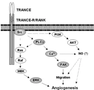
The present study demonstrates the TRANCE signaling pathway in endothelial cells (Fig. 8). We have delineated the expression and functionality of the cognate TRANCE receptor RANK in human endothelial cells. RANK expression in HUVECs and HMEC-1 was confirmed by RT-PCR, Western blot, and fluorescence-activated cell sorter analysis using fluo-rescein isothiocyanate-TRANCE (Fig. 1 and data not shown). As reported in osteoclasts and dendritic cells (16), activation of the receptor downstream signaling mediators, such as ERKs, Src, and AKT, was observed in HUVECs. Interestingly, we recently found that VEGF pretreatment increased RANK ex-pression on the surface of HUVECs and subsequently potenti-ated the effects of TRANCE on ERK activation and tube
Src blocks TRANCE-induced AKT activation in osteoclasts and dendritic cells in vitro and that the Src activity is required for TRANCE-mediated osteoclast survival (16, 35). Additionally, we found that Src and PLC were required for TRANCE-induced HUVEC migration. The roles of these signaling molecules in angiogenesis are implicated in other systems. It has been shown that activation of Src is essential for VEGF-induced angiogenesis and that PLC is required for S1P-induced cell migration (36, 37). Several lines of evidence also noted the significant role of Ca2⫹in endothelial cell migration (27, 37). In
particular, a recent study showed that VEGF increases intra-cellular Ca2⫹leading to the generation of nitric oxide (NO) by activation of endothelial NO synthase, and the resulting endo-thelial NO production is required for VEGF-induced angiogen-esis including endothelial cell proliferation, migration, and net-work formation (27). Our results indicate that TRANCE-induced endothelial cell motility requires increases in intracellular Ca2⫹. Therefore, our findings raise the possibility
that NO production via the Src-PLC-Ca2⫹signaling pathway may be an essential step in TRANCE-induced angiogenesis. Alternatively, AKT can mediate endothelial cell survival and lead to increases in NO production via activation of endothelial NO synthase (38). Since we observed that TRANCE activated AKT in HUVECs (data not shown), the AKT signaling pathway may in part contribute to the angiogenic processes triggered by TRANCE. These possibilities are under investigation.
New blood vessel formation is required for endochondral bone formation by serving as the conduit that allows a variety of cells essential for bone morphogenesis (endothelial cells, chondroclasts, and osteoblasts) to migrate into the growth plate. A recent study demonstrates that VEGF generated from hypertrophic chondrocytes in the epiphyseal growth plate cou-ples hypertrophic cartilage remodeling, ossification, and angio-genesis during endochondral bone formation (19). Since the angiogenic processes in vivo are regulated by coordinative ac-tion of various angiogenic factors in addiac-tion to VEGF, another factor generated from cells inside or in the close vicinity of cartilage seems to be involved in the progression of angiogen-esis during bone formation. Therefore, our findings strongly suggest that TRANCE produced by osteoblasts may play a cooperative role with VEGF in the proper vascular formation during bone growth and remodeling. Angiogenesis is also an essential requirement for tumor growth and metastasis and is regulated by a complex network of factors that interact be-tween stromal cells, endothelial cells, and neoplastic cells within solid tumor. Cytokines, such as IL-1, IL-6, IL-11, IL-17, and TNF␣, generated by tumor-associated macrophages, increase the expression of TRANCE with decrease of OPG expression in stromal cells (39). This evidence suggests that TRANCE produced by stromal cells may be involved in the formation of microvessels in the tumor site. Furthermore, in-flammatory cytokines TNF␣ and IL-1␣ elevate TRANCE ex-pression in human microvascular endothelial cells (40). These results together with our observation suggest that the timely generation of TRANCE from endothelial cells by other
inflam-2Y.-M. Kim and Y.-G. Kwon, unpublished observations.
FIG. 8. Schematic illustration of TRANCE-induced angiogenic
signaling pathway in endothelial cells.
at Ewha Medical Library on October 26, 2016
http://www.jbc.org/
matory cytokines may in part contribute to new vasculature formation in an inflammatory site. Thus, we propose that TRANCE can act as an important modulator of angiogenesis under physiological and pathological conditions.
Acknowledgment—We thank Dr. Yun Soo Bae for valuable advice on
analyzing IP3concentration.
REFERENCES
1. Folkman, J. (1995) Nat. Med. 1, 27–31
2. Jackson, J. R., Seed, M. P., Kircher, C. H., Willoughby, D. A., and Winkler, J. D. (1997) FASEB J. 11, 457– 465
3. Risau, W. (1997) Nature 386, 671– 674
4. Bussolino, F., Mantovani, A., and Persico, G. (1997) Trends. Biochem. Sci. 22, 251–256
5. Anderson, D. M., Maraskovsky, E., Billingsley, W. L., Dougall, W. C., Tometsko, M. E., Roux, E. R., Teepe, M. C., DuBose, R. F., Cosman, D., and Galibert, L. (1997) Nature 390, 175–179
6. Green, E. A., and Flavell, R. A. (1999) J. Exp. Med. 189, 1017–1020 7. Kong, Y. Y., Boyle, W. J., and Penninger, J. M. (2000) Immunol. Today 10,
495–502
8. Kong, Y. Y., Yoshida, H., Sarosi, I., Tan, H. L., Timms, E., Capparelli, C., Morony, S., Oliveirados-Santos, A. J., Van, G., Itie, A., Khoo, W., Wakeham, A., Dunstan, C. R., Lacey, D. L., Mak, T. W., Boyle, W. J., and Penninger, J. M. (1999) Nature 397, 315–323
9. Wong, B. R., Josien, R., Lee, S. Y., Sauter, B., Li, H. L., Steinman, R. M., and Choi, Y. (1997) J. Exp. Med. 186, 2075–2080
10. Bachmann, M. F, Wong, B. R., Josien, R., Steinman, R. M., Oxenius, A., and Choi, Y. (1999) J. Exp. Med. 189, 1025–1031
11. Li, J., Sarosi, I., Yan, X. Q., Morony, S., Capparelli, C., Tan, H. L., McCabe, S., Elliott, R., Scully, S., Van, G., Kaufman, S., Juan, S. C., Sun, Y., Tarpley, J., Martin, L., Christensen, K., McCabe, J., Kostenuik, P., Hsu, H., Fletcher, F., Dunstan, C. R., Lacey, D. L., and Boyle, W. J. (2000) Proc. Natl. Acad.
Sci. U. S. A. 97, 1566 –1571
12. Hsu, H., Lacey, D. L., Dunstan, C. R., Solovyev, I., Colombero, A., Timms, E., Tan, H. L., Elliott, G., Kelley, M. J., Sarosi, I., Wang, L., Xia, X. Z., Elliott, R., Chiu, L., Black, T., Scully, S., Capparelli, C., Morony, S., Shimamoto, G., Bass, M. B., and Boyle, W. J. (1999) Proc. Natl. Acad. Sci. U. S. A. 96, 3540 –3545
13. Kong, Y. Y., Feige, U., Sarosi, I., Bolon, B., Tafuri, A., Morony, S., Capparelli, C., Li, J., Elliott, R., McCabe, S., Wong, T., Campagnuolo, G., Moran, E., Bogoch, E. R., Van, G., Nguyen, L. T., Ohashi, P. S., Lacey, D. L., Fish, E., Boyle, W. J., and Penninger, J. M. (1999) Nature 402, 304 –309 14. Lacey, D. L., Timms, E., Tan, H. L., Kelley, M. J., Dunstan, C. R., Burgess, T.,
Elliott, R., Colombero, A., Elliott, G., Scully, S., Hsu, H., Sullivan, J., Hawkins, N., Davy, E., Capparelli, C., Eli, A., Qian, Y. X., Kaufman, S.,
Sarosi, I., Shalhoub, V., Senaldi, G., Guo, J., Delaney, J., and Boyle, W. J. (1998) Cell 93, 165–176
15. Wong, B. R., Rho, J., Arron, J., Robinson, E., Orlinick, J., Chao, M., Kalachikov, S., Cayani, E., Bartlett, F. S., III, Frankel, W. N., Lee, S. Y., and Choi, Y. (1997) J. Biol. Chem. 272, 25190 –25194
16. Wong, B. R., Besser, D., Kim, N., Arron, J. R., Vologodskaia, M., Hanafusa, H., and Choi, Y. (1999) Mol. Cell 4, 1041–1049
17. Wong, B. R., Josien, R., Lee, S. Y., Vologodskaia, M., Steinman, R. M., and Choi, Y. (1998) J. Biol. Chem. 273, 28355–28359
18. Harper, J., and Kalgsbrun, M. (1999) Nat. Med. 5, 617– 618
19. Gerber, H. P., Vu, T. H., Ryan, A. M., Kowalski, J., Werb, Z., and Ferrara, N. (1999) Nat. Med. 5, 623– 628
20. Jaffe, E. A., Nachman, R. L., Becker, C. G., and Minick, C. R. (1973) J. Clin.
Invest. 52, 2745–2756
21. Lee, O. H., Kim, Y. M., Lee, Y. M., Moon, E. J., Lee, D. J., Kim, J. H., Kim, K. W., and Kwon, Y. G. (1999) Biochem. Biophys. Res. Commun. 264, 743–750
22. Kim, M. S., Lee, Y. M., Moon, E. J., Kim, S. E., Lee, J. J., and Kim, K. W. (2000)
Int. J. Cancer 87, 269 –275
23. Wong B. R., Josien R., and Choi Y. (1999) J. Leukoc. Biol. 65, 715–724 24. Abedi, H., and Zachary, I. (1995) Cardiovasc. Res. 30, 544 –556
25. Schaller M. D., and Parsons, J. T. (1994) Curr. Opin. Cell Biol. 6, 705–710 26. Abedi, H., and Zachary, I. (1997) J. Biol. Chem. 272, 15442–15451 27. He, H., Venema, V. J., Gu, X., Venema, R. C., Marrero, M. B., and Caldwell,
R. B. (1999) J. Biol. Chem. 274, 25130 –25135
28. Zachary, I. (2001) Am. J. Physiol. Cell Physiol. 280, C1375–C1386 29. Kuzuya, M., Satake, S., Ramos, M. A., Kanda, S., Koike, T., Yoshino, K., Ikeda,
S., and Iguchi, A. (1999) Exp. Cell. Res. 248, 498 –508 30. Yu, Y., and Sato, J. D. (1999) J. Cell. Physiol. 178, 235–246
31. Groskopf, J. C., Syu, L. J., Saltiel, A. R., and Linzer, D. I. (1997) Endocrinology
138, 2835–2840
32. Shono, T., Kanetake, H., and Kanda, S. (2001) Exp. Cell Res. 264, 275–283 33. Liu, F., Verin, A. D., Wang, P., Day, R., Wersto, R. P., Chrest, F. J., English,
D. K., and Garcia, J. G. (2001) Am. J. Respir. Cell Mol. Biol. 24, 711–719 34. Lynch, C. N., Wang, Y. C., Lund, J. K., Chen, Y. W., Leal, J. A., and Wiley, S. R.
(1999) J. Biol. Chem. 274, 8455– 8459
35. Xing, L., Venegas, A. M., Chen, A., Garrett-Beal, L., Boyce, B. F., Varmus, H. E., and Schwartzberg, P. L. (2001) Genes Dev. 15, 241–253
36. Schlessinger, J. (2000) Cell 100, 293–296
37. Lee, O. H., Lee, D. J., Kim, Y. M., Kim, Y. S., Kwon, H. J., Kim, K. W., and Kwon, Y. G. (2000) Biochem. Biophys. Res. Commun. 268, 47–53 38. Dimmeler, S., Fleming, I., Fisslthaler, B., Hermann, C., Busse, R., and Zeiher,
A. M. (1999) Nature 399, 601– 605
39. Nakashima, T., Kobayashi, Y., Yamasaki, S., Kawakami, A., Eguchi, K., Sasaki, H., and Sakai, H. (2000) Biochem. Biophys. Res. Commun. 275, 768 –775
40. Collin-Osdoby, P., Rothe, L., Anderson, F., Nelson, M., Maloney, W., and Osdoby, P. (2001) J. Biol. Chem. 276, 20659 –20672
at Ewha Medical Library on October 26, 2016
http://www.jbc.org/
10.1074/jbc.M109434200
Access the most updated version of this article at doi:
Alerts:
When a correction for this article is posted
•
When this article is cited
•
to choose from all of JBC's e-mail alerts
Click here
http://www.jbc.org/content/277/9/6799.full.html#ref-list-1
This article cites 40 references, 15 of which can be accessed free at
at Ewha Medical Library on October 26, 2016
http://www.jbc.org/