DOI : 10.3341/jkos.2009.50.12.1847 = 증례보고 =
샤임플러그 사진기와 초음파 생체현미경을
이용한 전방 계측 비교
박용식1⋅안현석1⋅김나래1⋅마경탁2⋅홍사민1⋅성공제1⋅김찬윤1 연세대학교 의과대학 안과학 교실, 시기능 개발연구소1, 실로암 안과병원2목적: 원발폐쇄각 및 정상군에서 Oculus Pentacam system (Oculus Inc., Germany)과 Hi-scan ultrasound biomicroscopy (OPTIKON
2000, Rome, Italy)의 전방계측치를 비교함으로써 PentacamⓇ의 신뢰도를 평가하고자 했다.
대상과 방법: 2006년 6월부터 2007년 1월까지 내원한 원발폐쇄각군 26명(51안)과 정상군 20명(39안), 총 46명(90안)에 대해 전향적 연구
를 하였다. UBM과 PentacamⓇ의 전방계측치와 폐쇄각 진단능력을 비교하였다.
결과: 전방깊이는 정상군(r=0.821)과 환자군(r=0.957)에서 두 기계간의 강한 상관관계를, 전방각은 정상군(r=0.523)과 환자군(r=0.456)에서 중등도 상관관계와 양호한 일치도를 보였다. ROC curve로써 평가한 전방각 측정값의 폐쇄각 진단능력(AUC of UBM, 0.942; AUC of PentacamⓇ, 0.931)은 비슷하였다. 결론: 두 도구의 전방계측치는 고도 또는 중등도의 상관관계, 양호한 일치도 그리고 비슷한 폐쇄각 진단능력을 보이므로 폐쇄각의 진단 에 PentacamⓇ의 역할이 기대된다. <대한안과학회지 2009;50(12):1847-1852> ■ 접 수 일: 2009년 5월 4일 ■ 심사통과일: 2009년 8월 17일 ■ 책 임 저 자: 김 찬 윤 서울시 서대문구 신촌동 134 연세대학교 신촌세브란스병원 안과 Tel: 02-2228-3570, Fax: 02-312-0541 E-mail: [email protected] 전방의 구조적 변화를 동반하는 원발폐쇄각녹내장의 진 단에 있어서 전방각이나 전방 깊이를 측정하는 것은 임상적 으로 중요한 의미를 지닌다.1-3전방각의 측정시, 과거에는
전방각경을 사용한 검사를 통해 Van Herick and Shaffer grade, 또는 Spaeth grading system 등으로 표현하는 방법이 주로 사용되었고, 전방 깊이의 측정에는 고식적인 초음파를 사용한 A-scan (applanation ultrasound)이 주로 사용되어 왔다.4 그러나 전방각경을 사용한 검사는 검사자의 주관적인 요소가 많이 작용하며 결과를 정량적으로 비교하기 어려운 단점이 있으며 고식적 A-scan의 경우에는 각막에 가해지는 압력에 의한 측정값의 오차가 발생할 수 있고, 측정 위치에 따라 측정값의 변화가 있을 수 있다는 단점이 있다.5-8따라서 근래 에는 이들 외에도 전방의 구조를 좀더 정량적으로 정확하게 측정하려는 시도가 많이 이루어지고 있으며 그 결과 초음파 생체현미경(ultrasound biomicroscope, UBM)이나 샤임플
러그 사진기 원리를 사용하여 전방을 계측하는 Pentacam®과 같은 검사장비들이 개발되어 사용되고 있다. 초음파생체현미경은 고주파의 초음파를 이용하여 전안부의 여러 구조에 대한 고해상도의 이미지를 얻을 수 있으며 내장 된 프로그램을 사용하여 전방각 및 전방 깊이를 정량적으로 측정할 수 있어 최근 널리 사용되고 있다. 그러나 초음파생체 현미경은 눈을 물에 잠기게 한 상태로 검사를 시행해야 하 므로 시간이 많이 걸리며 환자와 검사자에게 불편을 야기 하는 측면이 있으며, 일정 수준 이상의 숙련된 검사자를 필요 로 하는 등 외래에서 빈번하게 사용하기에는 불편한 점이 많다.9,10 최근에 개발되어 소개되고 있는 Pentacam®은 회전하는 샤임 플러그 사진기를 사용한 광학적 화상 측정 도구로서, 약 2초 가량의 스캔을 통해 얻은 화상을 통해 전방의 구조를 3차원적 으로 재구성하게 되며 내장된 프로그램을 통해 정량적인 측정 이 가능하다.11-13Pentacam®은 비접촉식 측정 방식을 채택 하고 있으므로 접촉으로 인해 생길 수 있는 오차를 줄일 수 있으며 짧은 측정 시간 및 측정 방법의 용이성이라는 장점이 있다. 본 연구에서는 전향적 연구를 통해 원발폐쇄각녹내장 환자 및 정상군에서 Pentacam®과 초음파생체현미경을 사용하여 전방각 및 전방 깊이를 측정 비교함으로써 새롭게 소개된 진단 도구인 Pentacam®의 임상적 신뢰도를 평가하고자 한다.
대상과 방법
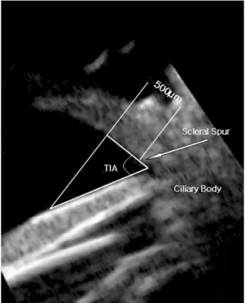
본 연구는 2006년 6월부터 2007년 1월까지 녹내장 외래에 내원한 환자 중 연구참여에 동의한 원발폐쇄각 또는 원발폐쇄 각녹내장 환자를 대상으로 하였고 역시 같은 기간에 비슷한Figure 1. Trabecular-iris angle (TIA) depicted on an ultrasound biomicroscopic (UBM) image of a normal eye.
Table 1. Characteristics of the study population
Age (yrs) Sex
M F
Normal (n=20) 46.7±21.2 10 10
Angle closure glaucoma (n=26) 70.5±7.4 21 5
나이 성별을 가지는 정상 전방각을 가지고 있는 환자를 대상 군으로 설정하였다. 본 연구는 기관 윤리위원회의 승인하에 연구가 진행되었다. 환자군 26명(51안)과 정상군 20명(39안), 총 46명의 환자(90안)가 연구에 포함되었다. 환자군은 원발 폐쇄각 또는 원발폐쇄각녹내장 환자를 대상으로 하였고, 레이 저를 제외한 녹내장 수술의 병력이 있는 환자는 제외하였다. 원발폐쇄각의 진단은 다른 병적인 선행요인 없이 비공막함입 전방각경검사상 270도 이상에서 섬유주 구조물이 보이지 않는 폐쇄각(occludable angle) 소견이 있으면서 급성폐쇄각 전력, 약물치료전 안압이 21 mmHg 이상이거나 전방각경상 홍채전 유착 소견이 있는 경우로 정의하였다. 원발폐쇄각녹내장 환자 의 경우에는 비공막함입 전방각경검사상 270도 이상에서 섬유 주 구조물이 보이지 않는 폐쇄각(occludable angle) 소견이 있으면서 시신경, 시경섬유, 시야검사상 녹내장성 시신경손상 소견이 동반된 경우로 정의하였다. 이후 모든 서술에서 편의 상 원발폐쇄각 또는 원발폐쇄각녹내장군은 원발폐쇄각군으로 서술하고자 한다. 정상 대조군은 다른 안과적 질환의 병력이 있거나, 안구 외상, 포도막염, 레이저 치료, 안과 수술(굴절 교정수술 포함)의 과거력이 있는 환자는 제외하였으며, 비공 막함입 전방각경검사상 360도에서 섬유주가 관찰 가능한 개방 각 소견을 가지는 환자를 대상으로 하였다. 초음파생체현미경 측정은 동일 검사자에 의해 이루어졌다. 전방 깊이의 측정시에는 25 MHz, 전방각의 측정을 위해서는 50 MHz의 해상도를 사용하였고, 수평면의 두 방향에서 얻은 이미지를 사용하였다. 전방각의 측정 지표로는 공막극과 이로 부터 500 um 떨어진 섬유주 상의 지점을 연결하는 선과, 이곳 에서 홍채에 수직으로 대응되는 점에서 홍채면에 접하는 선이 이루는 각 TIA (trabecular-Iris Angle)이 사용되었다(Fig. 1). 통계처리를 위해 수평면의 두 방향의 값을 평균하여 사용 하였다. 전방 깊이의 측정은 25 MHz의 해상도를 사용하여 얻은 영상을 바탕으로 내장된 소프트웨어를 사용하여 계산 하였다. Pentacam®도 역시 동일 검사자에 의해 측정이 이루어졌다. UBM 측정시의 manipulation에 의한 측정값 변화의 가능성을 배제하기 위해, 모든 검사자는 우선적으로 Pentacam®측정 후 초음파생체현미경 측정을 시행하였다. 전방각 및 전방 깊이의 측정값은 360도 방위로 스켄된 이미지 중 수평면에서 얻어진 화상을 바탕으로 내장된 소프트웨어에 의해 mm 및 degree 단위로 자동 계산되었다. 각 측정값에 대한 평균값 및 표준편차가 계산되었으며, 두 진단 도구로 얻은 측정값 사이의 통계적 연관성은 Pearson correlation coefficient에 의해 계산되었다. 원발폐쇄각녹내 장의 진단에 두 진단 도구의 각 지표들의 민감도와 특이도는 Receiver operating characterisitic (ROC) curve를 그린 후 Areas under the ROCs curves (AUC)를 구하여 판단하였다. p-value<0.05를 통계적으로 의미 있는 결과로 간주하였으며 통계 분석에는 SPSS (Ver. 13.0 for window; SPSS, Inc., Chicago, IL, USA)를 사용하였다.
결
과
환자의 나이는 정상군은 46.7±21.2세, 원발폐쇄각군은 70.5±7.4세였으며 성별은 정상군은 남자 10명 여자 10명, 원발폐쇄각군은 남자 5명 여자 21명이었다. 원발폐쇄각군 중
Figure 2. Bland-Altmann plots of anterior biometry measurements between ultrasound biomicroscopy (UBM) and Pentacam®. Solid line: 95% confidence interval of the difference between the methods.
원발폐쇄각은 40안, 원발폐쇄각녹내장은 11안이었다(Table 1). 전방 깊이는 정상군에서 초음파생체현미경과 Pentacam® 으로 측정한 값은 각각 2.74±0.63 mm, 3.00±0.60 mm로 통계적으로 유의한 차이가 없었고(p<0.05 by paired t-test) 원발폐쇄각군에서는 각각 1.70±0.27 mm, 1.80±0.30 mm로 역시 유의한 차이가 없었다(p<0.05). Pearson correlation coefficient상 상관계수는 정상군과 환자군 각각 0.821, 0.957 로 두 진단 도구간의 강한 상관관계가 있었다(Table 2). 전방 각은 정상군에서 초음파생체현미경과 Pentacam®으로 각각 38.00±7.00°, 37.80±8.82°이었고 환자군에서는 각각 24.10± 5.50°, 21.38±5.01°이었으며 Pearson correlation coefficient
상 상관계수는 정상군과 환자군 각각 0.523, 0.456로 두 진단 도구간의 중등도의 상관관계가 있었다(Table 3). 두 기계간의 측정치의 일치도(agreement)는 Figure 2에 나타난 것과 같이 전방 깊이는 정상군, 폐쇄각군에서 UBM 측정치에서 Pentacam® 측정치를 뺀 차가 각각 0.36 mm, 0.08 mm의 95% 신뢰구간내 편차를 보였고 전방각의 경우에는 각각 7.02°, 7.68°의 95% 신뢰구간내 편차를 보였다. 폐쇄각 진단에 대한 초음파 생체 현미경의 전방각 측정값의 AUC는 0.942이고 Pentacam®의 전방각 측정값의 AUC는 0.931이며 95% confidence interval 은 UBM은 0.899~0.985이고 pentacam®은 0.871~0.992로 비슷한 진단력을 나타내었다(Fig. 3).
Figure 3.Comparison of ultrasound biomicroscopy (UBM) and Pentacam®measurements of the anterior chamber angle for the diagnosis of primary angle-closure or primary angle-closure glaucoma.
Table 2. Correlation analysis between ultrasound biomicroscopy (UBM) and Pentacam®measurements of anterior chamber depth of normal and primary angle-closure patients
Anterior chamber depth (mm) Pearson’s correlation coefficient
UBM Pentacam® Coefficeint p-value*
Normal 2.74±0.63 3.00±0.60 0.821 <0.01
Patients 1.70±0.27 1.80±0.30 0.957 <0.01
*In normal and primary angle-closure patients, anterior chamber depth was measured by ultrasound biomicroscopy (UBM) and
Pentacam®; Pearson’s correlation coefficient was computed between the two groups (p<0.05).
Table 3. Correlation analysis between ultrasound biomicroscopy (UBM) and Pentacam® measurements of anterior chamber angle of normal and primary angle-closure patients
Anterior chamber angle (°) Pearson’s correlation coefficient
UBM Pentacam® Coefficeint p-value*
Normal 38.00±7.00 37.80±8.82 0.523 0.01
Patients 24.10±5.50 21.38±5.01 0.456 0.01
*In normal and primary angle-closure patients, anterior chamber angle was measured by ultrasound biomicroscopy (UBM) and
Pentacam®; Pearson’s correlation coefficient was computed between the two groups (p<0.05).
고
찰
본 연구에서는 UBM과 Pentacam®을 이용하여 원발폐쇄각 녹내장과 정상군의 전방 깊이, 전방각을 측정하고 측정 방법 에 따른 측정값의 상관관계와 각 진단 방법의 민감도와 특이 도를 알아보았다. 폐쇄각녹내장의 위험인자에는 인종, 나이, 여성, 가족력, 작은 각막 직경, 얕은 전방 깊이, 좁은 전방각, 수정체의 전방 이동 및 두께, 작은 안축장 등이 있으며14-18그러므로 조기 진단에 전방각 및 전방 깊이 측정이 중요하다. Pentacam®은 환자가 앉은 상태에서 신속하게 비접촉식으로 전안부를 계측 함으로써 환자에게 마취가 필요하지 않으며 각막 미란의 위험 이 없고 전방각과 전방 깊이 등의 전안부 계측이 한번에 이루 어진다는 장점이 있다. Pentacam®의 전방 깊이 측정의 정확성은 IOLMaster에 견줄 만 하다는 보고19가 있으며 본 연구 결과에서도 Pentacam® 과 UBM간의 전방 깊이 측정값은 강한 상관관계를 보였다. Oka et al20은 폐쇄각녹내장 환자와 원발개방각녹내장 환자 의 주변부 전방 깊이 및 전방 부피를 Pentacam®을 이용하여 측정한 후 폐쇄각녹내장에서 주변부 전방 깊이 및 전방 부피가 유의하게 작음을 보고하였는데 본 연구에서는 Pentacam®과 UBM의 전방각의 측정값이 중등도의 상관관계가 있었으며 원발성폐쇄각녹내장 진단 능력에 있어서 Pentacam® (AUC= 0.931)과 UBM (AUC=0.942)은 비슷하게 우수한 능력을 보여주었다. 본 연구에서는 Pentacam®과 UBM의 전방각과 전방 깊이측정값을 비교하였는데 다른 기계와의 측정값과 비교한 문헌 을 찾아보면 Buehl et al21은 정상인에서 각막의 두께와 전방 의 깊이를 알기 위해 Pentacam®과 Orbscan, AC-Master를 이용하였으며 각 기계간에 중심각막두께 및 전방 깊이에서 매우 높은 상관관계가 있음을 보고 하였고(r=0.92 to 0.97), 이 중 Pentacam® (rotating Scheimpflug camera)과 AC- Master (partial coherence interferometry)에서 Orbscan에 비하여 측정값의 신뢰도가 높았다고 하였다. 또 Lackner et al22은 전방 깊이의 측정에서 초음파보다 Pentacam®으로 측정한 값이 더 깊게 나타나지만 임상적으로 받아들일만한 정도라고 보고 하였다. Pentacam®과 전방 optical coherence tomography (OCT)를 비교한 연구에서도 역시 정상인, 폐쇄 각 환자 모두에서 Pentacam®이 OCT와 유사한 폐쇄각 진단 능력과 재현성을 보여 주었다.23,24 본 연구 결과 두 가지 측정 도구에 의한 전방각 측정값이 중등도의 상관관계를 보였는데 UBM으로 전방각을 측정할 때에는 수평면의 전방각 측정값의 평균으로 하였고 Pentacam® 의 내장된 프로그램에서는 수평면의 두 전방각 중 작은 측정 값을 선택하였기 때문으로 보인다. 또한 UBM은 누운 자세 에서 시행하며 Pentacam®은 앉은 자세에서 시행하므로 전방 각 측정시 자세의 차이도 영향이 있었을 것이다. 폐쇄각녹내장은 시신경이나 섬유주의 비가역적인 변화가 발생하기 전에 발견하여 예방할 수 있는 병이므로 조기 진단 이 중요한데 전방각 폐쇄를 관찰하는 가장 정확한 방법은 전방 각경검사와 UBM으로 알려져 있다.25전방각경검사는 밝은 환경에서 시행하므로 축동을 일으켜 부정확한 검사가 될 수 있는데 반해 UBM은 빛 대신 초음파를 사용하므로 어두운 환경에서 정확한 검사를 할 수 있으나 검사 시간이 오래 걸리 고 눈을 침수시켜야 하며 점안마취가 필요하고 숙련자에 의해 검사를 시행해야 하는 등의 불편함과 한계가 있다. 본 연구 에서 Pentacam®은 UBM과 전방 깊이 측정치의 강한 상관관계 를 보였고 전방각 측정값이 폐쇄각녹내장을 진단하는데 높은 민감도와 특이도를 보였다. Pentacam®은 어두운 환경에서 시행 가능하며 시행 시간이 짧고 비숙련 검사자가 시행할 수 있는 등의 장점이 있어 폐쇄각의 선별검사에 유용할 것으로 생각된다. 하지만 폐쇄각 선별검사로써의 구체적인 기준과 방법에 대하여서는 추가 연구가 필요할 것으로 사료된다.
참고문헌
1) Kim YY, Jung HR. Clarifying the nomenclature for primary angle closure glaucoma. Surv Ophthalmol 1997;42:125-36.
2) Ang MH, Baskaran M, Kumar RS, et al. National survey of oph-thalmologists in Singapore for the assessment and management of asymptomatic angle closure. J Glaucoma 2008;17:1-4.
3) Lavanya R, Wong TY, Friedman DS, et al. Determinants of angle
closure in older Singaporeans. Arch Ophthalmol 2008;126:686-91. 4) Koranyi G, Lydahl E, Norrby S, Taube M. Anterior chamber depth
measurement: A-scan versus optical methods. J Cataract Refract Surg 2002;28:243-7.
5) Reddy AR, Pande MV, Finn P, El-Gogary H. Comparative esti-mation of anterior chamber depth by ultrasonography, Orbscan II, and IOL master. J Cataract Refractive Surg 2004;30:1268-71. 6) Vetrugno M, Cardascia N, Cardia L. Anterior chamber depth
mea-sured by two methods in myopic and hyperopic phakic IOL im-plant. Br J Ophthalmol 2000;84:1113-6.
7) Foster PJ, Devereux JG, Alsbirk PH. Detection of gonioscopically occludable angles and primary angle closure glaucoma by estimation of limbal chamber depth in Asians: modified grading scheme. Br J Ophthalmol 2000;84:186-92.
8) Congdon NG, Spaeth GL, Augsburger J, et al. A proposed simple method for measurement in the anterior chamber angle: biometric gonioscopy. Ophthalmology 1999;106:2161-7.
9) Jang JW, Choe YJ, Hong YJ. Measurement of the depth and angle of the peripheral anterior chamber and iris thickness by ultrasono-graphic biomicroscopy. J Korean Ophthalmol Soc 1995;36:1179-84. 10) Lee SC, Jin KH. Changes in Ciliary Sulcus Size, Anterior chamber
depth and angle during accommodation using ultrasound biomicro-scopy. J Korean Ophthalmol Soc 2005;46:1809-14.
11) Elbaz U, Barkana Y, Gerber Y, et al. Comparison of different tech-niques of anterior chamber depth and keratometric measurements. Am J Ophthalmol 2007;143:48-53.
12) Emre S, Doganay S, Yologlu S. Evaluation of anterior segment para-meters in keratoconic eyes measured with the Pentacam system. J Cataract Refract Surg 2007;33:1708-12.
13) Friedman DS, Gazzard G, Foster P, et al. Ultrasonographic biomicro-scopy, Scheimpflug photography, and novel provocative tests in contralateral eyes of Chinese patients initially seen with acute angle closure. Arch Ophthalmol 2003;121:633-42.
14) Sihota R, Lakshmaiah NC, Agarwal HC, et al. Ocular parameters in the subgroups of angle closure glaucoma. Clin Exp Ophthalmol 2000;28:253-8.
15) Sihota R, Gupta V, Agarwal HC, et al. Comparison of symptomatic and asymptomatic, chronic, primary angle-closure glaucoma, open- angle glaucoma, and controls. J Glaucoma 2000;9:208-13. 16) Marchini G, Pagliarusco A, Toscano A, et al. Ultrasound
biomicro-scopic and conventional ultrasonographic study of ocular dimensions in primary angle-closure glaucoma. Ophthalmology 1998;105:2091-8. 17) Chen HB, Kashiwagi K, Yamabayashi S, et al. Anterior chamber
angle biometry: quadrant variation, age change and sex difference. Curr Eye Res 1998;17:120-4.
18) Salmon JF. Predisposing factors for chronic angle-closure glaucoma. Prog Retin Eye Res 1999;18:121-32.
19) Reuland MS, Reuland AJ, Nishi Y, Auffarth GU. Corneal radii and anterior chamber depth measurements using the IOLmaster versus the Pentacam. J Refract Surg 2007;23:368-73.
20) Oka N, Otori Y, Okada M, et al. Clinical study of anterior ocular segment topography in angle-closure glaucoma using the three- dimensional anterior segment analyzer Pentacam. Nippon Ganka Gakkai Zasshi 2006;110:398-403.
21) Buehl W, Stojanac D, Sacu S, et al. Comparison of three methods of measuring corneal thickness and anterior chamber depth. Am J Ophthalmol 2006;141:7-12.
=ABSTRACT=
Comparison of Measurement of Anterior Segment Parameters
Between Scheimpflug Camera and Ultrasound Biomicroscopy
Yong-sik Park, MD1, Hyunseok Ahn, MD1, Na Rae Kim, MD1, Kyoung Tak Ma, MD2, Samin Hong, MD1, Gong Je Seong, MD, PhD1, Chan Yun Kim, MD, PhD1
The Institute of Vision Research, Department of Ophthalmology, Yonsei University College of Medicine1, Seoul, Korea
Siloam Eye Hospital2, Seoul, Korea
Purpose: To evaluate the clinical reliability of PentacamⓇ by comparing anterior chamber parameters measured by Oculus Pentacam system (Oculus Inc., Germany) and Hi-scan ultrasound biomicroscopy (OPTIKON 2000, Rome, Italy) in primary angle closure (PAC) and normal patients.
Methods: A prospective study was performed from June 2006 to January 2007. Fifty-one eyes in 26 primary angle-closure patients and 39 eyes in 20 normal control patients, for a total of 90 eyes of 46 patients were recruited from glaucoma out-patient clinics. The correlation and agreement of both measurements of anterior chamber depth and anterior chamber angle measured by UBM and PentacamⓇ were calculated. Sensitivity and specificity of each tool were also compared and the diagnostic value of angle closure was examined.
Results: Anterior chamber depth measured by UBM and PentacamⓇ showed strong correlation in the normal control group (r=0.821) and PAC group (r=0.957). Anterior chamber angle showed moderate correlation in the normal control group (r=0.523) and PAC group (r=0.456) while good agreement was also observed. In diagnosing angle closure, anterior chamber measurements appear similar in UBM and PentacamⓇ using the ROC curve (AUC of UBM, 0.942; AUC of PentacamⓇ, 0.931).
Conclusions: PentacamⓇ shows high to moderate correlation of anterior chamber measurements and good agreement compared with UBM. PentacamⓇ shows similar ability in diagnosing angle closure compared with UBM and thus is expected to be a good tool in diagnosing angle closure.
J Korean Ophthalmol Soc 2009;50(12):1847-1852
Key Words: Anterior chamber angle, Anterior chamber depth, Pentacam, Primary angle-closure glaucoma (PACG), Ultrasound biomicroscope (UBM)
Address reprint requests to Chan Yun Kim, MD, PhD
Department of Ophthalmology, Severance Hospital, College of Medicine, Yonsei University #134 Sinchon-dong, Seodaemun-gu, Seoul 120-752, Korea
Tel: 82-2-2228-3570, Fax: 82-2-312-0541, E-mail: [email protected] of anterior chamber depth measurements with Pentacam and Orb-scan. Optom Vis Sci 2005;82:858-61.
23) Yi JH, Hong S, Seong GJ, et al. Anterior chamber measurements by pentacam and AS-OCT in eyes with normal open angles. Korean J Ophthalmol 2008;22:242-5.
24) Yi J, Hong S, Seong GJ, et al. Detection of occludable angles with
the Pentacam and the anterior segment optical coherence tomo-graphy. Yonsei Med J (in press).
25) Kunimatsu S, Tomidokoro A, Mishima K, et al. Prevalence of appo-sitional angle closure determined by ultrasonic biomicroscopy in eyes with shallow anterior chambers. Ophthalmology 2005;112: 407-12.