Therapeutic strategy using DNA repair
enzyme after ischemic stroke in mice
Hyun Woo Kim
Department of Medical Science
The Graduate School, Yonsei University
Therapeutic strategy using DNA repair
enzyme after ischemic stroke in mice
Hyun Woo Kim
Department of Medical Science
The Graduate School, Yonsei University
Therapeutic strategy using DNA repair
enzyme after ischemic stroke in mice
Directed by Professor Byung In Lee
The Doctoral Dissertation
submitted to the Department of Medical Science,
the Graduate School of Yonsei University
in partial fulfillment of the requirements for the degree of
Doctor of Philosophy
Hyun Woo Kim
This certifies that the Doctoral Dissertation
of Hyun Woo Kim is approved
Thesis Supervisor : Byung-In Lee
Thesis Committee Member : Young-Soo Ahn
Thesis Committee Member : Jin-Woo Chang
Thesis Committee Member : Yun-Seon Song
Thesis Committee Member : Chul-Hee Choi
The Graduate School
Yonsei University
ACKNOWLEDGEMENTS
어느덧 길었던 시간이 지나고 박사과정을 마치게 되었
습니다. 그 동안 많은 관심과 격려로 늘 변함 없이 지도
하여 주신 이병인 교수님께 진심으로 감사 드립니다. 또
한 실험과 연구방법론에 있어 많은 도움을 주신 김경환
교수님께 마음 깊이 감사 드립니다. 바쁘신 가운데 저의
논문을 위해 조언과 심사를 해주신 안영수 교수님, 장진
우 교수님, 송윤선 교수님, 최철희 교수님께도 감사인사
드립니다.
무엇보다 저를 낳아주시고, 지금까지 키워주신 어머니,
하늘에 계신 아버지, 나의 동생 윤정이, 현수, 지금껏 하
지 못한 말, 이 지면을 통해 정말로 많이 사랑한다고 말
씀 드리고 싶습니다. 그리고 아버지의 빈자리를 채워주신
할아버지와 하늘에 계신 할머니께도 사랑과 감사 드립니
다.
연구실 생활하는데 격려와 조언을 아낌없이 주신 조경
주 선생님, 많은 기술을 가르쳐 주신 이두재 선생님, 짧
지만 든든했던 선배 용현이형, 대학원 입학동기인 똘똘이
현정이, 오랜 친구 같은 재완이, 울산 아가씨 후배 소영
이, 깜찍한 막내 수경이, 연구실의 기쁨조 수연이, 듬직
한 성호, 따뜻한 마음을 가지신 최희승 선생님, 쿨하신
배혜경 선생님들과 함께한 시간은 영원히 즐거운 추억으
로 남을 것입니다.
마지막으로 항상 저를 위해 기도해 주는 사랑하는 아내
와 조만간 만나게 될 호야, 처가 식구들에게 사랑과 고마
움을 전합니다.
저자 씀
TABLE OF CONTENTS
ABSTRACT ….………..……… 1
I. INTRODUCTION ….………..…………4
II. MATERIALS AND METHODS.………8
1. Animals and transient focal cerebral ischemia ….……….………8
2. Physiological and regional cerebral blood flow (RCBF) parameters….9 3. Western blot analysis after ischemia/reperfusion (I/R)………..…9
4. Immunofluorescent labeling and TUNEL staining ……….…10
5. Detection of superoxide radicals ….………...…………...…….…...11
6. Quantitative analysis of AP sites in nuclear DNA ….……….……….12
7. Adenoviral vectors ….………..12
8. In vivo gene transfer and cerebral ischemia ….………13
9. APE peptide synthesis and treatment………...….14
10. APE peptide activity assay ………14
11. APE peptide to DNA binding analysis by modified gel mobility shift assay .……….……...……….…16
12. Quantification of oxidative damage (8-OHdG)………..……...…….17
13. Immunofluorescence for single-stranded DNA (ssDNA) ………….18
15. Cell death assay ……….………..……..19
16. Measurement of infarct size ….……….19
17. Statistical analysis ………..20
III. RESULTS...….………..………...22
1. Physiological data and regional cerebral blood flow (RCBF) parameters………..……22
2. Spatial and temporal relationship between APE/Ref-1 and AP sites after focal cerebral ischemia/reperfusion ……….22
3. Adenovirus-mediated gene expression ………..……….24
4. Overexpression of APE/Ref-1 using an adenoviral vector after I/R ...27
5. Superoxide-radical production and oxidative damage after I/R …….29
6. Detection of AP sites and 8-OHdG following ischemia ………..31
7. Reduction of DNA fragmentation in the adv-APE/Ref-1-treated group after I/R ……….31
8. Reduction of cerebral infarction volume after I/R due to overexpression of APE/Ref-1 ………...33
9. APE peptide functional test ……….36
10. Analysis of AP sites and ssDNA immunohistochemistry …………..39
IV. DISCUSSION ………44
V. CONCLUSIONS ………...51
REFFERENCES ………..……….…….53
ABSTRACT (In Korean) ……….….….65
LIST OF FIGURES
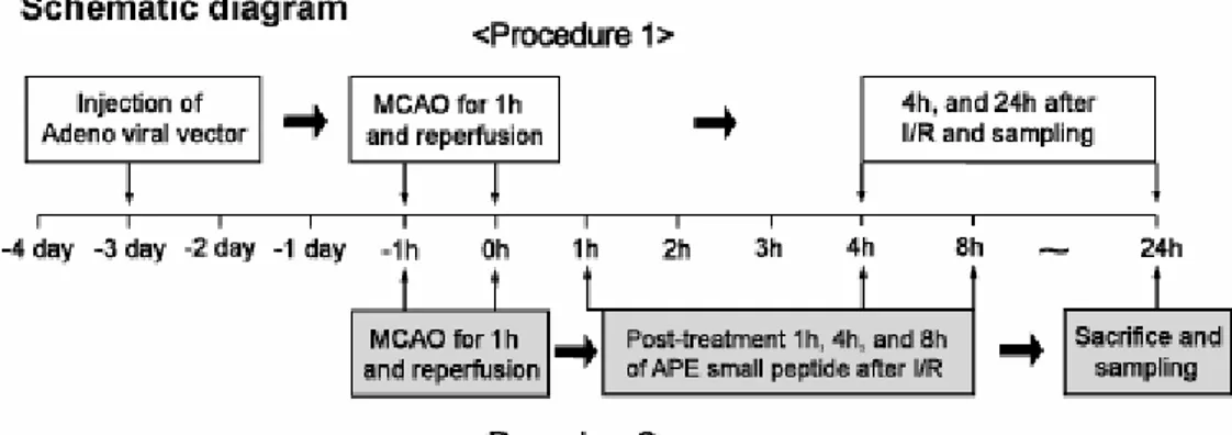
Figure 1. The experimental procedures .………...15
Figure 2. Temporal relationship between AP sites and APE/Ref-1
………...…...23
Figure 3. Immunohistochemical analysis of the adenovirus-treated
group………...25
Figure 4. Temporal profiles of APE/Ref-1 levels after transfection
of an adenovirus harboring the APE/Ref-1 gene…………..……26
Figure 5. Western blot analysis of APE/Ref-1 4 hours after focal
cerebral I/R in Adv-APE/Ref-1-injected mice..…...28
Figure 6. Superoxide-radical production after I/R….…………...30
Figure 7. Oxidative DNA damage after I/R..…...32
Figure 8. Effect of increased levels of APE/Ref-1 on DNA after
I/R.………34
Figure 9. Effect of increased levels of APE/Ref-1 on infarct
volume after I/R ...35
Figure 10. Functional tests for synthetic APE peptide..…………37
Figure 11. Detection for APE peptide in brain ………38
Figure 12. Analyses of AP site and ssDNA immunohistochemistry
after post-ischemic administration of APE peptide ……….40
Figure 13. Caspase-3 activity assay at various times………42
Figure 14. Reduction of ischemic injury by post-ischemic
administration of APE peptide ……….………43
LIST OF TABLES
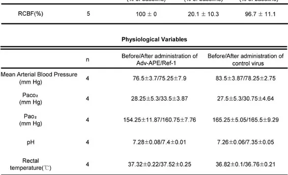
Table 1. Regional cerebral blood flow and physiological variables
in mice... ………..……….………21
ABSTRACT
Therapeutic strategy using DNA repair enzyme after ischemic stroke in mice
Hyun Woo Kim
Department of Medical Science The Graduate School, Yonsei University
(Directed by Professor Byung-In Lee)
Previous investigations demonstrated a nice correlation between the level of the DNA base excision repair protein, apurinic/apyrimidinic endonuclease/redox factor-1 (APE/Ref-1), and the degree of oxidative DNA damage. However, there has been no attempt undertaken yet to directly address the neuroprotective effects of modulating DNA repair functions in an
in vivo model of cerebral ischemia.
This study was performed to test the hypothesis that the augmentation of APE/Ref-1 is an effective neuroprotective measure in the ischemic neuronal
death and the role of APE/Ref-1 on neuroprotection is mainly related to its DNA-repairing function. I also investigated the temporal relationship of the therapeutic strategy using the DNA repairing mechanisms to the postischemic state in a model of cerebral Ischemia/Reperfusion (I/R)
Mice were subjected to intraluminal suture occlusion of the middle cerebral artery for 1 hour followed by reperfusion. Pre-ischemic treatment of the adenoviral vector harboring an entire APE/Ref-1 gene sequence and post-ischemic treatment of the small peptide containing only the APE functional domain were introduced intracerebroventricularly. Western blot analysis and immunohistochemistry were performed. Assays in AP sites, single-strand DNA breaks, caspase-3 activity, and apoptotic cell death were performed and quantified.
The reduction of APE/Ref-1 occurred before DNA fragmentation after I/R, which was shown by the temporal and spatial analysis of their correlations. Overexpression of APE/Ref-1 significantly decreased oxidative DNA damages and the volume of cerebral infarction after I/R. I found that
post-ischemic administration of the APE peptide up to 4 hours after I/R significantly inhibits the induction of apoptotic DNA fragmentation and subsequent infarct volume at 24 hours after I/R.
In conclusion, augmentation or restoration of decreased APE/Ref-1 exerted significant protective effects on ischemic stroke in both pre- and post-ischemic treatment, which was encouraging for the clinical development of APE peptide as a novel therapeutic strategy.
Key Words : stroke, cerebral ischemia, DNA repair, adenoviral vector, Apurinic/apyrimidinic endonuclease/redox factor-1
Therapeutic strategy using DNA repair enzyme after ischemic stroke in mice
Hyun Woo Kim
Department of Medical Science The Graduate School, Yonsei University
(Directed by Professor Byung-In Lee)
I. INTRODUCTION
Oxidative DNA damage after cerebral ischemia/reperfusion (I/R) was reported to occur at the early stage of reperfusion preceding cellular injury or DNA fragmentation.1-6 During oxidative DNA damage by I/R, the DNA repair enzyme prevents ongoing cell death by repairing damaged DNA, but continuous or severe damage beyond the capacity of the repair enzyme may trigger a cell-death transduction pathway,2, 7-10 suggesting that the repair enzyme may have a critical role in the deterrence of the cell-death pathway
after I/R.
The base excision repair (BER) pathway takes a charge in the repair of oxidative DNA lesions in the brain.11-15 Apurinic/apyrimidinic endonuclease/redox effector factor-1 (APE/Ref-1) is a multifunctional enzyme in the BER pathway responsible for repairing apurinic/apyrimidinic (AP) sites in DNA, which are generated by oxidative stress after I/R.5, 11, 14,
16-20 The BER pathway is initiated by removing the damaged base of a DNA
glycosylase, which generates an AP site. APE/Ref-1 then cleaves the AP site 5′ to phosphodeoxyribose, thus generating a free 3′-hydroxyl required for DNA polymerase to fill the resulting gap.16, 21 AP sites are toxic to the cell, most likely through initiating various cell-killing signaling pathways.15, 22 AP sites, upon accumulation, prevent DNA synthesis or gene transcription in cells at the lesion site, directly causing cell death.23 Thus, accumulation of AP sites is an important contributing factor to ischemic neuronal cell death.22 AP sites occur in the early stage after transient cerebral ischemia, and they are repaired by the enzyme APE/Ref-1 to prevent or reverse ongoing cell death.1,
2, 23 However, continuous or severe damage beyond the repair capabilities of
the repair enzyme ultimately results in DNA fragmentation and cell death.7, 8,
24
A number of previous in vivo studies have shown that APE/Ref-1 is markedly decreased in the brain after severe focal ischemia which is followed by DNA fragmentation. The temporal profile suggests that the reduction of APE/Ref-1 results in the failure to repair DNA, deteriorating the neuronal ability to cope with oxidative DNA damage, thus contributing to irreversible tissue injury.1, 2, 9, 23-28 On the contrary, sub-lethal levels of oxidative stress may induce upregulation of APE/Ref-1, conferring a protective effect against lethal doses of oxidative stress and DNA damage.22, 27, 29, 30 However, it is still not clear that an increase of APE/Ref-1, which may increase DNA repair activity, truly protects from inducing neuronal cell death after ischemic insult.16 Although several studies have investigated the upregulation of DNA repair performed in vitro,31-34 there have been no reports in vivo showing the direct evidence for neuronal rescue by enhanced DNA repair activity after
cerebral ischemia.
I hypothesized that upregulation of APE/Ref-1 would be able to rescue cells from oxidative DNA damage after cerebral ischemia. To test the hypothesis, I upregulated the expression of APE/Ref-1 using an adenovirus, a well-known DNA transfer vector,35-38 and investigated whether adenoviral-vector-mediated APE/Ref-1 (Adv-APE/Ref-1) could inhibit the induction of oxidative DNA damage and prevent neuronal cell death after I/R in mice. Next, I focused on APE/Ref-1 as a DNA repair activity and attempted to establish a therapeutic strategy by using synthetic APE peptide containing only the repair functional domain. I investigated whether the post-treatment of synthetic APE peptide prevents neuronal DNA damage after I/R in mice along with the exploration of the therapeutic time window of the novel therapeutic strategy during post-ischemic period.
II. MATERIALS AND METHODS
1. Animals and transient focal cerebral ischemia
Animal experiments were performed in accordance with Yonsei University’s Guide to the Care and Use of Laboratory Animals and approved by the Association for Assessment and Accreditation of Laboratory Animal Care (AAALAC). Adult male C57BL/6J mice weighing 25~30 g (3 months, Daehan Biolink Co., Chunbuk, Korea) were used in this study. Mice were anesthetized with 1.5~2.0% isoflurane in 30% oxygen and 70% nitrous oxide using a face mask. Rectal temperature was maintained at 37±0.5℃ using a homeothermic blanket during ischemia. MCA occlusion was produced by the intraluminal blockade technique, as described previously 39-41. An 11.0-mm 5/0 surgical monofilament nylon suture was inserted into the left internal carotid artery through the external carotid artery stump. After 60 min of MCA occlusion, blood flow was restored by suture removal.
2. Physiological and regional cerebral blood flow (RCBF) parameters
Cannulation of the femoral artery allowed the monitoring of blood pressure (Research BP transducer, Harvard Apparatus, Inc., Holliston, MA, USA) and arterial blood gases (Critical Care Analyzer, AVL Scientific Corporation, Roswell, GA, USA), and blood samples for analysis were taken immediately after cannulation. The regional cerebral blood flow (rCBF) was monitored by a Laser Doppler Flowmeter (Transonic Systems, Inc., Ithaca, NY, USA).
3. Western blot analysis after ischemia/reperfusion (I/R)
Western blot analysis was performed as described previously.41, 42 Experimental animals were decapitated at various times after the start of reperfusion and samples from the ischemic lesions were taken. Mice without MCA occlusion were used as normal controls. Homogenized samples were added to the sample buffer (125 mM Tris/HCl, 2% SDS, 10% glycerin, 1 mM DTT, and 0.002% bromophenol blue, pH 6.9) and boiled for 5 min. Proteins
were resolved in 10% SDS-polyacrylamide gels and blotted to polyvinylidine difluoride membranes (PVDF, Millipore, Bedford, MA, U.S.A.). Transmembranes were incubated with rabbit anti-APE/Ref-1 (1:500; Santa Cruz Biotechnology, Santa Cruz, CA, USA) or rabbit anti-type 5 hexon (1:500; Abcam, 332 Cambridge Science Park, Cambridge, CB4 0fw, UK)..Membranes were then incubated with horseradish peroxidase-conjugated anti rabbit IgG (1:5,000; Roche Diagnostics Co., Indianapolis, IN, USA). The bands were visualized with an enhanced chemiluminescence reagent (ECL plus; Amersham Biosciences, Piscataway, NJ, USA).
4. Immunofluorescent labeling and TUNEL staining
Immunofluorescent staining was performed with type 5 hexon, APE/Ref-1, and the terminal deoxynucleotidyl transferase-mediated uridine 5'-triphosphate-biotin nick end labeling (TUNEL) assay as reported previously.41, 43 Rabbit anti-APE/Ref-1 (1:200) or rabbit anti-type 5 hexon (1:200) were added to the fixed sections, after which they were incubated
with fluorescence-conjugated secondary antibody (Jackson Immuno Research, West Grove, PA, USA) and counterstained with PI (red). For TUNEL staining, a commercially-available kit (Roche Diagnostics GmbH, Penzberg, Germany) was used to detect DNA fragmentation in situ. The sections were observed under a LSM 510 confocal laser scanning microscope (Carl Zeiss, Thornwood, NY, USA).
5. Detection of superoxide radicals
To confirm the occurrence of O2-after I/R, in situ detection of oxidized
hydroethidine (HEt) was performed at 4 hours after ischemia as described previously.40, 41, 44, 45 A total of 200 ㎕ of HEt (stock solution 100 mg/mL in dimethyl sulfoxide was finally diluted to 1 mg/mL with phosphates-buffered saline; Molecular Probes) was administrated intravenously 30 minutes before scarifying, and prepared samples were observed with a microscope and computerized digital camera system under fluorescent light (Olympus). Intensity (optical density [OD]) in the high-magnification field and
expression patterns of the oxidized HEt were analyzed with a computerized analysis system and program (MetaMorpho Imaging; Molecular Devices).42
6. Quantitative analysis of AP sites in the nuclear DNA
The nuclear DNA isolated from the ischemic and sham brain tissues were subjected to quantitative analysis of AP sites using a colorimetric assay according to the manufacturer’s protocol (Dojindo Molecular Technologies, Gaithersburg, MD, USA). Purified DNA was dissolved at a concentration of 100 ㎍/mL in TE, and 10 ㎕ of DNA solution was incubated with the ARP solution. ARP in the labeled DNA was measured using an ELISA-like assay in a microtiter plate. AP sites per 105 nucleotides were calculated based on the linear calibration curve generated for each experiment using ARP-DNA standard solutions.
7. Adenoviral vectors
sequence (Adv-APE/Ref-1) was purchased from Lab Frontier (Lab Frontier, GyeongGi-Do, Korea). An adenovirus vector without the APE/Ref-1 sequence was used as a control virus. The adenoviruses were purified to ensure that viral suspensions were free from wild-type viruses. Titers of the purified adenovirus were assessed by plaque assays.
8. In vivo gene transfer and cerebral ischemia
Mice under anesthesia were placed in a stereotaxic frame and injected intracerebrally with either Adv-APE/Ref-1 or control virus (4.0 x 10⁹ pfu/ml) into the left cortex and striatum (cortex, 1 ㎕, 0.7 mm anterior to the bregma, 3.0 mm lateral to the midline, 1.3 mm beneath the dura; striatum, 2 ㎕, 0.7 mm anterior to the bregma, 2.0 mm lateral to the midline, 3.3 mm beneath the dura). A total volume was injected over 10 minutes using a Hamilton microsyringe (Hamilton Co., Nevada, USA). The needle was left in place for an additional 10 minutes and removed slowly. Then, the scalp was sutured and the mice were returned to standard housing. Three days after the injection
of adenovirus, transient focal cerebral ischemia was performed as previously described above (Fig 1.).
9. APE peptide synthesis and treatment
The APE peptide (TLK ICS WNV DGL RAW IKK KG [61~80]) 16 was covalently linked at its C terminus to a 10 amino acid carrier peptide derived from the HIV-TAT peptide (GRK KRR QRR R). To monitor peptide delivery, the synthetic peptide was conjugated with fluorescein isothiocyanate (FITC) and injected intracerebroventricularly (2 ㎕, bregma; 0.2 mm posterior, 1 mm lateral, 3.1 mm deep).
10. APE peptide activity assay
APE activity was assayed by using depurinated ccc form plasmid DNA as following the previouly reported method (ref Seki).17 Supercoiled pUC18 DNA was depurinated by treating it with 3 volumes of 50 mM sodium citrate
the depurinated DNA solution was used to measure APE activity. It has been reported that acid treatment produces approximately 6 alkali-sensitive sites per pUCI8 DNA molecule.17 The assay mixture (15 ㎕ final vol.) contained 0.25 ㎍ (0.14 pmol) acid-depurinated pUCI8 DNA and 10 ng of APE peptide in Triton-buffer B (0.0175% Triton X-100, 0.25 M sucrose, 10 mM Tris-HCl, 4 mM MgCl2, 1mM EDTA, and 6 mM 2-mercaptoethanol, pH 8.0 adjusted at 25°C). The assay mixture was incubated at 37°C for 20 min then chilled to stop the reaction. The mixture was loaded on a 0.8% agarose gel. Electrophoretic analysis of the conformation of pUC18 DNA was conducted as described previously.17
11. APE peptide to DNA binding analysis by modified gel mobility shift
assay
The binding of APE peptide with DNA containing AP sites was analyzed by modified gel shift mobility assay. For the DNA binding reaction, intact DNA was extracted from normal mouse brain. The FITC-APE peptide was
incubated with depurined DNA, which was depurinated in 50 mM sodium citrate (pH 3.5), in the gel shift binding buffer consisting of 20% glycerol, 5 mM MgCl2, 2.5 mM EDTA, 2.5 mM DTT, 250 mM NaCl, and 50 mM Tris-HCl (pH 7.5). The binding mixture was analyzed on a non-denaturing 4% acrylamide gel to detect the DNA-peptide complex via intensity of FITC fluorescence. The assay was performed without DNA (no input) or with intact DNA as a control.
12. Quantification of oxidative damage (8-OHdG)
Fixed sections were incubated with the mouse monoclonal primary antibody anti-8-hydroxyguanosine (8-OHdG) (QED Bioscience, San Diego, CA, U.S.A.) for 60 min at RT. For 8-OHdG staining, I followed the manufacturer’s protocol (MOM; Dako, Carpinteria, CA, U.S.A.). Nuclear localization was accomplished using a methyl green staining solution (Sigma) for 30 min at RT. Samples were then washed two times in PBS, once in double-distilled water (ddH2O), and were mounted on glass slides using
mounting medium (Vector Laboratories).
13. Immunofluorescence for single-stranded DNA (ssDNA)
Single-stranded DNA (ssDNA) was detected by a monoclonal IgM antibody (MAb-F7-26; Bender MedSystems, Vienna, Austria) using a modified method described by Zhu et al..46, 47 Slides were incubated with formamide at 56℃ for 20 min and then immersed in ice-cold PBS. The prepared sections were exposed to primary antibody diluted 1:10 (10 mg/ml) and incubated with the secondary antibody, biotinylated goat anti-mouse IgM 120 using a MOM kit (Vector Laboratories Ltd., 30 Ingold Road, Burlingame, CA, USA) to block endogenous mouse immunoglobulins in mouse tissue.
14. Caspase-3 activity
To quantify caspase-3 activation, a commercial enzyme immunoassay (Chemicon International, Temecula, CA, USA) was used.48 The cytosolic samples were prepared per the manufacturer’s protocol.
15. Cell death assay
To quantify apoptosis-related DNA fragmentation, a commercial enzyme immunoassay was used to determine cytoplasmic histone-associated DNA fragments (Roche Diagnostics), which detect apoptotic oligo-DNA fragmentation.
16. Measurement of infarct size
To verify the reproducibility of the ischemic lesions in the brain after I/R, anesthetized animals were euthanized at 24 h after I/R. The brains were quickly removed and processed for brain cutting. Serial coronal sections of 1 mm thickness were obtained. To compare infarction volume, tissue sections were stained with 2,3,5-triphenyltetrazolium chloride (TTC)40 and scanned. The volumes of the lesions were measured using a computerized image analysis tool. Infarct sizes were expressed as contralateral hemisphere (mm3) minus undamaged ipsilateral hemisphere (mm3) to correct for brain edema.49
17. Statistical analysis
The data are expressed as mean ± S.D.. The statistical comparisons between two groups were performed by unpaired t-tests and multiple groups were compared statistically with the one-way ANOVA analysis of variance followed by the Student-Newman-Keuls Method (StatView, SAS Institute Inc., Cary, NC. USA). Significance was assigned at p < 0.05.
Table 1. Regional cerebral blood flow1 and physiological variables2 in mice
III. RESULTS
1. Physiological data and regional cerebral blood flow (RCBF)
parameters
Changes in RCBF were measured indirectly by laser Doppler flowmetry (Transonic Systems Inc., Ithaca NY 14850, USA); RCBF decreased after ischemia compared with preischemia and the contralateral side. There were no statistically significant differences in RCBF during ischemia between the group before occlusion and the group after reperfusion (Table 1.). Physiological values were as follows in Table 1. There was no deviation from these values over the period of assessment. Intracerebral administration of adenoviral vector harboring an entire APE/Ref-1 gene sequence (Adv-APE/Ref-1) does not alter the physiological parameters of mice.
2. Spatial and temporal relationship between APE/Ref-1 and AP sites
Figure 2. Temporal relationship between AP sites and APE/Ref-1. (A)
Western blot analysis of APE/Ref-1, with ß-actin as an internal control. APE/Ref-1 was detected as a single band in the whole fractionated brain after I/R. Optical density values of APE/Ref-1 are shown in a time-dependent manner. (B) AP sites were analyzed. Nor., normal control; A, n=4; B, n=3; values are mean ± S.D.; *p<0.05 compared with the normal control group.
APE/Ref-1 immunoreactivity was evident as a single band of 37 kDa, peaking at 30 minutes and decreasing from 1 to 24 hours in mice brains after I/R, as compared to control mice. In contrast, ß-actin immunoreactivity remained constant (Fig. 2A). Statistical analysis confirmed the significant decrease of APE/Ref-1 starting 1 hour after I/R (O.D. of the APE/Ref-1; Nor., 11563.8 ± 287.7; 30 min to 24 h, 12880.3 ± 2039.2, 6711.3 ± 2822.4, 4706.5 ± 1986.1, 4226.5 ± 2208.1 respectively). As shown in figure 2B, the number of AP sites per 100,000 nucleotides at each time point was counted and analyzed. At 30 min and 1 h after I/R, the number of AP sites was maintained, but significantly increased at 4 hours with further elevation at 24 hours after I/R (AP sites, Nor., 1.82 ± 1.72; 30 min, 3.54 ± 1.11; 1 h, 4.24 ± 0.81; 4 h, 13.84 ± 1.72; 8 h, 24.24 ± 0.91; 24 h, 26.67 ± 1.11).
3. Adenovirus-mediated gene expression
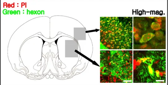
Adenovirus particles were detected by double staining with type 5 hexon (green), one of the major structural components of the capsid protein, and PI
Figure 3. Immunohistochemical analysis of the adenovirus-treated group.
Regional expression of the adenovirus was detected by double staining with type 5 hexon (green) and PI (red) in the cortex and striatum under a confocal microscope. High-mag., high magnification; Scale bar = 50 and 20 ㎛.
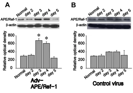
Figure 4. Temporal profiles of APE/Ref-1 levels after transfection of an
adenovirus harboring the APE/Ref-1 gene. After injection of Adv-APE/Ref-1 or control virus, the amount of APE/Ref-1 was measured by western blot analysis. n=3; values are mean ± S.D.; *p<0.05 compared with the normal control group.
(red) in the adenoviral vector-injected hemisphere, cortex, and striatum of mice, suggesting that the adenovirus was well-distributed in target areas (Fig. 3). After injection of Adv-APE/Ref-1, the amount of APE/Ref-1 was measured by western blot analysis of proteins extracted from the brains of mice (Fig. 4A, O.D. Normal, 300 ± 23; day 2, 320 ± 25; day 3, 680 ± 45; day 4, 610 ± 39; day 5, 250 ± 35). APE/Ref-1 was markedly increased at days 3 and 4, and returned to baseline level at day 5, while the level of APE/Ref-1 in the control virus-treated group was not different from that of normal control group (Fig. 4B). These results show that maximum expression of APE/Ref-1 is achieved at days 3 and 4 after injection of Adv-APE/Ref-1.
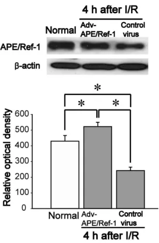
4. Overexpression of APE/Ref-1 using an adenoviral vector after I/R
The Adv-APE/Ref-1 or control virus was transfected in mice brains, and thereafter, I/R injury was performed on day 3 (Fig. 1). Western blot analysis demonstrated that APE/Ref-1 expression increased in the Adv-APE/Ref-1 transfected mice compared with the control virus-infused group 4 hours after
Figure 5. Western blot analysis of APE/Ref-1 4 hours after focal cerebral I/R
in Adv-APE/Ref-1-injected mice. Normal, normal control; Adv-APE/Ref-1 or Control virus, transfection of APE/Ref-1 or no carrying APE/Ref-1 mediated by adenoviral vector; I/R, ischemia/reperfusion; n=3; values are mean ± S.D.; *p<0.05 compared with the normal control group.
I/R (Fig. 5, O.D. Normal, 430 ± 36; Adv-APE/Ref-1, 523 ± 27; Control virus, 242 ± 22). These results suggest that Adv-APE/Ref-1 overexpresses APE/Ref-1 effectively and inhibits the loss of APE/Ref-1 after I/R.
5. Superoxide-radical production and oxidative damage after I/R
To investigate the effect of overexpressed APE/Ref-1 on DNA damage caused by ischemic injury, brain tissues from the cortex and striatum of the Adv-APE/Ref-1 and control virus group were taken and O2-,
8-hydroxyguanosine (8-OHdG), and AP sites were measured quantitatively. The production of O2- was expressed by oxidized hydroethidine (HEt) signals
as red particles, which were measured 4 hours after I/R (Fig. 6). A quantitative assay showed that HEt signals were significantly increased in both Adv-APE/Ref-1 and control virus groups without any differences between them (Fig. 6).
Figure 6. Superoxide-radical production after I/R. Superoxide-radical
production was measured 4 h after I/R using hydroethidine. Oxidized hydroethidine signals as appears as red spots at perinuclear sites. Scale bar = 10 ㎛.
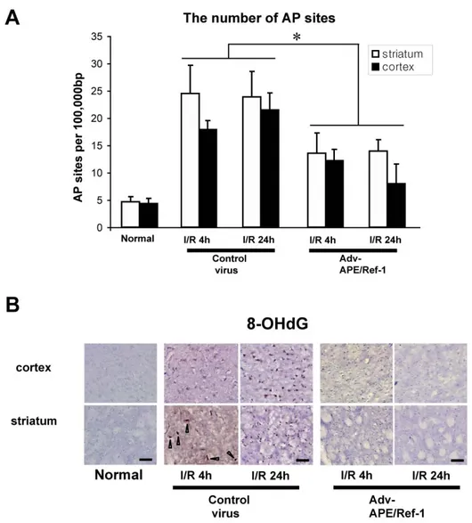
6. Detection of AP sites and 8-OHdG following ischemia
To investigate the effects of Adv-APE/Ref-1 on oxidative DNA damage after I/R, AP site lesions and 8-OHdG in the cortex and striatum were quantitated (Fig. 7). Figure 6 illustrates the profile of AP site and 8-OHdG induction, respectively, in the cortex and striatum 4 and 24 hours after I/R. In the control virus group, the number of AP sites and 8-OHdG increased in both the cortex and striatum at 4 and 24 hours after I/R. In contrast, in the Adv-APE/Ref-1 group, the number of AP site lesions and 8-OHdG in the cortex and striatum were significantly less than that of the control virus group. These results reflect the inhibitory role of Adv-APE/Ref-1 treatment on the nuclear accumulation of AP site lesions and 8-OHdG in the cortex and striatum after I/R.
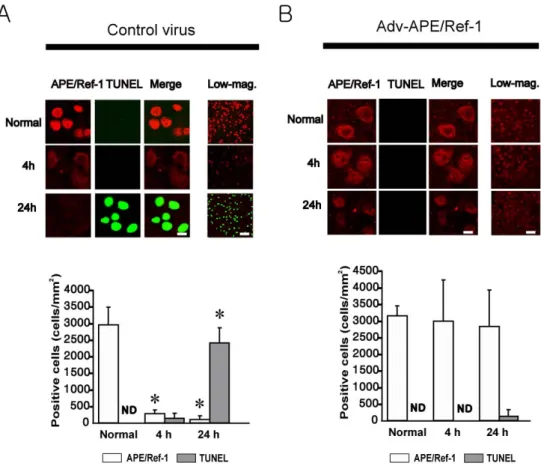
7. Reduction of DNA fragmentation in the Adv-APE/Ref-1-treated group
after I/R
Figure 7. Oxidative DNA damage after I/R (A) Quantitative measurement of
nuclear contents of AP site lesions from the cortex and striatum at 4 and 24 hours after I/R. n=3, mean ± S.D.; *p<0.05 (B) Immunohistochemistry of 8-OHdG (brown positive cells and arrow heads) and counter staining of nucleus (Methyl green) at 4 and 24 hours after I/R. Scale bar=50 ㎛
expression and DNA fragmentation, double labeling with an antibody against APE/Ref-1 (red) and TUNEL (green) staining was performed in mice brains from the control virus (Fig. 8A) and Adv-APE/Ref-1 (Fig. 8B) -treated groups. In the Adv-APE/Ref-1 group, there was no significant decrease of APE/Ref-1 immunopositive cells and no remarkable TUNEL-positive cells were detected at 4 and 24 hours after I/R compared with the normal. These results were in contrast to those from the control virus group, in while the number of APE/Ref-1 positive cells was markedly decreased at 4 and 24 hours and the number of TUNEL positive cells was markedly increased at 24 hours after I/R.
8. Reduction of cerebral Infarction volume after I/R due to
overexpression of APE/Ref-1
To investigate the effect of Adv-APE/Ref-1 on infarct volumes, both the Adv-APE/Ref-1 and control virus groups underwent TTC staining 24 hours
Figure 8. Effect of increased levels of APE/Ref-1 on DNA after I/R. (A)
Double-staining and semi-quantitative analysis of APE/Ref-1 (red) and TUNEL (green) (ND, Not Detected). Right side layers are lower magnifications of left side layers. A, n=8; B, n=5; Scale bars = 10 and 50 ㎛.
Figure 9. Effect of increased levels of APE/Ref-1 on infarct volume after I/R.
Coronal brain sections stained with TTC and analyzed for infarct volume. n=3; mean ± S.D.; *p<0.05 compared with control virus; Scale bar=4 mm
showed a significantly decreased infarct volume compared to that of the control virus group (Fig. 9). The infarct volume in the Adv-APE/Ref-1 group was 4.2 ± 7.2 mm3 and in thecontrol virus group it was 138.8 ± 20.9 mm3.
9. APE peptide functional test
An ex vivo assay was performed to test the endonuclease activity of the synthetic APE peptide, which contained only the repair functional domain (Fig. 10A). The APE peptide showed nicking activity on supercoiled, acid-depurinated pUC18 DNA containing AP sites, but showed no activity on native pUC18 DNA. The binding of APE peptide and DNA was evaluated with modified gel mobility shift assay (Fig. 10B). FITC-APE bound with DNA including AP sites was shift upper than control. In addition, cellular transduction of the APE peptide was investigated with conjugated FITC (Fig. 11). Co-labeling of FITC with PI counter-stain was confirmed by orthogonal views with confocal and optical sectioning. In animals treated with FITC- labeled APE peptide, FITC-positive cells were detected in the striatum and
Figure 10. Functional tests for synthetic APE peptide. (A) The endonuclease
activity of the APE peptide was measured by agarose gel electrophoresis. NC, nicked circular pUC18 DNA; SC, supercoiled pUC18 DNA; MCAO, middle cerebral artery occlusion; I/R, ischemia/reperfusion. (B) Modified gel shift mobility assay was analyzed for the binding of APE peptide with DNA containing AP sites. Arrow head indicates upper shifted band
Figure 11. Detection for APE peptide in brain. Mouse brain was treated with
FITC-conjugated APE peptide and stained using PI. Co-labeling of APE peptide with PI was confirmed by confocal optical sectioning with orthogonal views in the xz and yz planes. right panel, Scale Bars=10 ㎛.
cortex.
10. Analysis of AP sites and ssDNA immunohistochemistry
Compared with the vehicle control, the number of AP sites in DNA 24 hours after I/R was significantly reduced by treatment with APE peptide 4 hours after I/R (4-hour APE post-treatment), but not by 8-hour post-treatment (Fig. 12A; AP sites, Vehicle, 27.07 ± 0.20; Post 4 h, 12.22 ± 1.52; Post 8 h, 22.83 ± 0.3; n = 3; Values are mean ± SD). Upon ssDNA staining, red ssDNA-positive cells in mice treated with APE 4 hours after I/R were barely visible 24 hours after I/R, while red cells were intensively detected in mice that received 8-hour APE post-treatment (Fig. 12B; Vehicle, 2986.7 ± 337.2; Post 4 h, 425.0 ± 354.4; Post 8 h, 1975.0 ± 758.7; n = 3; Values are mean ± SD).
11. Reduction of ischemic injury after application of APE peptide
Figure 12. Analyses of AP site and ssDNA immunohistochemistry after
post-ischemic administration of APE peptide. (A) APE peptide or vehicle was administered 4 and 8 h after ischemia/reperfusion; ischemic brain was sampled 24 h after reperfusion. Nuclear DNA was subjected to quantitative analysis of AP sites. Post 4 h / 8 h, APE peptide treated at 4 h / 8 h after ischemia/reperfusion. (B) Temporal profiles of ssDNA (red) were analyzed by immunohistochemistry in low and high magnification (lower line). *p<0.001 compared with vehicle, Scale Bars=50 and 10 ㎛.
1 hour, peaked at 4 hours and was sustained until 24 hours after I/R (Fig. 13A; Optical density [OD]; nor., 0.66 ± 0; 30min, 0.558 ± 0.06; 1 h, 1.298 ± 0.07; 4 h, 1.626 ± 0.11; 8 h, 1.338 ± 0.05; 24 h, 1.445 ± 0.09;
n = 4; Values are mean ± SD). Caspase-3 activity 24 hours after I/R was
significantly decreased by 4-hour APE post-treatment compared with vehicle-treated mice (Fig. 13B; OD; Vehicle, 1.42 ± 0.081; Post 4 h, 0.585 ± 0.08; Post 8 h, 1.703 ± 2.282; n = 4; Values are mean ± SD). Cell death 24 hours after I/R was significantly reduced by 4-hour APE post-treatment compared with the vehicle control (Fig. 14A; OD; Vehicle, 21.5 ± 4.66; Post 4 h, 4.18 ± 2.36; Post 8 h, 12.9 ± 1.73; n = 3, values are mean ± SD). The mice that received APE peptide 4 hours after I/R also showed a significant reduction of infarct volume 24 hours after I/R compared with vehicle-treated mice. The infarct volume 24 hours after I/R of mice treated with APE peptide 8 hours after reperfusion was not significantly reduced compared with vehicle-treated mice (Fig. 14B).
Figure 13. Caspase-3 activity assay at various times. (A) Caspase-3 activity
assay was performed from 1 to 24 h after ischemia/reperfusion. APE peptide or vehicle was administered 4 and 8 h after ischemia/reperfusion, and (B) Capsase-3 activity assay was performed on ischemic brain samples 24 h after ischemia/reperfusion. vehicle, vehicle-treated; Post 4 h / 8 h APE peptide treated at 4 h / 8 h after ischemia/reperfusion; Nor., normal control; *p<0.001 compared with normal control; #p<0.001 compared with vehicle.
Figure 14. Reduction of ischemic injury by post-ischemic administration of
APE peptide. APE peptide or vehicle was administered 4 and 8 h after ischemia/reperfusion. Subsequently, (A) cell death assay and (B) infarct volume analysis were performed on ischemic brain samples 24 h after ischemia/reperfusion. vehicle, vehicle-treated; Post 4 h / 8 h APE peptide treated at 4 h / 8 h after ischemia/reperfusion; Nor., normal control; *p<0.001 compared with normal control; #p<0.001 compared with vehicle.
IV. DISCUSSION
The results of study were summarized as follows; First, there was a close relationship between loss of APE/Ref-1 and DNA fragmentation in the ischemic lesion. Second, overexpression of APE/Ref-1 by using intracranial administration of adenovirus vector infected significantly reduced oxidative DNA damage and TUNEL-positive cells and infarct volume. Finally, post-ischemic administration of synthetic APE peptide within 4 hours after I/R significantly diminished infarct volume at 24 hours compared to vehicle-treated mice with concomitant attenuation of single stranded DNA (ssDNA)-positive cells, AP sites, and DNA fragmentation in ischemic brain at 24 hours compared to vehicle-treated mice.
As shown in the present results, many previous in vivo studies have reported that the loss of APE/Ref-1 induced DNA fragmentation, and thereby subsequent neuronal cell death.24-26, 41, 50-54 Thus, augmentation of APE/Ref-1 may have a beneficial role in rescuing cells from oxidative DNA damage
after ischemic injury.18, 22, 23, 27, 29, 55, 56
This concept is supported by circumstantial in vitro and in vivo evidences that a sublethal level of reactive oxygen species (ROS) or ischemic preconditioning by brief ischemia is known to activate the APE/Ref-1 gene and increase the expression of APE/Ref-1, thereby helping to prevent neuronal damage produced by further lethal levels of ROS or prolonged-cerebral ischemia.22, 23, 29, 57 Also in the current study (Fig. 2A), even though not statistically significant, western blot analysis showed slightly increased APE/Ref-1 at 30 minutes after I/R when the cells are under the transiently sublethal ischemic injury. This agrees with previous report that APE/Ref-1 immunoreactivity temporally increased at an early time point, in photochemical induced-cortical infarction, and decreased after that.25
To test the hypothesis that upregulation of APE/Ref-1 may prevent neuronal cell death by enhancing the repair function against endogenous oxidative DNA damage, APE/Ref-1 levels artificially increased by using an adenoviral vector encoding the entire APE/Ref-1 gene (Adv-APE/Ref-1). The
blood brain barrier, however, limits diffusion of blood-derived molecules into the brain.58, 59 Therefore, in many previous in vivo reports, various regions of the brain were transfected directly with a recombinant adenovirus instead of using intraventricular injections.36-38, 60 Based on these reports, adenovirus was introduced into the both striatum and cortex stereotaxically. These regions were selected to examine the effects of a selective and temporal increase of APE/Ref-1 in the mouse brain. Type 5 hexon, one of the major structural components of the capsid protein, was observed in the injected hemisphere, indicating that the infected virus was well distributed (Fig. 3). In the western blot analysis of the Adv-APE/Ref-1 treatment group, the temporal profile of APE/Ref-1 (Fig. 4) and expression of APE/Ref-1 were determined to demonstrate the increasing amount of APE/Ref-1 protein during ischemic injury (Fig. 5). The data suggests that Adv-APE/Ref-1 increases APE/Ref-1 expression and compensates for the loss of APE/Ref-1 after I/R.
22, 61, 62 and it is induced mainly by direct attack of the DNA by ROS
overproduced during I/R.5, 63 In previous studies, loss of DNA repair activity caused by decrease of APE/Ref-1 protein levels has been suggested to be a possible mechanism for the accumulation of AP sites and oxidative DNA damage.22, 23 Oxidized hydroethidine (HEt) signals, a possible index of ROS production, were seen as red small particles in the cytosol and perinuclear area of ischemic lesions after I/R. In this study, ROS were increased and were not significantly different in the ischemic lesions of either the Adv-APE/Ref-1 or control virus groups at 4 hours after I/R (Fig. 6). In the control virus group, the number of early AP sites and subsequent immunoreactivity of 8-OHdG increased significantly from 4 hours to 24 hours after I/R. However, in the Adv-APE/Ref-1 treatment group, there were significant decreases in the levels of oxidative DNA damage, indicated by the number of early AP sites and immunoreactivity of 8-OHdG compared to the control virus group (Fig. 7). These results suggest that increased levels of APE/Ref-1 may repair oxidative DNA damage in ischemic lesions effectively.
The number of APE/Ref-1 positive cells in the Adv-APE/Ref-1 treated-mice was still detected at 4 or 24 hours after I/R up to near the normal level, while the control virus treated-mice showed significant loss of APE/Ref-1 positive cells over time (Fig. 8A). Corresponding to the result of APE/Ref-1 staining, TUNEL-positive cells were not detected 24 hours after I/R in the Adv-APE/Ref-1 treated-mice (Fig. 8B). Moreover, a significant decrease in infarct volume was also observed in the APE/Ref-1-treated mice compared with the control virus-treated mice (Fig. 9).
Taken together, these results clearly suggest that increasing APE/Ref-1 rescued ischemic neurons from their demise after I/R, probably via the preserved DNA repair capacity during postischemic period. Present data agree with previous reports that overexpressing APE/Ref-1 using adenoviral vector results in a significant increase in cell viability by exposure to various concentrations of H2O2 or spontaneous DNA damage in cell culture
systems.31, 32 Furthermore, Adv-APE/Ref-1 suppressed activation of the TNF-α cascade in vitro; this cascade mediates acute cell death and neurological
dysfunction.33, 64
In this next study, we tried to prove that the neuroprotective effect of APE/Ref-1 was mediated by its DNA repairing mechanisms. APE peptide with the only repair activity was synthesized. APE/Ref-1 has two major functions: repair of faulty base excision and transcriptional control.16 The synthetic APE peptide was covalently linked with HIV-TAT peptide, a carrier peptide, at C-terminus for its transduction function as previously described,
65-68 and monitored with an FITC label (Fig. 11). The TAT linked APE peptide
was effectively penetrated into the neuronal cells. It is consistent with the previous report that the Tat-APE/Ref-1 protein inhibited activation of the TNF-α cascade in vitro.34
The endonuclease and DNA-binding activity of the synthetic APE peptide containing only the repair functional domain were confirmed ex vivo (Fig. 10 and 11). The number of AP sites was quantified and analyzed in vivo (Fig. 12A). Collectively these results clearly suggest that the synthesized APE peptide functioned as repairing oxidative damaged DNA even in the
post-ischemic DNA damage. These results also demonstrated that the administration of APE peptide at 1 and 4 hours after I/R significantly reduced ssDNA breaks, caspase-3 activity, cell death, and infarction at 24 hours after I/R, compared with vehicle-treated mice (Fig. 12B, 13 and 14). This suggested that treatment with the synthetic APE peptide effectively inhibited the induction of post-ischemic DNA damage and subsequent cerebral infarction. The significant neuroprotective effect by the post-treatment APE peptide indicates that DNA repairing mechanisms are crucial for preventing the initiation of ischemic cascades and provide a novel therapeutic strategy for treating ischemic damage after stroke.34, 69, 70 These findings have additional clinical relevance in that the administration of synthetic APE peptide may expand the time window of current therapies, providing damaged cells extra time to initiate endogenous DNA repair.
V. CONCLUSION
In recent years, oxidative DNA damage and repair have been drawing more attention in the field of ischemic stroke. Enhancement of DNA repair activity may provide one of major mechanisms for neuroprotection after cerebral ischemia, however, no studies have been conducted yet to address its neuroprotective effects directly in vivo. The investigation was conducted to test whether augmentation of APE/Ref-1 has any protective role on ischemic stroke in pre- and post-treatment.
This study showed that: first, there was a close relationship between loss of APE/Ref-1 and DNA fragmentation after I/R. Second, the overexpression of adenoviral-vector-mediated APE/Ref-1 reduced the oxidative DNA damages and subsequent induction of apoptotic DNA fragmentation in the ischemic brain after I/R. Finally, the activity of synthetic APE peptide containing the repair functional domain was confirmed, and the post-ischemic administration of the synthetic APE peptide reduced post-ischemic DNA
lesions and thereby reduced cerebral infarction after I/R.
Augmentation of DNA repair-capability seems to be an important neuroprotective strategy, which can be accomplished by direct administration of APE peptide during post-ischemic period. The therapeutic time window of 4 hours in I/R model provides a strong hope for the development of practical therapeutic strategy by augmenting DNA repair activity in human.
REFERENCES
1. Liu PK, Hsu CY, Dizdaroglu M, Floyd RA, Kow YW, Karakaya A, et al. Damage, repair, and mutagenesis in nuclear genes after mouse forebrain ischemia-reperfusion. J Neurosci. 1996;16:6795-806
2. Chen J, Jin K, Chen M, Pei W, Kawaguchi K, Greenberg DA, et al. Early detection of DNA strand breaks in the brain after transient focal ischemia: Implications for the role of DNA damage in apoptosis and neuronal cell death. J Neurochem. 1997;69:232-45
3. Chan PH. Oxygen radicals in focal cerebral ischemia. Brain Pathol. 1994;4:59-65
4. Chan PH. Role of oxidants in ischemic brain damage. Stroke. 1996;27:1124-9
5. Chan PH. Reactive oxygen radicals in signaling and damage in the ischemic brain. J Cereb Blood Flow Metab. 2001;21:2-14
6. Dirnagl U, Iadecola C, Moskowitz MA. Pathobiology of ischaemic stroke: An integrated view. Trends Neurosci. 1999;22:391-7
7. Rich T, Allen RL, Wyllie AH. Defying death after DNA damage. Nature. 2000;407:777-83
checkpoints in perspective. Nature. 2000;408:433-9
9. Chopp M, Chan PH, Hsu CY, Cheung ME, Jacobs TP. DNA damage and repair in central nervous system injury: National institute of neurological disorders and stroke workshop summary. Stroke. 1996;27:363-9
10. Danial NN, Korsmeyer SJ. Cell death: Critical control points. Cell. 2004;116:205-19
11. Fishel ML, Vasko MR, Kelley MR. DNA repair in neurons: So if they don't divide what's to repair? Mutat Res. 2006
12. Izumi T, Mitra S. Deletion analysis of human ap-endonuclease: Minimum sequence required for the endonuclease activity. Carcinogenesis. 1998;19:525-7
13. Izumi T, Brown DB, Naidu CV, Bhakat KK, Macinnes MA, Saito H, et al. Two essential but distinct functions of the mammalian abasic endonuclease. Proc Natl Acad Sci U S A. 2005;102:5739-43
14. Bernstein C, Bernstein H, Payne CM, Garewal H. DNA repair/pro-apoptotic dual-role proteins in five major DNA repair pathways: Fail-safe protection against carcinogenesis. Mutat Res. 2002;511:145-78 15. Hanna M, Chow BL, Morey NJ, Jinks-Robertson S, Doetsch PW,
Xiao W. Involvement of two endonuclease iii homologs in the base excision repair pathway for the processing of DNA alkylation damage
in saccharomyces cerevisiae. DNA Repair (Amst). 2004;3:51-9
16. Evans AR, Limp-Foster M, Kelley MR. Going ape over ref-1. Mutat Res. 2000;461:83-108
17. Seki S, Ikeda S, Watanabe S, Hatsushika M, Tsutsui K, Akiyama K, et al. A mouse DNA repair enzyme (apex nuclease) having exonuclease and apurinic/apyrimidinic endonuclease activities: Purification and characterization. Biochim Biophys Acta. 1991;1079:57-64
18. Zhao B, Grandy DK, Hagerup JM, Magenis RE, Smith L, Chauhan BC, et al. The human gene for apurinic/apyrimidinic endonuclease (hap1): Sequence and localization to chromosome 14 band q12. Nucleic Acids Res. 1992;20:4097-8
19. Yang G, Chan PH, Chen J, Carlson E, Chen SF, Weinstein P, et al. Human copper-zinc superoxide dismutase transgenic mice are highly resistant to reperfusion injury after focal cerebral ischemia. Stroke. 1994;25:165-70
20. Barzilay G, Hickson ID. Structure and function of apurinic/apyrimidinic endonucleases. Bioessays. 1995;17:713-19
21. Tell G, Damante G, Caldwell D, Kelley MR. The intracellular localization of ape1/ref-1: More than a passive phenomenon? Antioxid Redox Signal. 2005;7:367-84
22. Li W, Luo Y, Zhang F, Signore AP, Gobbel GT, Simon RP, et al. Ischemic preconditioning in the rat brain enhances the repair of endogenous oxidative DNA damage by activating the base-excision repair pathway. J Cereb Blood Flow Metab. 2006;26:181-98
23. Lan J, Li W, Zhang F, Sun FY, Nagayama T, O'Horo C, et al. Inducible repair of oxidative DNA lesions in the rat brain after transient focal ischemia and reperfusion. J Cereb Blood Flow Metab. 2003;23:1324-39
24. Kawase M, Fujimura M, Morita-Fujimura Y, Chan PH. Reduction of apurinic/apyrimidinic endonuclease expression after transient global cerebral ischemia in rats: Implication of the failure of DNA repair in neuronal apoptosis. Stroke. 1999;30:441-8; discussion 449
25. Chang YY, Fujimura M, Morita-Fujimura Y, Kim GW, Huang CY, Wu HS, et al. Neuroprotective effects of an antioxidant in cortical cerebral ischemia: Prevention of early reduction of the apurinic/apyrimidinic endonuclease DNA repair enzyme. Neurosci Lett. 1999;277:61-4
26. Walton M, Lawlor P, Sirimanne E, Williams C, Gluckman P, Dragunow M. Loss of ref-1 protein expression precedes DNA fragmentation in apoptotic neurons. Brain Res Mol Brain Res. 1997;44:167-70
27. Grosch S, Fritz G, Kaina B. Apurinic endonuclease (ref-1) is induced in mammalian cells by oxidative stress and involved in clastogenic adaptation. Cancer Res. 1998;58:4410-6
28. Fujimura M, Morita-Fujimura Y, Sugawara T, Chan PH. Early decrease of xrcc1, a DNA base excision repair protein, may contribute to DNA fragmentation after transient focal cerebral ischemia in mice. Stroke. 1999;30:2456-62; discussion 2463
29. Ramana CV, Boldogh I, Izumi T, Mitra S. Activation of apurinic/apyrimidinic endonuclease in human cells by reactive oxygen species and its correlation with their adaptive response to genotoxicity of free radicals. Proc Natl Acad Sci U S A. 1998;95:5061-6
30. Sugawara T, Noshita N, Lewen A, Kim GW, Chan PH. Neuronal expression of the DNA repair protein ku 70 after ischemic preconditioning corresponds to tolerance to global cerebral ischemia. Stroke. 2001;32:2388-93
31. Fung H, Demple B. A vital role for ape1/ref1 protein in repairing spontaneous DNA damage in human cells. Mol Cell. 2005;17:463-70 32. Vasko MR, Guo C, Kelley MR. The multifunctional DNA
repair/redox enzyme ape1/ref-1 promotes survival of neurons after oxidative stress. DNA Repair (Amst). 2005;4:367-79
33. Kim CS, Son SJ, Kim EK, Kim SN, Yoo DG, Kim HS, et al. Apurinic/apyrimidinic endonuclease1/redox factor-1 inhibits monocyte adhesion in endothelial cells. Cardiovasc Res. 2006;69:520-6
34. Song YJ, Lee JY, Joo HK, Kim HS, Lee SK, Lee KH, et al. Tat-ape1/ref-1 protein inhibits tnf-alpha-induced endothelial cell activation. Biochem Biophys Res Commun. 2008;368:68-73
35. Kaspar BK, Vissel B, Bengoechea T, Crone S, Randolph-Moore L, Muller R, et al. Adeno-associated virus effectively mediates conditional gene modification in the brain. Proc Natl Acad Sci U S A. 2002;99:2320-25
36. Tenenbaum L, Chtarto A, Lehtonen E, Velu T, Brotchi J, Levivier M. Recombinant aav-mediated gene delivery to the central nervous system. J Gene Med. 2004;6 Suppl 1:S212-22
37. Cressant A, Desmaris N, Verot L, Brejot T, Froissart R, Vanier MT, Maire I, Heard JM. Improved behavior and neuropathology in the mouse model of sanfilippo type iiib disease after adeno-associated virus-mediated gene transfer in the striatum. J Neurosci. 2004;24:10229-39
38. Yenari MA, Dumas TC, Sapolsky RM, Steinberg GK. Gene therapy for treatment of cerebral ischemia using defective herpes simplex
viral vectors. Neurol Res. 2001;23:543-52
39. Tsuchiya D, Hong S, Kayama T, Panter SS, Weinstein PR. Effect of suture size and carotid clip application upon blood flow and infarct volume after permanent and temporary middle cerebral artery occlusion in mice. Brain Res. 2003;970:131-9
40. Kim GW, Kondo T, Noshita N, Chan PH. Manganese superoxide dismutase deficiency exacerbates cerebral infarction after focal cerebral ischemia/reperfusion in mice: Implications for the production and role of superoxide radicals. Stroke. 2002;33:809-15
41. Kim GW, Noshita N, Sugawara T, Chan PH. Early decrease in dna repair proteins, ku70 and ku86, and subsequent DNA fragmentation after transient focal cerebral ischemia in mice. Stroke. 2001;32:1401-7
42. Fujimura M, Morita-Fujimura Y, Kawase M, Copin JC, Calagui B, Epstein CJ, et al. Manganese superoxide dismutase mediates the early release of mitochondrial cytochrome c and subsequent DNA fragmentation after permanent focal cerebral ischemia in mice. J Neurosci. 1999;19:3414-22
43. Kim GW, Sugawara T, Chan PH. Involvement of oxidative stress and caspase-3 in cortical infarction after photothrombotic ischemia in mice. J Cereb Blood Flow Metab. 2000;20:1690-701
44. Kim GW, Gasche Y, Grzeschik S, Copin JC, Maier CM, Chan PH. Neurodegeneration in striatum induced by the mitochondrial toxin 3-nitropropionic acid: Role of matrix metalloproteinase-9 in early blood-brain barrier disruption? J Neurosci. 2003;23:8733-42
45. Murakami K, Kondo T, Kawase M, Li Y, Sato S, Chen SF, Chan PH. Mitochondrial susceptibility to oxidative stress exacerbates cerebral infarction that follows permanent focal cerebral ischemia in mutant mice with manganese superoxide dismutase deficiency. J Neurosci. 1998;18:205-13
46. Zhu C, Wang X, Hagberg H, Blomgren K. Correlation between caspase-3 activation and three different markers of DNA damage in neonatal cerebral hypoxia-ischemia. J Neurochem. 2000;75:819-29 47. Fujita K, Kawarada Y, Terada K, Sugiyama T, Ohyama H, Yamada T.
Quantitative detection of apoptotic thymocytes in low-dose x-irradiated mice by an anti-single-stranded DNA antibody. J Radiat Res (Tokyo). 2000;41:139-49
48. Lee BI, Chan PH, Kim GW. Metalloporphyrin-based superoxide dismutase mimic attenuates the nuclear translocation of apoptosis-inducing factor and the subsequent DNA fragmentation after permanent focal cerebral ischemia in mice. Stroke. 2005;36:2712-7 49. Vakili A, Kataoka H, Plesnila N. Role of arginine vasopressin v1 and
v2 receptors for brain damage after transient focal cerebral ischemia. J Cereb Blood Flow Metab. 2005;25:1012-9
50. Fujimura M, Morita-Fujimura Y, Kawase M, Chan PH. Early decrease of apurinic/apyrimidinic endonuclease expression after transient focal cerebral ischemia in mice. J Cereb Blood Flow Metab. 1999;19:495-501
51. Lewen A, Sugawara T, Gasche Y, Fujimura M, Chan PH. Oxidative cellular damage and the reduction of ape/ref-1 expression after experimental traumatic brain injury. Neurobiol Dis. 2001;8:380-90 52. Morita-Fujimura Y, Fujimura M, Kawase M, Chan PH. Early
decrease in apurinic/apyrimidinic endonuclease is followed by DNA fragmentation after cold injury-induced brain trauma in mice. Neuroscience. 1999;93:1465-73
53. Roos WP, Kaina B. DNA damage-induced cell death by apoptosis. Trends Mol Med. 2006;12:440-50
54. Fujimura M, Morita-Fujimura Y, Narasimhan P, Copin JC, Kawase M, Chan PH. Copper-zinc superoxide dismutase prevents the early decrease of apurinic/apyrimidinic endonuclease and subsequent DNA fragmentation after transient focal cerebral ischemia in mice. Stroke. 1999;30:2408-15
apurinic/apyrimidinic endonuclease (ape, ref-1) by oxidative stress requires creb. Biochem Biophys Res Commun. 1999;261:859-63 56. Gillardon F, Bottiger B, Hossmann KA. Expression of nuclear redox
factor ref-1 in the rat hippocampus following global ischemia induced by cardiac arrest. Brain Res Mol Brain Res. 1997;52:194-200
57. Barlow C, Treuner K. DNA instability in the brain: Survival of the 'fittest'. Nat Med. 2005;11:474-5
58. Wolburg H, Lippoldt A. Tight junctions of the blood-brain barrier: Development, composition and regulation. Vascul Pharmacol. 2002;38:323-37
59. Shen F, Su H, Fan Y, Chen Y, Zhu Y, Liu W, et al. Adeno-associated viral-vector-mediated hypoxia-inducible vascular endothelial growth factor gene expression attenuates ischemic brain injury after focal cerebral ischemia in mice. Stroke. 2006;37:2601-6
60. Hermann DM, Kilic E, Kugler S, Isenmann S, Bahr M. Adenovirus-mediated gdnf and cntf pretreatment protects against striatal injury following transient middle cerebral artery occlusion in mice. Neurobiol Dis. 2001;8:655-66
61. Huang D, Shenoy A, Cui J, Huang W, Liu PK. In situ detection of ap sites and DNA strand breaks bearing 3'-phosphate termini in ischemic mouse brain. Faseb J. 2000;14:407-17
62. Lin LH, Cao S, Yu L, Cui J, Hamilton WJ, Liu PK. Up-regulation of base excision repair activity for 8-hydroxy-2'-deoxyguanosine in the mouse brain after forebrain ischemia-reperfusion. J Neurochem. 2000;74:1098-105
63. Kumura E, Yoshimine T, Iwatsuki KI, Yamanaka K, Tanaka S, Hayakawa T, et al. Generation of nitric oxide and superoxide during reperfusion after focal cerebral ischemia in rats. Am J Physiol. 1996;270:C748-52
64. Bermpohl D, You Z, Lo EH, Kim HH, Whalen MJ. Tnf alpha and fas mediate tissue damage and functional outcome after traumatic brain injury in mice. J Cereb Blood Flow Metab. 2007;27:1806-18
65. Borsello T, Clarke PG, Hirt L, Vercelli A, Repici M, Schorderet DF, et al. A peptide inhibitor of c-jun n-terminal kinase protects against excitotoxicity and cerebral ischemia. Nat Med. 2003;9:1180-6
66. Kilic U, Kilic E, Dietz GP, Bahr M. Intravenous tat-gdnf is protective after focal cerebral ischemia in mice. Stroke. 2003;34:1304-1310 67. Kaneto H, Nakatani Y, Miyatsuka T, Kawamori D, Matsuoka TA,
Matsuhisa M, et al. Possible novel therapy for diabetes with cell-permeable jnk-inhibitory peptide. Nat Med. 2004;10:1128-32
68. Kilic E, Kilic U, Hermann DM. Tat fusion proteins against ischemic stroke: Current status and future perspectives. Front Biosci.
2006;11:1716-21
69. Fink K, Zhu J, Namura S, Shimizu-Sasamata M, Endres M, Ma J, et al. Prolonged therapeutic window for ischemic brain damage caused by delayed caspase activation. J Cereb Blood Flow Metab. 1998;18:1071-6
70. Fisher M. Characterizing the target of acute stroke therapy. Stroke. 1997;28:866-72
ABSTRACT (IN KOREAN) 허혈성 뇌졸중 후 DNA 복구효소를 이용한 치료기술 개발 <지도교수 이 병 인> 연세대학교 대학원 의과학과 김 현 우 뇌졸중은 많은 나라에서 주요 사망원인의 하나로 알려져 있으며, 국내에서도 전체 사망원인 2위, 단일장기 질환으로는 사망원인 1위로 사망률이 높은 질환이다. 한국사회가 점차 고령화 되면서 뇌졸중 발생빈도의 증가에 따른 사회경제적인 부담이 높아지게 될 것으로 예상된 바 뇌졸중의 사망위험을 낮추는 인자를 찾아내는 등 치료방법 개발이 매우 절실히 요구 되고 있다. 뇌졸중의 세포손상 기전은 뇌졸중으로 과도하게 발생된 활성산소종이 뇌신경세포에 다양한 손상을 일으키는 것으로 알려져 있다. 그러나 아직 직접적인 작용기전은 확실히 밝혀지지 않고 있으며, 뇌졸중 후 산화적 손상이 대뇌의 전반에 미치는 영향에 대한 체계적인 연구가 필요한 실정이다. 이 연구에서는 활성산소종으로 야기되는 DNA의 산화적 손상 및 DNA 복구효소의
역할에 대해 연구하였으며, 더 나아가 DNA 복구 효소를 이용한 뇌졸중 치료의 가능성을 타진하기 위한 목적으로 수행되었다.
산화적 손상에 의한 DNA 병변은 주로 base excision repair (BER) pathway를 통하여 복구되며, 이 과정의 초기역할을 담당하는 DNA 복구효소인 APE/Ref-1은 손상 받은 DNA를 복구하여 세포사멸의 진행을 막는다. APE/Ref-1은 DNA의 apurinic/apyrimidinic (AP) site를 복구하는 BER pathway에 작용하는 다 기능성 효소이다. 이전의 동물실험 연구들에 따르면, 대뇌허혈조직에서 APE/Ref-1의 소실이 DNA 절편화를 야기한다고 보고되었다. 그러나 대뇌허혈 후 소실된 APE/Ref-1 회복을 통하여 DNA 절편화를 막고 이에 따른 세포고사를 막는지에 대한 연구는 되어 있지 않다. 본 연구는 실험용 쥐(mouse)에 대뇌허혈을 유도한 후, 대뇌 신경세포에서 AP site와 8-hydroxyguanosine 등 DNA의 산화적 손상을 확인하였으며, 시간적 추이에 따라 APE/Ref-1 효소의 감소와 세포고사의 증가를 확인하였다. APE/Ref-1이 융합된 adenoviral vector를 이용하여 DNA 복구 효소인 APE/Ref-1을 증가시킴으로 뇌졸중에 의한 뇌세포의 DNA 절편화와 세포고사를 억제 할 수 있음을 확인하였다. 또한 합성 APE 펩타이드의 처치 역시 신경세포사멸 보호효과 및 병변크기 감소에 효과가 있었다. 뇌졸중 발생 이후 처치한 합성 APE 펩타이드의 신경세포 보호 효과는 APE/Ref-1 효소를 이용한 뇌졸중 치료기술의 가능성을
제시하였고, 뇌졸중 발병이후 APE/Ref-1 효소를 이용한 치료가능 시간을 확립하였다. 본 결과들은 감소된 APE/Ref-1 효소의 복구가 뇌졸중 치료 및 뇌졸중에 의한 뇌신경 보호에 효과가 있음을 보여주었다. 뇌졸중은 신경세포의 DNA 손상이 동반되므로, DNA 복구에 관한 본 연구 결과들을 더욱 발전시킨다면 뇌졸중의 예방과 치료에 기여 할 것으로 사료된다. 핵심 되는 말: 뇌졸중, 대뇌허혈, DNA 복구, 유전자치료, Apurinic/apyrimidinic endonuclease/redox factor-1
PUBLICATION LIST
1. Kim HW, Cho KJ, Lee BI, Kim HJ, Kim GW. Post-ischemic
administration of peptide with apurinic/apyrimidinic endonuclease activity inhibits induction of cell death after focal cerebral ischemia/reperfusion in mice. Neurosci Lett. 2009;460:166-169
2. Kim HW, Cho KJ, Park SC, Kim HJ, Kim GW. The adenoviral
vector-mediated increase in apurinic/apyrimidinic endonuclease inhibits the induction of neuronal cell death after transient ischemic stroke in mice. Brain Res. 2009;1274:1-10
3. Cho KJ, Lee BI, Cheon SY, Kim HW, Kim HJ, Kim GW. Inhibition of apoptosis signal-regulating kinase 1 reduces endoplasmic reticulum stress and nuclear huntingtin fragments in a mouse model of huntington disease. Neuroscience. 2009;163:1128-1134
4. Kim GW, Kim HJ, Cho KJ, Kim HW, Cho YJ, Lee BI. The role of mmp-9 in integrin-mediated hippocampal cell death after pilocarpine-induced status epilepticus. Neurobiol Dis. 2009;36:169-180
5. Minn Y, Cho KJ, Kim HW, Kim HJ, Suk SH, Lee BI, Kim GW. Induction of apoptosis signal-regulating kinase 1 and oxidative stress mediate age-dependent vulnerability to 3-nitropropionic acid in the mouse striatum. Neurosci Lett. 2008;430:142-146
6. Heo K, Cho YJ, Cho KJ, Kim HW, Kim HJ, Shin HY, Lee BI, Kim GW. Minocycline inhibits caspase-dependent and -independent cell death pathways and is neuroprotective against hippocampal damage after treatment with kainic acid in mice. Neurosci Lett. 2006;398:195-200