Korean J Intern Med 2016;31:660-668 http://dx.doi.org/10.3904/kjim.2015.007
INTRODUCTION
Elevation of serum lipase activity is included in the diag-nostic criteria for acute pancreatitis [1]. However, non-pan-creatic elevations of serum lipase levels, such as increases due to renal insufficiency, bowel obstructions, diabetic ketoacidosis, inflammatory bowel disease, and intracranial
hemorrhage and even idiopathic cases have been report-ed, and differential diagnosis is needed for clinical prac-tice [2-5]. Because the interpretation of elevated serum lipase concentrations is complex, cross-sectional imag-ing studies, includimag-ing computed tomography (CT) or magnetic resonance imaging, are usually recommended. However, these modalities have the limitations of radia-Department of Internal Medicine,
Hallym University College of Medicine, Chuncheon, Korea
Received : January 11, 2015 Revised : May 23, 2015 Accepted : June 9, 2015 Correspondence to Jin Bong Kim, M.D.
Department of Internal Medicine, Hallym University College of Medicine, 77 Sakju-ro, Chuncheon 24253, Korea
Tel: +82-33-240-5811 Fax: +82-33-241-8064
E-mail: [email protected] *These authors contributed equal-ly to this work.
Background/Aims: Non-pancreatic elevations of serum lipase have been reported, and differential diagnosis is necessary for clinical practice. This study aimed to evaluate the clinical efficacy of serum lipase subtype analysis for the differential diagnosis of pancreatic and non-pancreatic lipase elevation.
Methods: Patients who were referred for the serum lipase elevation were prospec-tively enrolled. Clinical findings and serum lipase subtypes were analyzed and compared by dividing the patients into pancreatitis and non-pancreatitis groups.
Results: A total of 34 patients (12 pancreatitis vs. 22 non-pancreatitis cases) were enrolled. In univariate analysis, the fraction of pancreatic lipase (FPL) in the total amount of serum lipase subtypes was statistically higher in patients with pancre-atitis ([median, 0.004; interquartile range [IQR], 0.003 to 0.011] vs. [median, 0.002; IQR, 0.001 to 0.004], p = 0.04). Based on receiver operating characteristic curve analysis for the prediction of acute pancreatitis, FPL was the most valuable pre-dictor (area under the receiver-operating characteristic curve [AUROC], 0.72; 95% confidence interval [CI], 0.54 to 0.86; sensitivity, 83.3%; specificity, 63.6%; positive predictive value, 55.6%; negative predictive value, 97.5%). In multivariate analysis, a cut-off value higher than 0.0027 for the FPL was associated with acute pancre-atitis (odds ratio, 8.3; 95% CI, 1.3 to 51.7; p = 0.02).
Conclusions: The results did not support that serum lipase subtype analysis could replace standard lipase measurement for the diagnosis of acute pancreati-tis. However, the test demonstrated adequate sensitivity for use in triage or as an add-on test for serum lipase elevation.
Keywords: Lipase; Lipase subtypes; Pancreatitis
Clinical efficacy of serum lipase subtype
analy-sis for the differential diagnoanaly-sis of pancreatic and
non-pancreatic lipase elevation
Chang Seok Bang*, Jin Bong Kim*, Sang Hyun Park, Gwang Ho Baik, Ki Tae Su, Jai Hoon Yoon,
tion exposure or high expense.
Pancreatic lipase, also known as pancreatic triacyl-glycerol lipase, is secreted into the duodenum, and the serum level of this enzyme is low in patients without pancreatic diseases. However, under conditions of pan-creatic injury, panpan-creatic autolysis induces elevation of the serum pancreatic lipase level. In the measurement of serum lipase levels, the triglyceride lipase gene sub-family, including lipoprotein lipase, hepatic lipase, and endothelial lipase, is also measured [6]. Thus, measure-ment of only serum pancreatic lipase concentrations is more specific and more rational for the differential diagnosis of serum lipase elevation. This study aimed to evaluate the clinical efficacy of serum lipase subtype analysis for the differential diagnosis of pancreatic and non-pancreatic serum lipase elevation.
METHODS
Ethics statementThis study was conducted according to the principles expressed in the Declaration of Helsinki. Voluntary participation was requested, and written informed con-sent was obtained from each participant. This study was approved by the Institutional Review Board of Hallym University Chuncheon Sacred Heart Hospital.
Study design
This study was conducted at Hallym University Chun-cheon Sacred Heart Hospital, a teaching hospital in the Korea. From July 2012 through February 2014, consecu-tive patients who were referred to the pancreatobiliary department for serum lipase elevation were prospective-ly enrolled. Clinical findings and serum lipase subtypes (pancreatic, endothelial, lipoprotein, and hepatic lipase) were analyzed and compared after division into pancre-atitis and non-pancrepancre-atitis groups.
The diagnosis of acute pancreatitis was made accord-ing to the revised definition of Atlanta 2012 —two of the following three features: (1) acute onset of persistent, severe, epigastric pain often radiating to the back; (2) serum lipase activity (or amylase activity) at least three times greater than the upper limit of normal; and (3) characteristic findings of acute pancreatitis on con-trast-enhanced computed tomography (CECT) [1].
Clinical findings were recorded for the following vari-ables: age, sex, alcohol use, smoking history, body mass index (BMI), presence of intracranial hemorrhage, and laboratory results, including serum amylase, lipase, as-partate aminotransferase (AST), alanine aminotransfer-ase (ALT), alkaline phosphataminotransfer-ase (ALP), gamma glutamyl transpeptidase (GGT), and C-reactive protein (CRP). Se-rum amylase and lipase levels were measured by enzy-matic colorimetric assay (Cobas 8000 C702 Chemistry autoanalyzer, Roche-Hitachi Corp., Basel, Switzerland), which is a form of spectrophotometric assay. Among the serial laboratory outcomes, values at the time of the highest lipase level were selected and recorded. All of the laboratory variables were measured and reported by the Laboratory Medicine Department of Hallym Uni-versity Chuncheon Sacred Heart Hospital (2013-82).
Enzyme-linked immunosorbent assay
Blood samples obtained from the enrolled patients were allowed to clot for 30 minutes at room temperature. Af-ter centrifugation, serum was collected and stored at –80°C. The activity of serum lipase subtypes was mea-sured using an enzyme-linked immunosorbent assay (ELISA) analysis kit (Cloud-Clone Corp., Houston, TX, USA) for pancreatic lipase, endothelial lipase, lipopro-tein lipase, and hepatic lipase, according to the manu-facturer’s protocol. The minimum detectable doses the lipase subtypes were typically less than 0.239 ng/mL for pancreatic lipase, less than 27 pg/mL for endothelial li-pase, less than 0.247 ng/mL for lipoprotein lili-pase, and less than 33 pg/mL for hepatic lipase. The coefficient of variation (CV) was calculated using the following equa-tion, and the values of the intra- and interassay CVs were as follows; CV (%) = standard deviation / mean × 100; intra-assay, CV < 10%; inter-assay, CV < 12%. A bio-chemical analyzer assessed the lipase subtype levels in the serum. Absorbance (A) was detected at 450 nm. The content of each sample was estimated using a standard curve.
Statistical analysis
Continuous variables are expressed as the medians and interquartile ranges (IQRs) because they were not nor-mally distributed. Categorical variables are expressed as numbers and percentages. The Mann-Whitney test and Fisher exact test were used to compare two variables.
The diagnostic performance of serum lipase subtypes was assessed using the receiver-operating characteristic (ROC) curve, which plots sensitivity over 1-specificity. To detect the best cut-off value associated with serum lipase subtype analysis for the prediction of acute pancreatitis, a maximum of the Youden index was selected. Multi-variate logistic regression analysis was performed to assess the independent risk factors associated with the detection of acute pancreatitis. A p < 0.05 (2-tailed) was adopted as the threshold of statistical significance for all of the tests. The analyses were performed using SPSS version 18.0 (SPSS Inc., Chicago, IL, USA), and Medcalc version 13.3.3 (Medcalc Software, Ostend, Belgium).
RESULTS
Characteristics of patients
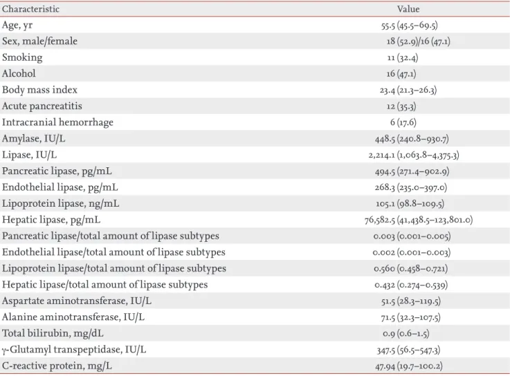
Of the 34 eligible patients initially enrolled in this study, no patients were excluded due to refusal to participate; as a result, a total of 34 patients (18 male and 16 female) participated, and 12 patients (35.3%) were diagnosed with acute pancreatitis (CECT was performed for all of the enrolled patients). The characteristics of the enrolled patients are summarized in Table 1. The median age of enrolled patients was 55.5 years old (IQR, 45.5 to 69.5). The proportions of smokers and alcoholics were 32.4% (n = 11) and 47.1% (n = 16), respectively. The median BMI was 23.4 (IQR, 21.3 to 26.3). Among the enrolled population, the proportion of patients with intracranial hemorrhage was 17.6% (n = 6).
Laboratory values
The median levels of serum amylase and lipase were 448.5 IU/L (IQR, 240.8 to 930.7) and 2,214.1 IU/L (IQR, 1,063.8 to 4,375.3), respectively. In the subtype analysis of lipase, the median levels of serum lipase subtypes were as follows: pancreatic lipase (494.5 pg/mL; IQR, 271.4 to 902.9), endothelial lipase (268.3 pg/mL; IQR, 235 to 397), lipoprotein lipase (105.1 ng/mL; IQR, 98.8 to 109.5), and hepatic lipase (76,582.5 pg/mL; IQR, 41,438.5 to 123,801).
To determine the comparative activity of serum lipase subtypes, the fraction of each lipase in the total amount of lipase subtypes was calculated. The fraction of each lipase in the total amount of serum lipase subtypes were as follows: pancreatic lipase, median 0.003 (IQR, 0.001
to 0.005); endothelial lipase, median 0.002 (IQR, 0.001 to 0.003); lipoprotein lipase, median 0.560 (IQR, 0.458 to 0.721); and hepatic lipase, median 0.432 (IQR, 0.274 to 0.539). The detailed laboratory values of the enrolled pa-tients are described in Table 1.
The distributions of each lipase in the total amount of serum lipase subtypes were as follows: lipoprotein li-pase, 62%; hepatic lili-pase, 37%; pancreatic lili-pase, 0.6%; and endothelial lipase, 0.5% (the relative proportion of the mean value of each lipase subtype was calculated) (Fig. 1).
Univariate analysis for the associations of acute pan-creatitis
Among the clinical variables, there were no statistical-ly significant differences in age, sex, smoking history, BMI, presence of intracranial hemorrhage, or laboratory results, including serum amylase, lipase, AST, ALT, ALP, GGT, and CRP, between the pancreatitis and non-pan-creatitis groups. Only alcohol consumption differed, being noted in nine of 12 patients (75%) in the acute pan-creatitis group and seven of 12 (31.8%) in the non-pan-creatitis group (p = 0.03).
Among the laboratory values, there were no statisti-cally significant differences in serum amylase, lipase, pancreatic lipase, endothelial lipase, lipoprotein lipase, or hepatic lipase between the pancreatitis and non-pan-creatitis groups. The level of serum pancreatic lipase in the patients with acute pancreatitis was higher than in
Lipoprotein lipase 62% Hepatic lipase
37%
Pancreatic lipase 0.6% Endothelial lipase 0.5%
Figure 1. Distribution of each lipase in the total amount of serum lipase subtypes. The relative proportion of mean val-ues in each lipase subtypes are demonstrated.
the patients without acute pancreatitis, although the dif-ference was statistically insignificant ([median, 626 pg/ mL; IQR, 465 to 1,664.2] vs. [median, 407.8 pg/mL; IQR, 200 to 766.5], p = 0.17).
Among the fractions of each lipase in the total amount of lipase subtypes, there were no statistically significant differences in the fractions of endothelial lipase, lipo-protein lipase, or hepatic lipase. Only the fraction of pancreatic lipase in the total amount of serum lipase subtypes (FPL) showed a higher value in the acute pan-creatitis group than in the non-panpan-creatitis group ([me-dian, 0.004; IQR, 0.003 to 0.011] vs. [me([me-dian, 0.002; IQR, 0.001 to 0.004], p = 0.04). The detailed content of the uni-variate analysis for the associations of acute pancreatitis is demonstrated in Table 2.
Prediction of acute pancreatitis
In the ROC curve analysis for the prediction of acute pancreatitis, FPL was the most valuable predictor (AU-ROC, 0.72; 95% confidence interval [CI], 0.54 to 0.86; p = 0.04). The best cut-off value associated with FPL was 0.0027. The sensitivity and specificity of this test using the cut-off value of 0.0027 were 83.3% (95% CI, 51.6 to 97.9) and 63.6% (95% CI, 40.7 to 82.8), respectively. The positive and negative likelihood ratios were 2.3 (95% CI, 1.3 to 4.2) and 0.3 (95% CI, 0.1 to 0.9), respectively. The positive and negative predictive values were 55.6% (95% CI, 30.8 to 78.5) and 87.5% (95% CI, 61.7 to 98.5), respec-tively. Other tests, including serum amylase, lipase, and lipase subtypes other than FPL showed, statistically non-significant results (Table 3, Fig. 2).
Table 1. Characteristics of enrolled population (n = 34)
Characteristic Value
Age, yr 55.5 (45.5–69.5)
Sex, male/female 18 (52.9)/16 (47.1)
Smoking 11 (32.4)
Alcohol 16 (47.1)
Body mass index 23.4 (21.3–26.3)
Acute pancreatitis 12 (35.3) Intracranial hemorrhage 6 (17.6) Amylase, IU/L 448.5 (240.8–930.7) Lipase, IU/L 2,214.1 (1,063.8–4,375.3) Pancreatic lipase, pg/mL 494.5 (271.4–902.9) Endothelial lipase, pg/mL 268.3 (235.0–397.0) Lipoprotein lipase, ng/mL 105.1 (98.8–109.5) Hepatic lipase, pg/mL 76,582.5 (41,438.5–123,801.0)
Pancreatic lipase/total amount of lipase subtypes 0.003 (0.001–0.005)
Endothelial lipase/total amount of lipase subtypes 0.002 (0.001–0.003)
Lipoprotein lipase/total amount of lipase subtypes 0.560 (0.458–0.721)
Hepatic lipase/total amount of lipase subtypes 0.432 (0.274–0.539)
Aspartate aminotransferase, IU/L 51.5 (28.3–119.5)
Alanine aminotransferase, IU/L 71.5 (32.3–107.5)
Total bilirubin, mg/dL 0.9 (0.6–1.5)
γ-Glutamyl transpeptidase, IU/L 347.5 (56.5–547.3)
C-reactive protein, mg/L 47.94 (19.7–100.2)
Table 2. Univariable analysis for the association of acute pancreatitis
Characteristic acute pancreatitis (n = 12)Patients with acute pancreatitis (n = 22)Patients without p value
Age, yr 53 (39.5–60.3) 62.5 (48–72) 0.12
Sex, male/female 7 (58.3)/5 (41.7) 11 (50.0)/11 (50.0) 0.73
Smoking 6 (50.0) 5 (22.7) 0.14
Alcohol 9 (75.0) 7 (31.8) 0.03
Body mass index 25.4 (20.1–27.8) 23.3 (21.3–26.1) 0.53
Intracranial hemorrhage 0 6 (27.3) 0.07 Amylase, IU/L 693 (230.0–1004.1) 429.5 (240.8–878) 0.71 Lipase, IU/L 3,064 (1,047.8–10,770.8) 1,523 (1,063.8–3,256.5) 0.47 Pancreatic lipase, pg/mL 626 (465.0–1,664.2) 407.8 (200.0–766.5) 0.17 Endothelial lipase, pg/mL 304.1 (239.8–602.2) 261.4 (223.7–357.9) 0.55 Lipoprotein lipase, ng/mL 101.9 (93.1–109) 106.2 (99.9–110.7) 0.28 Hepatic lipase, pg/mL 45,875.7 (37,136.2–95,269.5) 96,270.2 (54,139.8–124,710.0) 0.10 Pancreatic lipase/total amount of
lipase subtypes
0.004 (0.003–0.011) 0.002 (0.001–0.004) 0.04
Endothelial lipase/total amount of lipase subtypes
0.002 (0.001–0.004) 0.001 (0.001–0.002) 0.14
Lipoprotein lipase/total amount of lipase subtypes
0.702 (0.488–0.733) 0.526 (0.452–0.645) 0.16
Hepatic lipase/total amount of lipase subtypes
0.294 (0.261–0.485) 0.472 (0.352–0.545) 0.11
Aspartate aminotransferase, IU/L 76 (31.0–175.5) 46.5 (25.5–92.3) 0.39
Alanine aminotransferase, IU/L 64 (25.5–191.8) 71.5 (35.5–103.8) 0.84
Total bilirubin, mg/dL 1.3 (0.6–1.8) 0.9 (0.6–1.4) 0.47
γ-Glutamyl transpeptidase, IU/L 441 (43.0–1173.3) 323.5 (104.3–472.5) 0.31
C-reactive protein, mg/L 50.45 (21.10–96.66) 45.83 (16.88–107.65) 0.51
Values are presented as number (%) or median (interquartile range).
Table 3. AUROC for the prediction of acute pancreatitis
Test AUROC
OR CI p value
Pancreatic lipase/total accumulation of subtype lipase 0.72 0.54–0.86 0.04
Hepatic lipase 0.67 0.47–0.88 0.10 Pancreatic lipase 0.65 0.47–0.80 0.17 Lipoprotein lipase 0.62 0.42–0.81 0.27 Endothelial lipase 0.56 0.35–0.78 0.54 Serum lipase 0.58 0.40–0.75 0.51 Serum amylase 0.54 0.36–0.71 0.72
Table 4. Multivariable analysis for the prediction of acute pancreatitis
Variable β Standard error Wald OR (95% CI) p value
Pancreatic lipase/total accumulation of subtype lipasea 2.12 0.93 5.19 8.3 (1.3–51.7) 0.02
Serum amylase < 0.001 0.001 0.06 1.0 (0.9–1.0) 0.81
Serum lipase < 0.001 < 0.001 0.001 1.0 (1.0–1.0) 0.98
Pancreatic lipase < 0.001 0.001 0.006 1.0 (0.9–1.1) 0.94
Intracranial hemorrhage –20.68 15,237.40 < 0.001 < 0.001 0.32
Alcohol 1.21 1.05 1.33 3.4 (0.4–26.3) > 0.99
OR, odds ratio; CI, confidence interval. aCut-off > 0.0027.
A B
100-Specificity
0 20 40 60 80 100
Sensitivity
Pancreatic lipase/total accumulation of subtype lipase 100 80 60 40 20 0 100-Specificity 0 20 40 60 80 100 Sensitivity Pancreatic lipase 100 80 60 40 20 0 100-Specificity 0 20 40 60 80 100 Sensitivity Serum lipase 100 80 60 40 20 0 100-Specificity 0 20 40 60 80 100 Sensitivity Serum amylase 100 80 60 40 20 0
Figure 2. Receiver-operating characteristic curves for the prediction of acute pancreatitis. (A) Area under the receiver-operat-ing characteristic curve (AUROC) of pancreatic lipase/total accumulation of subtype lipase 0.72 (95% confidence interval [CI], 0.54 to 0.86; p = 0.04). (B) AUROC of pancreatic lipase 0.65 (95% CI, 0.47 to 0.80; p = 0.17). (C) AUROC of serum lipase 0.58 (95% CI, 0.40 to 0.75; p = 0.51). (D) AUROC of serum amylase 0.54 (95% CI, 0.36 to 0.71; p = 0.72).
Multivariate analysis for the associations of acute pancreatitis
In multivariate analysis for the associations of acute pancreatitis, only FPL using a cut-off value of 0.0027 was associated with acute pancreatitis (OR, 8.3; 95% CI, 1.3 to 51.7; p = 0.02) (Table 4).
DISCUSSION
In the present study, the clinical efficacy of serum lipase subtype analysis for the differential diagnosis of pan-creatic and non-panpan-creatic serum lipase elevations was assessed. Lipase and amylase are released from acinar cells in patients with acute pancreatitis, and the mea-surements of serum concentrations of these enzymes are used to confirm diagnosis [7]. Serum lipase measure-ment is generally recommended because of its greater specificity, sensitivity, and durability than serum amy-lase measurement [8-11]. However, non-pancreatic ele-vations of serum lipase levels make it difficult to diag-nose acute pancreatitis, and radiologic imaging studies are inevitable. FPL showed adequate sensitivity (83.3%) and negative predictive value (87.5%) although relative-ly low specificity (63.6%) and positive predictive value (55.6%) in this study. However, considering that the total number of patients (n = 34) was small, and the prevalence of pancreatitis (32.4%) was low, these values could have been higher if applied in the larger cohort. The direct comparison of pancreatic lipase between patients with acute pancreatitis and those without showed statistically non-significant results, although the pancreatitis group showed higher levels (Table 2). The small sample size could be the reason for this study, which must be repli-cated with a larger sample size.
The sensitivity and specificity of serum lipase mea-surement for acute pancreatitis were reported as 55% to 100% and greater than 95%, respectively, at a cut-off value of 600 IU/L, showing greater specificity than FPL [7,10]. However, a major difference from our study was the target population. The reported diagnostic perfor-mance of serum lipase was based on a population with acute abdominal pain. However, this study includ-ed patients with serum lipase elevations regardless of symptoms. Thus, diagnostic performance could not be applied directly when comparing with serum lipase or
amylase. Considering the radiation exposure and high expense of CT, FPL testing could be used for triage or as an add-on test in patients with serum lipase eleva-tions and without definite symptoms suggesting acute pancreatitis. In particular, it could be useful in certain clinical situations, such as typical symptoms with a nor-mal range of serum lipase or an elevated level of serum lipase without typical symptoms.
Another outcome was the distribution of each lipase subtype in serum. Among the isoforms of lipase, the majority portion of the serum concentration was lipo-protein lipase or hepatic lipase in this study, and this finding was consistent independent of acute pancreati-tis (Tables 1 and 2, Fig. 1). Although we are not yet aware of the exact function of each lipase subtype, this result indicates the need for more specific measurement of lipase subtypes for different etiologies. Among the en-rolled population, patients with lithium toxicity or he-patocellular carcinoma showed higher concentrations of hepatic lipase than pancreatic lipase compared to the patients with acute pancreatitis.
Despite the potential diagnostic performance of lipase subtype analysis, there are several concerns that need to be clarified for widespread application. First, there is no reference assay or reference concentration range for the measurement of serum pancreatic lipase [12-16]. In this study, the authors also could not enroll an asymptomat-ic control group with normal serum lipase levels. We as-sessed the enzymatic activity using the ELISA technique; however, it would be cumbersome to do so in clinical practice. Second, the reference diagnostic method was not perfect. We used the revised definition of Atlan-ta 2012 as the gold sAtlan-tandard for the diagnosis of acute pancreatitis. Serum lipase activity (or amylase activity) at least three times greater than the upper limit of normal was generally accepted [1]. However, the diagnostic per-formance of the index test could change according to the diagnostic cut-off of the gold standard test and the study population [10]. Another issue was the lack of anal-ysis of pancreatic lipase-related protein 2 (PLRP2). From the three different mRNAs encoding human pancreatic lipases, there are three expressions of human pancreat-ic lipase, PLRP1, and PLRP2, respectively [17,18]. PLRP1 is known to have no lipase activity, whereas PLRP2 is known to have lipase activity in vitro and is known to be reduced in patients with chronic calcifying pancreatitis,
although statistically insignificantly [18,19]. Human gas-tric lipase, a member of the acid lipase gene family, is known to compensate partly for the loss of pancreatic lipase in situations of exocrine pancreatic insufficiency [20]. However, the activity of this enzyme was not mea-sured in this study. Finally, the small sample size was also a limitation of this study.
There have been efforts to determine laboratory bio-markers for the diagnosis of acute pancreatitis to re-place or compensate for traditional serum enzyme mea-surements, although measuring serum lipase activity remains the gold standard [10]. Non-pancreatic lipase elevation is complex with heterogeneous conditions to be clarified, and this study was the first clinical analy-sis of combined assessment of human pancreatic lipase isoforms. Through each of the etiological approaches to serum lipase subtypes, the characteristics and interac-tions of each lipase isoform could be elucidated. These results do not support that serum lipase subtype analy-sis could replace the standard lipase measurement for the diagnosis of acute pancreatitis. However, the test demonstrated adequate sensitivity to be used for triage or as an add-on test in serum lipase elevation, but these findings need to be replicated with a larger sample size.
Conflict of interest
No potential conflict of interest relevant to this article was reported.
REFERENCES
1. Banks PA, Bollen TL, Dervenis C, et al. Classification of acute pancreatitis: 2012: revision of the Atlanta
classifi-cation and definitions by international consensus. Gut 2013;62:102-111.
2. Liu KJ, Atten MJ, Lichtor T, et al. Serum amylase and li-pase elevation is associated with intracranial events. Am Surg 2001;67:215-219.
3. Gumaste VV, Roditis N, Mehta D, Dave PB. Serum lipase levels in nonpancreatic abdominal pain versus acute pan-creatitis. Am J Gastroenterol 1993;88:2051-2055.
4. Yadav D, Nair S, Norkus EP, Pitchumoni CS. Nonspecific hyperamylasemia and hyperlipasemia in diabetic keto-acidosis: incidence and correlation with biochemical ab-normalities. Am J Gastroenterol 2000;95:3123-3128. 5. Bokemeyer B. Asymptomatic elevation of serum lipase
and amylase in conjunction with Crohn’s disease and ul-cerative colitis. Z Gastroenterol 2002;40:5-10.
6. Yadav D, Agarwal N, Pitchumoni CS. A critical evaluation of laboratory tests in acute pancreatitis. Am J Gastroen-terol 2002;97:1309-1318.
7. Matull WR, Pereira SP, O’Donohue JW. Biochemical markers of acute pancreatitis. J Clin Pathol 2006;59:340-344.
8. Treacy J, Williams A, Bais R, et al. Evaluation of amylase and lipase in the diagnosis of acute pancreatitis. ANZ J Surg 2001;71:577-582.
9. Gomez D, Addison A, De Rosa A, Brooks A, Cameron IC. Retrospective study of patients with acute pancreatitis: is serum amylase still required? BMJ Open 2012;2:e001471. 10. Lippi G, Valentino M, Cervellin G. Laboratory diagnosis
of acute pancreatitis: in search of the Holy Grail. Crit Rev Clin Lab Sci 2012;49:18-31.
11. Shah AM, Eddi R, Kothari ST, Maksoud C, DiGiacomo WS, Baddoura W. Acute pancreatitis with normal serum lipase: a case series. JOP 2010;11:369-372.
12. Aoubala M, Ivanova M, Douchet I, De Caro A, Verger R. Interfacial binding of human gastric lipase to lipid monolayers, measured with an ELISA. Biochemistry 1995;34:10786-10793.
13. Beisson F, Tiss A, Riviere C, Verger R. Methods for lipase detection and assay: a critical review. Eur J Lipid Sci Technol 2000;102:133-153.
14. Lessinger JM, Parashou S, Arzoglou P, et al. Determina-tion of lipase catalytic activity in two reference materials: BCR 693 and BCR 694 by titrimetry at constant pH. Clin Chem Lab Med 2004;42:62-66.
15. Panteghini M. The never-ending search of an acceptable compromise for pancreatic lipase standardisation. Clin
KEY MESSAGE
1. The fraction of pancreatic lipase in the total amount of serum lipase subtypes was statisti-cally higher in patients with pancreatitis.
2. Although serum lipase subtype analysis cannot replace the standard lipase measurement for the diagnosis of acute pancreatitis, the test demon-strated adequate sensitivity for use in triage or as an add-on test for serum lipase elevation.
Chem Lab Med 2012;50:419-421.
16. Doolittle MH, Ben-Zeev O. Immunodetection of lipopro-tein lipase: antibody production, immunoprecipitation, and Western blotting techniques. Methods Mol Biol 1999;109:215-237.
17. Giller T, Buchwald P, Blum-Kaelin D, Hunziker W. Two novel human pancreatic lipase related proteins, hPLRP1 and hPLRP2: differences in colipase dependence and in lipase activity. J Biol Chem 1992;267:16509-16516.
18. Eydoux C, Aloulou A, De Caro J, et al. Human pancreatic
lipase-related protein 2: tissular localization along the di-gestive tract and quantification in pancreatic juice using a specific ELISA. Biochim Biophys Acta 2006;1760:1497-1504.
19. Lowe ME. Properties and function of pancreatic lipase related protein 2. Biochimie 2000;82:997-1004.
20. Carriere F, Grandval P, Renou C, et al. Quantitative study of digestive enzyme secretion and gastrointestinal lipoly-sis in chronic pancreatitis. Clin Gastroenterol Hepatol 2005;3:28-38.