Anti-Vascular Endothelial Growth Factor Treatment Augments Tumor Radiation
Response under Normoxic or Hypoxic Conditions
1Chang-Geol Lee, Marcus Heijn, Emmanuelle di Tomaso, Genevieve Griffon-Etienne, M. Ancukiewicz, Chieko Koike,
K. R. Park, Napoleone Ferrara, Rakesh K. Jain, Herman D. Suit,
2and Yves Boucher
Edwin L. Steele Laboratory, Department of Radiation Oncology, Massachusetts General Hospital and Harvard Medical School, Boston, Massachusetts 02114 [C. G. L., M. H., E. d. T., G. G-E., M. A., C. K., K. R. P., R. K. J., H. D. S., Y. B.] and Department of Cardiovascular Research, Genentech, Inc., South San Francisco, California 94080 [N. F.]
ABSTRACT
Recent studies in experimental animals have shown that combining antiangiogenic therapy with radiation can enhance tumor response. Whether this enhancement is mainly attributable to angiogenesis inhibi-tion, endothelial cell radiosensitivity, tumor cell apoptosis, or a decrease in the number of hypoxic cells (improved oxygenation) is not known. We designed this study to discern the role of tumor oxygenation. We chose an anti-vascular endothelial growth factor (anti-VEGF) monoclonal antibody (mAb) which has a known target, human VEGF. We also measured interstitial fluid pressure (IFP) to test the hypothesis that the decreased vascular permeability induced by the anti-VEGF mAb can lower IFP. The effect of anti-VEGF mAb on vascular density, partial oxygen tension (pO2), and apoptosis was also measured. Athymic NCr/Sed nu/nu mice bearing 6-mm xenograft of the human glioblastoma multiforme (U87), or colon adenocarcinoma (LS174T) were treated with anti-VEGF mAb in-jected i.p. on alternate days for a total of six injections at a dosage of 100
g/injection/mouse. For combined anti-VEGF and radiation, single
radi-ation doses were given under normal blood flow (20 and 30 Gy) or clamped hypoxic conditions (30 and 40 Gy) 24 h after the sixth injection of mAb. The inhibition of the growth of U87 and LS174T tumors by the anti-VEGF mAb was associated with a significant reduction in tumor vascular density and a relatively small increase in the number of apoptotic cells. Compared with size-matched controls, IFP decreased by 74% in LS174T, and 73% in U87 in mice treated with anti-VEGF mAb. After antibody treatment pO2increased significantly in U87, but did not change in LS174T tumors. Combined treatment induced in U87 tumors a tumor-growth delay (TGD) which was greater than additive; in LS174T except for the 40-Gy hypoxic group, the effect was only additive. In both U87 and LS174T the TGD induced by the antibody was independent of oxygen levels in the tumor at the time of radiation. The fact that the increase in TGD occurred under both normoxic and hypoxic conditions suggests that anti-VEGF mAb treatment can compensate for the resistance to radiation induced by hypoxia.
INTRODUCTION
Antiangiogenic agents can enhance the tumor response induced by radiation (1– 4). The inhibition of vessel formation is not the only mechanism by which angiogenesis inhibitors delay tumor growth. TNP-470, angiostatin, and anti-VEGF3
mAb can also increase tumor cell apoptosis (5– 8). Hypoxia, a major factor contributing to the radioresistance of tumor cells, was reduced by angiogenesis inhibitors (squalamine or the combination of TNP-470 and minocycline; 1, 9). The combination of radiation with TNP-470 and minocycline induced a greater TGD than radiation alone (1). Recent studies have shown
that angiostatin or anti-VEGF mAb radiosensitizes endothelial cells, and the combination of radiation with the anti-VEGF antibody or angiostatin increases the antitumor effects of radiation (2, 4). Thus, antiangiogenic agents enhance the effects of radiation via both direct and indirect mechanisms that target tumor and endothelial cells.
VEGF is a critical growth factor that promotes endothelial cell proliferation, angiogenesis, and tumor growth, and maintains the elevated vascular permeability of tumor vessels (10 –12). Pre-clinical studies have demonstrated that the growth of several tumor types is inhibited by blocking antibodies against human VEGF (13–16). Both the neutralization of VEGF action and the suppression of VEGF expression are associated with increases in the fraction of apoptotic tumor cells and reductions in vascular density and permeability (8, 11, 17). Grunstein et. al. (11) have recently shown that deletion of the VEGF gene in ras-transformed fibroblasts increased tumor hypoxia as measured by the formation of cellular adducts with the small molecule nitroimidazole EF5.
We designed the present study to determine whether tumor growth inhibition by the combination of radiation and anti-VEGF mAb is dependent on tumor oxygen levels. To discern the role of tumor oxygenation, treatment over 11 days with human anti-VEGF mAb was followed by a single dose of ionizing radiation that was given either under normoxic or clamped hypoxic conditions. To identify other mechanisms that could inhibit tumor growth induced by the antibody or the combination of antibody and radiation, the effects of anti-VEGF mAb on pO2, vascular density, and the fraction of
apo-ptotic cells were measured.
Tumor interstitial hypertension is a likely cause of the poor delivery of large therapeutic molecules to solid tumors (18). The high vascular permeability and vascular resistance are two mechanisms that con-tribute to the elevated IFP of solid tumors. VEGF increases vascular permeability (12) and the anti-VEGF mAb decreases vascular perme-ability (17). We, thus, tested the hypothesis that VEGF inhibition with anti-VEGF mAb would lower tumor IFP.
MATERIALS AND METHODS
Animal Studies. Athymic NCr/Sed nude (nu/nu) mice, male, 8 –10 weeks old, from our defined flora- and specific pathogen-free animal colony were used. The mice were further immunosuppressed 24 h before tumor transplan-tation by whole-body irradiation with 5 Gy using a Gammacell 137Cs unit (0.8 Gy/min). Tumor chunks of the human glioblastoma U87 and colon adenocar-cinoma LS174T were implanted s.c. in the legs of mice.
Anti-VEGF mAb. The murine antihuman VEGF mAb A.4.6.1. (anti-VEGF mAb; Genentech, South San Francisco, CA) was used. Mice bearing 6-mm diameter tumors (100 mm3) were given six i.p. injections (one every other day) of anti-VEGF mAb (100 g/mouse) diluted in saline. Control animals were injected with saline.
Irradiation Procedures. A single radiation dose under normal blood flow (20 Gy, 30 Gy) or hypoxic (30 Gy, 40 Gy) conditions was administered at a rate of about 5.6 Gy/min using a specially designed cesium irradiator. During irradiation the mice were immobilized using a plastic tube fixed on a brass plate, and the tumor was centered within the 3-cm diameter circular irradiation field. In the groups treated with radiation only, tumors with a diameter of 6 mm were irradiated. In the groups treated with anti-VEGF mAb and radiation,
Received 1/17/00; accepted 8/2/00.
The costs of publication of this article were defrayed in part by the payment of page charges. This article must therefore be hereby marked advertisement in accordance with 18 U.S.C. Section 1734 solely to indicate this fact.
1This work was supported by Outstanding Investigator Grant (R35-CA-56591; to R. K. J.) and a MERIT Grant (R37-CA-13111 to H. D. S.) from the National Cancer Institute.
2To whom requests for reprints should be addressed, at Department of Radiation Oncology, Massachusetts General Hospital, Harvard Medical School, Boston, MA 02114. Phone: (617) 726-8150; Fax: (617) 726-3603; E-mail: [email protected].
3The abbreviations used are: VEGF, vascular endothelial growth factor; pO 2, partial oxygen tension; IFP, interstitial fluid pressure; MVP, microvascular pressure; mAb, monoclonal antibody; TGD, tumor growth delay.
anti-VEGF treatment was initiated at a tumor size of 6 mm, and the single radiation dose was given 24 h after the sixth antibody injection.
Tumors were exposed to single doses under oxygenated (normal blood flow) or hypoxic (clamped tumors) conditions. In the latter case, a clamp was placed across the base of the tumor-bearing leg to occlude the tumor blood flow for 2 min before, and then during, irradiation. The mice were anesthetized with pentobarbital (50 mg/kg body weight i.p.) before clamping. The mice that were irradiated under oxygenated conditions were not anesthetized.
Tumor Volume. Tumor diameters in control and treated groups were measured every 2 days with a caliper. All experimental animals were observed on a daily basis, and tumor sizes were measured every other day. Tumor volume was calculated as V⫽ ab2/2, where a and b are the long and the short
axes, respectively. Mice were sacrificed with an i.p. injection of pentobarbital (200 mg/kg body weight) once the tumor reached 12 mm in diameter.
pO2Measurements. Oxygen measurements were performed in tumors using a polarographic pO2histograph (Eppendorf, Hamburg, Germany). The pO2was measured once, 24 h after the sixth injection of anti-VEGF mAb. pO2 was also measured in size-matched untreated tumors. The mice were anesthe-tized with ketamine/xylazine (100/10 mg/kg body weight) and placed on a heating pad to maintain the body temperature at 37°C. The skin overlying the tumor was cut and the cathode was placed through the cut to the surface of the tumor. The electrode was advanced automatically through the tumor, each forward step followed by a short backward step to reduce artifacts in pO2 measurements from compression of the tissue by the electrode. pO2 measure-ments were taken in four tumor regions, and 40 –50 measuremeasure-ments were taken in each tumor. The pO2values represent the median of eight mice per group. IFP Measurements. The wick-in-needle technique was used to measure IFP as described previously (19). IFP was measured in two locations in the center of s.c. tumors, 24 h after the sixth injection of anti-VEGF mAb. Because the IFP increases with tumor size, IFP was measured in untreated tumors that were size-matched with the volume of antibody-treated tumors.
Immunohistochemistry. After the mice were killed, tumors were imme-diately excised and placed in neutral-buffered formalin. The tumors were embedded in paraffin blocks from which 5m sections were cut. The degree of apoptosis was evaluated using Apop-Tag peroxidase kit (Oncor, Gaithers-burg, MD) according to the manufacturer’s recommendations. The apoptotic index was calculated as the percentage of positive nuclei— defined as stained with peroxidase combined with the presence of a nuclear halo or apoptotic bodies— observed in a minimum of 400 cells per histological section at a final magnification of⫻1240.
To estimate vessel density, tumor sections were incubated overnight at 4°C with rat antimouse CD31 (PharMingen, San Diego, CA) at a dilution of 1:30. Visualization of the antigen-antibody reaction was carried out using the Vec-tastain Elite ABC kit (Vector Laboratories, Burlingame, CA) according to the recommendations of the manufacturer. Vessel density was determined by counting the stained vessels in fields of 0.071 mm2at a final magnification of
⫻620. A number of 10 to 23 fields per histological section were included in the
analysis.
Western Analysis. Tissue samples were homogenized with lysis buffer containing 10 mMTris-HCl (pH 7.4), 150 mMNaCl, 1.0% Triton X-100, 10% NP40, 2 mMEDTA and protease inhibitor cocktail (Complete Mini EDTA-free, Boehringer Mannheim). 100g protein of each lysate were loaded into 12% SDS-PAGE gel, separated, and transferred onto the nitrocellulose mem-brane. One hundred ng of human recombinant VEGF protein was loaded as positive control. The membranes were subjected to immunoblotting with rabbit antimurine VEGF antibody, which was provided by Dr. Donald Senger of Beth Israel Deaconess Hospital (Boston, MA). The antibody cross-reacts with human VEGF. Immunodetections were carried out using Western blot chemi-luminescence reagent (New England Nuclear Life Science Products, Boston, MA).
Statistical Analysis. The TGD calculations were based on the median time in days for tumors to grow from 100 to 500 mm3. Some tumors did not reach 500 mm3during the experiment because either the experiment was interrupted before the tumors reached 500 mm3or the tumors regressed. In these cases, the duration of experiment was used as a censored measure of time to reach 500 mm3. Median times to grow from 100 to 500 mm3 were estimated with Kaplan-Meier curves. TGD attributable to antibody was estimated as a differ-ence of such medians in groups treated with anti-VEGF mAb and in corre-sponding controls (untreated tumors, radiation-treated tumors). The SEs of the
medians and of their differences were obtained with bootstrap methods (20). We used the log-rank test for statistical comparisons of times to grow from 100 to 500 mm3. Differences in apoptosis, vascular density, IFP, and pO
2between anti-VEGF mAb and control groups were tested with Student’s t test and ANOVA for normally distributed data, and with Mann-Whitney U test other-wise.
RESULTS
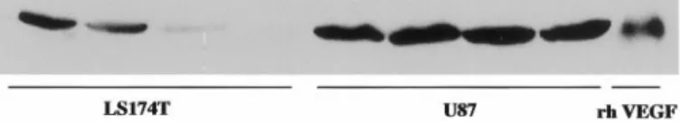
VEGF Levels in Tumors. The VEGF protein was detected by
Western analysis in both U87 and LS174T tumors. VEGF levels were higher in U87 than in LS174T tumors (Fig. 1).
Anti-VEGF mAb Inhibits Tumor Growth. The growth of
LS174T and U87 tumors was progressive for observations up to tumor volumes of 900-1000 mm3
(Fig. 2a). Anti-VEGF mAb retarded the growth of both U87 and LS174T, with a significantly greater effect on the growth of U87 (Table 1). The median TGD induced by the anti-VEGF mAb was 12 and 22 days for LS174T and U87, respec-tively. Furthermore, there were larger variations in times to grow to 900 mm3
for U87 tumors. In some U87 tumors the growth retardation during the 11 days of anti-VEGF mAb treatment was followed at irregular intervals by periods of several days without growth (Fig. 2a).
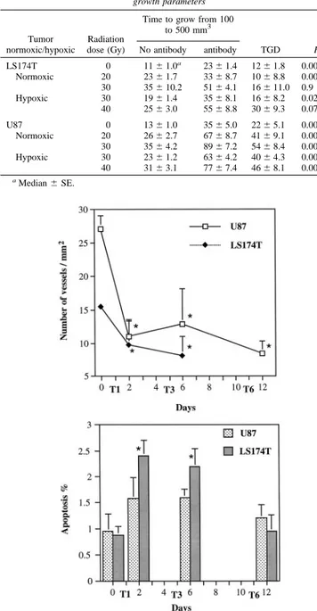
Anti-VEGF mAb Reduces Tumor Vessel Density. In both
LS174T and U87 tumors, 24 h after the first anti-VEGF treatment, microvessel density decreased significantly by 36 and 60%, respec-tively (Fig. 3a). The microvessel density after the third or sixth injection of anti-VEGF mAb was still significantly lower as compared with control tumors.
Induction of Neoplastic Cell Apoptosis by Anti-VEGF mAb.
Concomitant with the decrease in microvessel density, apoptosis was increased in U87 and LS174T. Compared with controls, apoptosis was significantly increased in LS174T 24 h after the first and third tion of anti-VEGF mAb (Fig. 3b). However, after six antibody injec-tions, the percentage of apoptotic cells was similar to control values. In U87, the increase in apoptosis was not significant.
Anti-VEGF mAb Reduces IFP. On the basis of the finding that
anti-VEGF reduces tumor vascular permeability and modifies the architecture of tumor vessels (17), we hypothesized that anti-VEGF treatment would reduce IFP. The mean IFP of LS174T and U87 dropped by 73 and 74%, respectively (Table 2). Some of the IFP values, especially in U87 were close to 0 mm Hg, thus comparable with the IFP in normal tissues. In four of eight treated U87 tumors, IFP varied between 1.0 and 3.0 mm Hg.
Differential Effect of Anti-VEGF mAb on pO2. The
modifica-tions in tumor vascular density suggest that anti-VEGF mAb could modify tumor blood flow and pO2. Anti-VEGF mAb increased the
median pO2 in U87 and significantly reduced the frequency of pO2
values below 5 mm Hg (Table 2; Fig. 4a). In LS174T, the antibody did not change the median pO2, and the decrease in frequency of pO2
values below 5 mm Hg was not significant (Table 2; Fig. 4b).
Anti-VEGF mAb Improves Radiation Response under Hypoxic and Normoxic Conditions. In comparison with radiation alone, the
combination of anti-VEGF mAb and radiation increased the time for U87 and LS174T to grow from 100 to 500 mm3
(Fig. 2, b and c; Table
Fig. 1. Western blots of VEGF levels in LS174T and U87 tumors. The quantity of VEGF protein is higher in U87 than in LS174T. Each lane, a different tumor; recombinant human VEGF (rh VEGF), control lane.
1). In most of the U87 tumors, the initial growth delay induced by radiation was followed by a steady and rapid growth phase. However, in U87 for both oxic (20 and 30 Gy) and hypoxic (40 Gy) conditions, anti-VEGF mAb plus radiation interrupted the growth or induced a steady regression of 10 – 43% of the tumors (Fig. 2c). In contrast, radiation alone (30 Gy oxic) induced the regression of only one U87 tumor. The combination of anti-VEGF mAb and radiation was
sig-nificantly less effective in LS174T than in U87 tumors, as shown by the time that it took to grow from 100 to 500 mm3
and the estimation of the TGD induced by the antibody (Fig. 5; Table 1).
Radiation alone delayed tumor growth in a dose-dependent manner; the growth delay was, as expected, less for radiation under hypoxic than that under aerobic conditions. Except for the significant TGD between 30 and 40 Gy hypoxic in LS174T tumors, the TGD induced Fig. 2. Effect of anti-VEGF mAb and the combination of radiation and anti-VEGF mAb on the growth of U87 and LS174T tumors. In A, the growth of both tumors was significantly inhibited by anti-VEGF mAb. B and C, effect of radiation or combined anti-VEGF mAb and radiation under oxic (Oxic) or hypoxic (Hypoxic) conditions on the growth of LS174T (B) and U87 (C) tumors. In both U87 and LS174T, there was a greater inhibition of tumor growth induced by the combination of radiation and anti-VEGF mAb as compared with radiation alone.
by the combined modality treatment was independent of radiation dose and tissue oxygenation at irradiation (Fig. 5). In LS174T, the combination of radiation (40 Gy hypoxic) and antibody induced a TGD that was greater than additive. At all other radiation doses, the effect was at best additive. In U87, at all radiation doses and for both aerobic and clamp hypoxia conditions, the TGD induced by the anti-VEGF mAb was greater than additive (Fig. 5). For U87, it was possible to calculate the TGD based on the time after radiation for the tumors to triple in size. The TGD calculations based on a 3-fold increase in tumor size (data not shown) or the time for the tumors to grow from 100 to 500 mm3
gave comparable results.
DISCUSSION
Vascular and Physiological Response to Anti-VEGF mAb.
Within 24 h of the initiation of anti-VEGF treatment there was a significant decrease in vessel density in U87 and LS174T tumors (Fig. 3a). Previous studies have also shown that the tumor vasculature regressed rapidly after VEGF down-regulation (21, 22). The reduction in tumor VEGF levels leads to endothelial cell detachment and apo-ptosis (21, 22). After VEGF withdrawal, Benjamin and Keshet (21) found that the marked decrease in the vessels of C6 glioma was associated with hemorrhage. In contrast, we found no evidence of hemorrhage. The reduction in tumor IFP is consistent with the de-crease in vascular permeability after treatment with anti-VEGF mAb or VEGF down-regulation (11, 17). Variations in vascular response after VEGF withdrawal or anti-VEGF mAb treatment could be attrib-utable to differences in the tumor levels of active VEGF, other survival factors for endothelial cells or the presence of periendothelial cells (e.g., pericytes). Benjamin et al. (23) have recently shown that tumor vessels devoid of periendothelial cells regress after the down-regulation of VEGF levels in the C6 glioma.
Fig. 3. Effect of anti-VEGF mAb on microvessel density (upper panel) and the apoptotic index (lower panel) in LS174T and U87 tumors. Microvessel density and the apoptotic index were quantified on histological sections of tumors that were resected 24 h after the first (T1), third (T3), and sixth (T6) injection of anti-VEGF mAb. There was a significant decrease in microvessel density (ⴱ) at all of the time points studied. The apoptotic index increased significantly after T1 and T3 in LS174T tumors (ⴱ) and returned to control values after T6. In U87, the increase in apoptosis was not significant. Data
points, the mean⫾ SD.
Fig. 4. Histograms of pO2values in U87 and LS174T tumors. White and black bars, the pO2 data in the control and anti-VEGF mAb groups, respectively. In U87, the anti-VEGF mAb induced a shift to the right of the pO2values as well as a significant decrease of the pO2values below 5 and 10 mm Hg. In LS174T, the shift to the right of the pO2was small, and there was no significant decrease of the pO2values below 5 or 10 mm Hg.
Table 1 Effect of the combination of the anti-VEGF mAb and radiation on tumor
growth parameters
Tumor normoxic/hypoxic
Radiation dose (Gy)
Time to grow from 100 to 500 mm3 TGD P No antibody antibody LS174T 0 11⫾ 1.0a 23⫾ 1.4 12⫾ 1.8 0.00002 Normoxic 20 23⫾ 1.7 33⫾ 8.7 10⫾ 8.8 0.0008 30 35⫾ 10.2 51⫾ 4.1 16⫾ 11.0 0.9 Hypoxic 30 19⫾ 1.4 35⫾ 8.1 16⫾ 8.2 0.025 40 25⫾ 3.0 55⫾ 8.8 30⫾ 9.3 0.07 U87 0 13⫾ 1.0 35⫾ 5.0 22⫾ 5.1 0.00001 Normoxic 20 26⫾ 2.7 67⫾ 8.7 41⫾ 9.1 0.0005 30 35⫾ 4.2 89⫾ 7.2 54⫾ 8.4 0.002 Hypoxic 30 23⫾ 1.2 63⫾ 4.2 40⫾ 4.3 0.00005 40 31⫾ 3.1 77⫾ 7.4 46⫾ 8.1 0.002 aMedian⫾ SE.
Table 2 Effect of anti-VEGF mAb on mean IFP and median pO2
Tx U87 LS174T IFP (mm Hg) Control 12.0⫾ 3.0 13.5⫾ 4.0 Anti-VEGF mAb 3.0⫾ 1.5a 3.5⫾ 1.0a pO2(mm Hg) Control 14 (8.0–18.5) 12.0 (8–16) Anti-VEGF mAb 20.5 (13–24)a 14.0 (11–14.5) aP⬍ 0.05. 5568
Antiangiogenic agents like squalamine, and TNP-470 combined with minocycline can reduce the degree of hypoxia in tumors (1, 9). Interestingly, in U87 the vessel regression induced by the anti-VEGF mAb was associated with an increase in median pO2and a decrease in
hypoxic values below 5.0 mm Hg. The increase in pO2 despite a
decrease in vascular density suggests that it is the quality of the vascular organization and not just the quantity of vessels that deter-mines the oxygen levels in a tumor. The improvement in tumor oxygenation could also be explained by a decrease in the number of tumor and endothelial cells consuming oxygen. Oxygen consumption by endothelial cells is significant, and it most likely influences the pO2
values in tissues (24, 25).
Elevated IFP is a hallmark of solid tumors (26). Anti-VEGF mAb reduced IFP from 12.0 to 3.0 mm Hg in U87 and from 13.5 to 3.5 mm Hg in LS174T, thus showing that VEGF is an important molecule responsible for the interstitial hypertension in solid tumors. Further-more, especially in some anti-VEGF-treated U87 tumors, the IFP was reduced to 1–2 mm Hg, which is comparable with the pressure in the subcutis and other normal tissues, which varies between 0 and⫺2.0 mm Hg (27). Two vascular factors contribute to interstitial hyperten-sion in solid tumors: the high permeability of tumor vessels and the tortuous nature including compression (reduction in vessel diameter) of vessels. The high vascular permeability and hydraulic conductivity of tumor vessels favor the equilibration of the MVP and IFP (28). The tortuous architecture of the vasculature and the vessel compression by tumor cells increase the vascular resistance and thus the MVP (29). Treatment with anti-VEGF mAb decreases vascular permeability (17) and could directly lower IFP. Furthermore, the loss of endothelial cells would reduce the tortuosity of vessels or eliminate vessels altogether. This modification in vascular architecture would reduce flow resistance and, hence, lower both MVP and IFP. Thus, VEGF contributes significantly to the high IFP in solid tumors, and normal-ization in vascular permeability and architecture is probably respon-sible for the lower IFP induced by the anti-VEGF mAb.
Antitumoral Effects of Anti-VEGF mAb. Similar to other
stud-ies, we found that the anti-VEGF mAb retards tumor growth and has differential effects dependent on the tumor type (13–16). Anti-VEGF mAb induced a TGD of only 6 days in a human sarcoma (data not shown), whereas the growth delays in LS174T and U87 were of 12
and 22 days, respectively. The differential tumor response to anti-VEGF mAb could be attributable to the relative levels of host (mu-rine) versus human VEGF, the absolute levels of VEGF, or other growth factors that stimulate angiogenesis. The mAb A.4.6.1 binds and neutralizes human VEGF and does not block murine VEGF. Fukumura et al. (30) have recently shown that host (mouse) stromal cells in human tumor xenografts can be a source of VEGF. The expression of VEGF varies from one tumor type to another. In accord with the finding that the levels of VEGF protein were higher in U87 than in LS174T tumors (Fig. 1), the anti-VEGF mAb did effect a greater TGD in U87 as compared with LS174T. However, other reports have found no correlation between the levels of VEGF pro-duced in vitro or in vivo and the antitumor response inpro-duced by the antihuman VEGF mAb MV833 (13, 15). Potential differences in antibody specificity or affinity could also affect antitumor response. In the present study, the mAb 4.6.1 against human-VEGF induced a significant delay in the growth of s.c. U87 tumors in nude mice. In contrast, mAb 293 against human VEGF did not inhibit the growth of U87 that was also implanted s.c. in nude mice (4).
In comparison with LS174T, the greater reduction in vascular density in U87 could be responsible for the larger TGD in that tumor. Tumor volume doubling times were comparable in untreated U87 (4.5 days) and LS174T (4.0 days), whereas the vessel density was of 27 and 15 vessels/mm2
, respectively. Vessel loss induced by the anti-VEGF mAb was 2.5 fold greater in U87 as compared with LS174T. Modifications in tumor cell proliferation or apoptosis induced by antiangiogenic agents could also explain the TGD induced by the anti-VEGF mAb. The in vitro proliferation of U87 tumor cells is not modified by the anti-VEGF mAb 4.6.1 (17). Yoshigi et al. (31) have shown that the in vivo inhibition of the VEGF signaling pathway in endothelial cells is associated with an increase in tumor cell apoptosis; however, the number of proliferating tumor cells was not modified (31). Increases in tumor cell apoptosis are also induced by various antiangiogenic agents (5–7, 32) including anti-VEGF mAb treatment and deletion of the VEGF gene in ras-transformed fibroblasts (8, 11). The small increase in apoptosis in U87 and LS174T probably plays a minor role in the inhibition of growth by the anti-VEGF mAb. Furthermore, the greater induction of apoptosis in LS174T was asso-ciated with a smaller antitumor response as compared with U87. We did not quantify necrosis; however, the examination of histological slides did not reveal obvious differences in necrosis between control and anti-VEGF mAb-treated tumors. Thus, based on the present results, the inhibition of angiogenesis is probably the dominant factor responsible for the TGD induced by the anti-VEGF mAb in both U87 and LS174T tumors.
Combination of Radiation and Anti-VEGF Improves Tumor Response. One of the aims of the present study was to determine
whether the increased TGD induced by the combination of anti-VEGF and radiation was dependent on the oxygenation status at the time of radiation. Similar to previous studies with other antiangiogenic agents (1, 9), the anti-VEGF mAb treatment reduced the hypoxia in U87 tumors. However, the TGD induced by the combination treatment was found to be essentially independent of tumor oxygenation. At the radiation dose of 30 Gy for both oxic and hypoxic conditions, the anti-VEGF mAb induced a TGD of 16 days in LS174T (Fig. 5). Similarly, in U87 tumors, there was no significant difference in TGD between aerobic and clamp hypoxia conditions. The increased TGD at 40 Gy hypoxic in LS174T also supports the interpretation that the oxygen level in a tumor is probably not the major cause of the increased radioresponse induced by the anti-VEGF mAb.
In U87, the combination of anti-VEGF mAb and radiation induced a TGD that was greater than additive. This enhancement in TGD could be attributable to vessel loss resulting from both antibody and Fig. 5. TGD induced by the anti-VEGF mAb in tumors treated either with anti-VEGF
alone or with the combination of radiation and anti-VEGF mAb. In both U87 and LS174T tumors, the TGD resulting from the anti-VEGF mAb was generally independent of the radiation dose or the oxygen levels in the tumor at the time of radiation. In LS174T treated under hypoxic conditions, a significant difference in TGD was found between 30 and 40 Gy (ⴱ). In U87 tumors and in LS174T, (only at a dose of 40 Gy under clamped hypoxia) the antibody induced a TGD that was greater than additive. Data points, the median⫾ SE.
radiation treatment. Radiation doses of 16 –21 Gy resulted in the loss of 50% of the vessels in a human melanoma implanted in nude mice (33). Furthermore, the cytotoxic effects of radiation could be en-hanced by the inhibition of VEGF in the tumor at the time of radiation. VEGF protects against radiation-induced endothelial cell killing and the addition of anti-VEGF mAb to endothelial cells in vitro increased the cytotoxic effects of radiation (4).
In conclusion, our results show that the anti-VEGF mAb signifi-cantly reduces tumor IFP and hypoxia. The improvement in tumor oxygenation was associated with a major decrease in vascular density in U87. The efficacy of the combined radiation and anti-VEGF mAb treatment was tumor-dependent. The TGD induced by the combina-tion treatment was greater than additive in U87, whereas in LS174T (except for 40 the Gy hypoxic group) the TGD was additive. In both U87 and LS174T, the enhanced TGD resulting from the anti-VEGF mAb was independent of the radiation dose and tumor oxygenation at the time of irradiation. The insignificant difference in TGD when radiation is given under oxic and hypoxic conditions suggests that the reduced hypoxia in U87 plays a minor role in the antitumor effect of the anti-VEGF mAb and radiation combination. The reduced vascular density induced by the anti-VEGF mAb is probably a significant factor that enhances the radiation effect under both oxic and hypoxic conditions.
REFERENCES
1. Teicher, B. A., Depuis, N. P., Kusumoto, T., Robinson, M. F., Liu, F., Menon, K., and Coleman, N. Anti-angiogenic agents can increase tumor oxygenation and response to radiation therapy. Radiat. Oncol. Investig., 2: 269 –276, 1995.
2. Mauceri, H. J., Hanna, N. N., Beckett, M. A., Gorski, D. H., Staba, M., Stellato, K. A., Biglow, K., Heimann, R., Gately, S., Dhanabal, M., Soff, G. A., Sukhatme, V. P., Kufe, D. W., and Weichselbaum, R. R. Combined effects of angiostatin and ionizing radiation in antitumor therapy. Nature (Lond.), 394: 287–291, 1998.
3. Gorski, D. H., Gately, S., Hellman, S., Beckett, M. A., Sukhatme, V. P., Soff, G. A., Kufe, D. W., and Weichselbaum, R. R. Potentiation of the antitumor effect of ionizing radiation by brief concomitant exposure to angiostatin. Cancer Res., 58: 5686 –5689, 1998.
4. Gorski, D. H., Beckett, M. A., Jaskowiak, N. T., Calvin, D. P., Mauceri, H. J., Salloum, R. M., Seetharam, S., Koons, A., Hari, D. M., Kufe, D. W., and Weichselbaum, R. R. Blockade of vascular endothelial growth factor stress response increases the antitumor effects of ionizing radiation. Cancer Res., 59: 3374 –3378, 1999.
5. O’Reilly, M. S., Holmgren, L., Chen, C., and Folkman, J. Angiostatin induces and sustains dormancy of human primary tumors in mice. Nat. Med., 2: 689 – 692, 1996. 6. Kirsch, M., Strasser, J., Allende, R., Bello, L., Zhang, J., and Black, P. M. Angiostatin
suppresses malignant tumor growth in vivo. Cancer Res., 58: 4654 – 4658, 1998. 7. Wassberg, E., Hedborg, F., Skoldenberg, E., Stridsberg, M., and Christofferson, R.
Inhibition of angiogenesis induces chromaffin differentiation and apoptosis in neu-roblastoma. Am. J. Pathol., 154: 395– 403, 1999.
8. Kamiya, K., Konno, H., Tanaka, T., Baba, M., Matsumoto, K., Sakaguchi, T., Yukita, A., Asano, M., Arai, T., and Nakamura, S. Antitumor effect on human gastric cancer and induction of apoptosis by vascular endothelial growth factor neutralizing anti-body. Jpn. J. Cancer Res., 90: 794 – 800, 1999.
9. Teicher, B. A., Williams, J. I., Takeuchi, H., Ara, G., Herbst, R. S., and Buxton, D. Potential of the aminosterol, squalamine in combination therapy in the rat 13,762 mammary carcinoma and the murine Lewis lung carcinoma. Anticancer Res., 18: 2567–2573, 1998.
10. Ferrara, N., and Davis-Smyth, T. The biology of vascular endothelial growth factor. Endocr. Rev., 18: 4 –25, 1997.
11. Grunstein, J., Roberts, W. G., Mathieu-Costello, O., Hanahan, D., and Johnson, R. S. Tumor-derived expression of vascular endothelial growth factor is a critical factor in tumor expansion and vascular function. Cancer Res., 59: 1592–1598, 1999.
12. Dvorak, H. F., Nagy, J. A., Feng, D., Brown, L. F., Dvorak, A. M. Vascular permeability factor/vascular endothelial growth factor and the significance of micro-vascular hyperpermeability in angiogenesis. Curr. Top. Microbiol. Immunol., 237: 97–132, 1999.
13. Kim, K. J., Li, B., Winer, J., Armanini, M., Gillett, N., Phillips, H. S., and Ferrara, N. Inhibition of vascular endothelial growth factor-induced angiogenesis suppresses tumor growth in vivo. Nature (Lond.), 362: 841– 844, 1993.
14. Kanai, T., Konno, H., Tanaka, T., Baba, M., Matsumoto, K., Nakamura, S., Yukita, A., Asano, M., Suzuki, H., and Baba, S. Anti-tumor and anti-metastatic effects of human-vascular-endothelial-growth factor-neutralizing antibody on human colon and gastric carcinoma xenotransplanted orthotopically into nude mice. Int. J. Cancer, 77: 933–936, 1998.
15. Asano, M., Yukita, A., Mastsumoto, T., Kondo, S., and Suzuki, H. Inhibition of tumor growth and metastasis by an immunoneutralizing monoclonal antibody to human vascular endothelial growth factor/vascular permeability factor. Cancer Res., 55: 5296 –5301, 1995.
16. Asano, M., Yukita, A., and Suzuki, H. Wide spectrum of antitumor activity of a neutralizing monoclonal antibody to human vascular endothelial growth factor. Jpn. J. Cancer Res., 90: 93–100, 1999.
17. Yuan, F., Chen, Y., Dellian, M., Safabakhsh, N., Ferrara, N., and Jain, R. K. Time-dependent vascular regression and permeability changes in established human tumor xenografts induced by an anti-vascular endothelial growth factor/vascular permeability factor. Proc. Natl. Acad. Sci. USA, 93: 14765–14770, 1996. 18. Netti, P. A., Hamberg, L. M., Babich, J. W., Kierstead, D., Graham, W., Hunter, G. J.,
Wolf, G. L., Fishman, A., Boucher, Y., and Jain, R. K. Enhancement of fluid filtration across tumor vessels: implication for delivery of macromolecules. Proc. Natl. Acad. Sci. USA, 96: 3137–3142, 1999.
19. Boucher, Y., Kirkwood, J. M., Opacic, D., Desantis, M., and Jain, R. K. Interstitial hypertension in superficial metastatic melanomas in humans. Cancer Res., 51: 6691– 6694, 1991.
20. Efron, B. Bootstrap methods: another look at the jackknife. Ann. Stat., 7: 1–26, 1979. 21. Benjamin, L., and Keshet, E. Conditional switching of vascular endothelial growth factor (VEGF) expression in tumors: induction of endothelial cell shedding and regression of hemangioblastoma-like vessels by VEGF withdrawal. Proc. Natl. Acad. Sci. USA, 94: 8761– 8766, 1997.
22. Jain, R. K., Safabakhsh, N., Sckell, A., Chen, Y., Jiang, P., Benjamin, L., Yuan, F., and Keshet, E. Endothelial cell death, angiogenesis, and microvascular function after castration in an androgen-dependent tumor: Role of vascular endothelial growth factor. Proc. Natl. Acad. Sci. USA, 95: 10820 –10825, 1998.
23. Benjamin, L. E., Golijanan, D., Itin, A., Pode, D., and Keshet, E. Selective ablation of immature blood vessels in established human tumors follows vascular endothelial growth factor withdrawal. J. Clin. Invest., 103: 159 –165, 1999.
24. Bruttig, S. P., and Joyner, W. L. Metabolic characteristics of cells cultured from human umbilical vessels: comparison with 3T3 fibroblasts. J. Cell Physiol., 116: 173–180, 1983.
25. Tsai, A. G., Friesenecker, B., Mazzoni, M. C., Kerger, H., Buerk, D. G., Johnson, P. C., and Intaglietta, M. Microvascular and tissue oxygen gradients in the rat mesentery. Proc. Natl. Acad. Sci. USA, 95: 6590 – 6595, 1998.
26. Jain, R. K. The next frontier of molecular medicine: delivery of therapeutics. Nat. Med., 4: 655– 657, 1998.
27. Aukland, K. Interstitial fluid balance in experimental animals and man. Adv. Micro-circ., 13: 110 –123, 1987.
28. Boucher, Y., and Jain, R. K. Microvascular pressure is the principal driving force for interstitial hypertension in solid tumors: implications for vascular collapse. Cancer Res., 52: 5110 –5114, 1992.
29. Griffon-Etienne, G., Boucher, Y., Brekken, C., Suit, H. D., and Jain, R. K. Taxane-induced apoptosis decompresses blood vessels and lowers interstitial fluid pressure in solid tumors: clinical implications. Cancer Res., 59: 3776 –3782, 1999.
30. Fukumura, D., Xavier, R., Sugiura, T., Chen, Y., Park, E. C., Lu, N., Selig, M., Nielsen, G., Taksir, T., Jain, R. K., and Seed, B. Tumor induction of VEGF promoter activity in stromal cells. Cell, 94: 715–725, 1998.
31. Yoshigi, H., Kuriyama, S., Ways, S., Yoshii, J., Miyamoto, Y., Kawata, Y., Ikenada, Y., Tsujinoue, H., Nakatani, T., Shibuya, M., and Fukui, H. Protein kinase C lies on the signaling pathway for vascular endothelial growth factor-mediated tumor devel-opment and angiogenesis. Cancer Res., 59: 4413– 4418, 1999.
32. Holmgren, L., Michael, S., O’Reilly, M., and Folkman, J. Dormancy of micrometas-tasis: balanced proliferation and apoptosis in the presence of angiogenesis suppres-sion. Nat. Med., 1: 149 –153, 1995.
33. Solesvik, O. V., Rofstad, E. K., and Brustad, T. Vascular changes in a human malignant melanoma xenograft after single-dose irradiation. Radiat. Res., 98: 115– 128, 1984.