S eung M in K im
1, K i W ha C hung
2B yung O k C hoi
3,4, E ui Soo Y oon
2Jung Y oung C hoi
2, K ee D uk P ark
3and Il N am Sunw oo
11Department of Neurology
College of Medicine, Yonsei University Seoul 120-752, Korea
2Department of Biological Science Kongju National University Kongju 314-701, Korea 3Department of Neurology and
Ewha Medical Research Center, College of Medicine Ewha Womens University
Seoul 110-783, Korea
4Corresponding author: Tel, 82-2-760-5257; Fax, 82-2-760-5008; E-mail, [email protected] Accepted 10 December 2003
Abbreviations: CMAP, compound muscle action potential; CMT, Charcot-Marie-Tooth disease; CMT1A, Charcot-Marie-Tooth disease type 1A; DML, distal motor latency; FDS, functional disability scale; HNPP, hereditary neuropathy with liability to pressure palsies; MNCV, motor nerve conduction velocity; NCV, nerve conduction velocities; SNAP, sensory nerve action potential; SNCV, sensory nerve conduction velocity
Abstract
Hereditary neuropathy w ith liability to pressure palsies (HNPP) is an autosom al dom inant inherited disorder characterized by recurrent pressure pal-sies. M ost HNPP patients have a 1.5 m b deletion in chrom osom e 17p11.2-p12. The present study aim ed at evaluating the deletion of the 17p11.2-p12 region in Korean subjects w ith fam ilies exhibiting HNPP phenotype, and to determine the clinical, elec-trophysiological and m orphological aspects speci-fically associated with this deletion in HNPP pa-tients. By genotyping six m icrosatellite m arkers (D17S921, D17S955, D17S1358, D17S839, D17S122 and D17S261), HNPP w ith the deletion was ob-served in 79% (19 of 24) of HNPP fam ilies. Nerve conduction studies were performed in 35 HNPP
pa-tients from these 19 families. The observed HNPP deletion frequency in Koreans is consistent with findings in other populations. Disease onset occur-red at a significantly earlier age in patients w ith recurrent pressure palsies than in those w ith a single attack (P < 0.01). Nerve conduction studies dem onstrated diffuse m ild to m oderate slowing of nerve conduction velocities that were worse over the com m on entrapm ent sites, regardless of the clinical m anifestations. A long duration of com -pound m uscle action potentials without a conduc-tion block or a tem poral dispersion is a character-istic of this disease. A sural nerve biopsy with teas-ing was perform ed in four patients, and tom acula of the m yelin sheath w as found in 56.4% . O ur findings appear to support the existence of a phe-notype/genotype correlation in HNPP patients of Korean ancestry w ith the deletion, and suggest that HNPP patients w ith earlier sym ptom onset face an increased chance of having recurrent at-tacks.
Keywords: Charcot-Marie-Tooth disease; hereditary
mo-tor and sensory neuropathies; hereditary sensory and autonomic neuroplathies; genotype; microsatellite repeats
Introduction
Hereditary neuropathy with liability to pressure palsies (HNPP) patients are characterized by recurrent pres-sure palsies and sausage-like swellings (tomacula) of the myelin sheaths by nerve biopsy (Behse et al., 1972). Deletion of the chromosome 17p11.2-p12 region includ-ing peripheral myelin protein 22 (PMP22) frequently provides the genetic basis of hereditary peripheral demyelinating neuropathy like HNPP (Verhagen et al., 1993; Guern et al., 1994). Mutations and the altered gene dosage of the PMP22 gene are regarded as the main reasons for hereditary peripheral neuropathies, and are found in approximately 80% of all cases (Ma-riman et al., 1994; Nelis et al., 1996; Timmerman et al., 1997). Deletion is the most frequent causative mu-tation, but is not found in all cases of HNPP (Chance et al., 1993). In rare cases, frame-shift mutations in the PMP22 gene lead to HNPP (Young et al., 1997). Myelin plays an important role in the saltatory
im-Hereditary neuropathy with liability to pressure palsies (HNPP)
patients of Korean ancestry with chromosome 17p11.2-p12
deletion
pulse transmission along neuronal extensions. Myelin- forming Schwann cells entrap large-caliber axons with their plasma membranes during the development of the peripheral nervous system, as part of the process of myelination (Martini et al., 2001). Communication defaults between Schwann cells and neurons, due to genetic defects, frequently lead to these peripheral neuropathies (Lobsiger et al., 2002).
Recently, the identification of the genetic causes of peripheral neuropathies has been undertaken in vari-ous ethnic groups (Georgiou et al., 2002; Yoshihara et al., 2002; Hattori et al., 2003). In the present study, the deletion of the 17p11.2-p12 region was deter-mined in Korean subjects with peripheral neuropathy- diagnosed families. To detect the deletion, DNA sam-ples were analyzed with six microsatellite markers, which were located within the 1.5 mb region. In iden-tified HNPP deletion families, clinical, electrophysio-logical and morphoelectrophysio-logical characteristics of the dis-ease were investigated.
M aterials and M ethods
Sam ples and clinical assessm ent
We performed mutational screening for the deletion in the chromosome 17p11.2-p12 region in 97 persons from 24 Korean HNPP families who were diagnosed clinically, electrophysiologically and pathologically. And we found 35 HNPP patients with chromosome 17p 11.2-p12 deletion in 19 families. HNPP with the
dele-tion was observed in 79% (19 of 24) of HNPP fam-ilies. To compare HNPP patients with this deletion, we applied the clinical data of 34 previously diagnosed CMT1A patients with chromosome 17p11.2-p12 dupli-cation.
The age of onset, duration of disease, functional disability scale (FDS), muscular atrophy, and foot de-formity were examined to compare phenotypic differ-ences between HNPP and CMT1A patients. Age of onset was determined by questioning HNPP patients about their age at first symptom onset, such as weak-ness, foot drop, wrist drop or sensory changes. We determined the disease severity of each patient ac-cording to a nine-point FDS, which was based on the following criteria: 0, normal; 1, normal but with cramps and fatigability; 2, inability to run; 3, walking difficulty but still possible unaided; 4, walk with cane; 5, walk with crutches; 6, walk with a walker; 7, wheelchair bound; and 8, bedridden.
Am plification of six m icrosatellite m arkers
DNA was extracted from collected whole blood or from buccal swabs. Informed consent was obtained from all individuals involved in this study. DNA extrac-tion from blood was carried out using a genomic DNA isolation kit (CoreBio System, Korea). To extract DNA from the buccal swabs, we used the phenol:chloro-form:isoamylalcohol method after proteinase K treat-ment at a final concentration of 0.3 mg/ml at 55oC for 3 h.
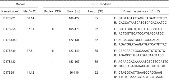
Table 1. Microsatellite markers and PCR conditions used for the analysis of HNPP patients with chromosome 17p11.2-p12 deletion.
ꠚꠚꠚꠚꠚꠚꠚꠚꠚꠚꠚꠚꠚꠚꠚꠚꠚꠚꠚꠚꠚꠚꠚꠚꠚꠚꠚꠚꠚꠚꠚꠚꠚꠚꠚꠚꠚꠚꠚꠚꠚꠚꠚꠚꠚꠚꠚꠚꠚꠚꠚꠚꠚꠚꠚꠚꠚꠚꠚꠚꠚꠚꠚꠚꠚꠚꠚꠚꠚꠚꠚꠚꠚꠚꠚꠚꠚꠚꠚꠚꠚꠚꠚꠚꠚꠚꠚꠚꠚꠚꠚꠚꠚꠚꠚꠚꠚꠚꠚꠚꠚꠚꠚꠚꠚꠚꠚꠚꠚꠚꠚ
Marker PCR condition
ꠏꠏꠏꠏꠏꠏꠏꠏꠏꠏꠏꠏꠏꠏꠏꠏꠏꠏꠏꠏꠏꠏꠏꠏꠏ ꠏꠏꠏꠏꠏꠏꠏꠏꠏꠏꠏꠏꠏꠏꠏꠏꠏꠏꠏꠏꠏꠏꠏꠏꠏꠏꠏꠏꠏꠏꠏꠏꠏꠏꠏꠏꠏꠏꠏꠏꠏꠏꠏꠏꠏꠏꠏꠏꠏꠏꠏꠏꠏꠏꠏꠏꠏꠏꠏꠏꠏꠏꠏꠏꠏꠏꠏꠏꠏꠏꠏꠏꠏꠏꠏꠏꠏꠏꠏꠏꠏꠏꠏ Name(Locus) Mapa(cM) Duplex PCR Size (bp) Temp. (oC) Primer sequences (5'→3') ꠏꠏꠏꠏꠏꠏꠏꠏꠏꠏꠏꠏꠏꠏꠏꠏꠏꠏꠏꠏꠏꠏꠏꠏꠏꠏꠏꠏꠏꠏꠏꠏꠏꠏꠏꠏꠏꠏꠏꠏꠏꠏꠏꠏꠏꠏꠏꠏꠏꠏꠏꠏꠏꠏꠏꠏꠏꠏꠏꠏꠏꠏꠏꠏꠏꠏꠏꠏꠏꠏꠏꠏꠏꠏꠏꠏꠏꠏꠏꠏꠏꠏꠏꠏꠏꠏꠏꠏꠏꠏꠏꠏꠏꠏꠏꠏꠏꠏꠏꠏꠏꠏꠏꠏꠏꠏꠏꠏꠏꠏꠏ D17S921 36.14 I 109-127 65 F: GTGTTGTATTAGGCAGAGTTCTCC R: CACCATAATCATGTCAGACAATCC D17S955 37.31 II 165-173 62 F: GGTTGGGTGTCCTTGGCCTAC R: ACTGGTGCATCCATGAGCATGC D17S1358 122-134 62 F: AGCACCATGCCGGGCCACAC R: AGATGGATAAGATGATCATGTTAC D17S839 37.8 II 123-143 65 F: CAACAACAGCGAAACTCTGTCTC R: AGACCCTGGAAGATCAACTACC D17S122 I 153-167 65 F: AGAACCACAAAAATGTCTTGCATTC R: GGCCAGACAGACCAGGCTCTGC D17S261 41.12 96-110 62 F: CTAGGCACTGAAGCCAGGAAG R: TTCTGGAAACCTACTCCTGAGC ꠏꠏꠏꠏꠏꠏꠏꠏꠏꠏꠏꠏꠏꠏꠏꠏꠏꠏꠏꠏꠏꠏꠏꠏꠏꠏꠏꠏꠏꠏꠏꠏꠏꠏꠏꠏꠏꠏꠏꠏꠏꠏꠏꠏꠏꠏꠏꠏꠏꠏꠏꠏꠏꠏꠏꠏꠏꠏꠏꠏꠏꠏꠏꠏꠏꠏꠏꠏꠏꠏꠏꠏꠏꠏꠏꠏꠏꠏꠏꠏꠏꠏꠏꠏꠏꠏꠏꠏꠏꠏꠏꠏꠏꠏꠏꠏꠏꠏꠏꠏꠏꠏꠏꠏꠏꠏꠏꠏꠏꠏꠏ a, Genetic distance (centi-Morgan) from the end of short arm.
The six (CA)n repeat microsatellite markers (D17S122, D17S921, D17S955, D17S839, D17S261 and D17S 1358) localized in the duplication/deletion region of chromosome 17p11.2-p12 were amplified by PCR. The primer sequences for PCR amplification were as described by Mersiyanova et al. (2000). The genetic distance, primer sequences, annealing temperatures and PCR sizes are summarized in Table 1. PCR was carried out in 20 ml of reaction mixture containing 10- 20 ng DNA, 10 pmol of each primer, 0.2 mM of each nucleotide, 2 mM MgCl2, 0.6 unit Taq DNA poly-merase and 1× buffer (Promega) using a thermal cycler (Perkin Elmer 2700). Two set of duplex PCRs (duplex 1: D17S122 and D17S921; duplex 2: D17S955 and D17S839) were performed. Other two markers (D17S261 and D17S1358) were amplified by the single PCR method.
Polyacrylam ide gel electrophoresis and silver staining
Electrophoresis was performed to genotype
microsatel-lites in a 5% denaturing polyacrylamide (acrylamide: bisacryl amide = 19:1) gel containing 7 M urea in 1× TBE buffer (T: 0.4 mm×L: 40 cm). PCR products were mixed with an equal volume of 2× STR loading buffer. Immediately after heating these mixtures at 95oC for 2 min, they were chilled by submersion in ice. Electrophoresis was carried out at a constant 1,600 V for 2-4 h.
DNA bands were visualized by the silver staining method described by Bassam et al. (1991) using a DNA silver staining kit (Promega). Gels were dipped into 10% ethanol for 20 min, followed by a 1% HNO3 solution for 10 min. After treatment with staining solu-tion (1 g AgNO3 and 1.5 ml of 37% formaldehyde/l) for 30 min, the gels were rinsed with deionized water briefly (less than 20 s), and then treated with a developer solution (30 g Na2CO3, 0.9 ml of 37% form-aldehyde and 0.5 ml of 1% Na2S2O3/l). The reaction was stopped using a stop solution (10% acetic acid) when the DNA bands appeared. The gels were gently agitated during each step.
Figure 1. Pedigree of FC-81 with HNPP deletion. The open symbols stand for unaffected males (□) and unaffected females (○). The filled
symbols represent affected males (■) and affected females (●). The diamonds (◆) indicate persons whose DNA was used for the analysis with 6 microsatellites. Alleles of the individuals within parentheses were inferred. The arrow indicates the proband. The deletion of the six-marker region responsible for HNPP deletion is indicated by the shadowed box.
Electrophysiological and pathological analysis
Nerve conduction studies were performed with surface stimulation and recording electrodes. Distal motor la-tency (DML), motor nerve conduction velocity (MNCV) and compound muscle action potential (CMAP) were recorded from the median, ulnar, peroneal and pos-terior tibial nerves in all patients. Sensory nerve action potential (SNAP) and sensory nerve conduction velo-city (SNCV) were recorded for all patients over a finger- wrist segment from the median and the ulnar nerves. These were also recorded for the sural nerve. Pathological examination of affected individuals in-cluded light and electron microscopic analysis and the teasing of a sural nerve biopsy from the left mal-leolus. Each specimen was fixed in buffered 2.5% glutaraldehyde, post-fixed in osmium tetroxide, and em-bedded in epoxy resin. A portion of the biopsy was cut into semithin sections (0.5 mm), stained with tolui-dine blue for light microscopy and cut into ultra thin sections for electron microscopy. About one hundred single myelinated fibers were teased from the remain-der of the nerve biopsy.
Statistical analysis
Percentages and means were compared using the Chi-square test and Student's t test, respectively. Dif-ferences were considered significant when P was < 0.05. Correlation studies were performed using single regression analysis, and correlations were considered significant when the correlation coefficient r was ≥ 0.4, and P was < 0.05. Analysis was performed using SPSS for Windows, version 11.0 (SPSS Inc., Chicago, Illinois).
R esults
Detection of deletion by genotyping of six m icrosatellites
In HNPP deletion patients, genotyping of the six (CA)n repeat microsatellites located on the 17p11.2-p12 re-gion was carried out by multiplex or single PCRs and denatured polyacrylamide gel electrophoresis. We con-sidered individuals to have the HNPP-deletion when they were hemizygous for all markers. The allele densities were also considered to determine the hemizygisity. In this study, D17S122 and D17S1358 were the most informative markers with heterozygosities of more than 0.7. The deletion was observed from 19 in 24 HNPP families, thus, the deletion frequency was cal-culated to 0.79.
Pedigree analysis of HNPP deletion
An example of a HNPP family (Family ID: FC-81) is shown in Figure 1. In the proband (III-3: indicated by
the arrow), only a single allele was detected for all the markers as the hemizygous state. The hemizygous haplotype of six markers in the proband was 6 (D17S921)-2 (D17S955)-2 (D17S1358)-2 (D17S839)- 11 (D17S122)-4 (D17S261). This haplotype was trans-mitted from his father, whereas no allele seemed to have originated from his mother. Hemizygosity was well matched with affected individuals (I-2, II-1, II-3 and III-3) by pedigree analysis of 10 family members. Unaffected members showed at least two markers having two different alleles. Members of the first generation (I-1 and I-2) were not analyzed, however, the deletion was
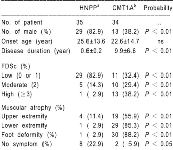
Table 2. Comparison of clinical aspects between 35 HNPP patients
with chromosome 17p11.2-p12 deletion and 34 CMTIA patients with the duplication. Values are mean±SD, and percentages are in parentheses. HNPPa, HNPP patients with chromosome 17p11.2-p12 deletion; CMT1Ab, CMT1A patients with chromosome 17p11.2-p12 duplication; FDSc, functional disability scale; ns, not significant. ꠚꠚꠚꠚꠚꠚꠚꠚꠚꠚꠚꠚꠚꠚꠚꠚꠚꠚꠚꠚꠚꠚꠚꠚꠚꠚꠚꠚꠚꠚꠚꠚꠚꠚꠚꠚꠚꠚꠚꠚꠚꠚꠚꠚꠚꠚꠚꠚꠚꠚꠚꠚꠚ
HNPPa CMT1Ab Probability ꠏꠏꠏꠏꠏꠏꠏꠏꠏꠏꠏꠏꠏꠏꠏꠏꠏꠏꠏꠏꠏꠏꠏꠏꠏꠏꠏꠏꠏꠏꠏꠏꠏꠏꠏꠏꠏꠏꠏꠏꠏꠏꠏ No. of patient 35 34 ... No. of male (%) 29 (82.9) 13 (38.2) P < 0.01 Onset age (year) 25.6±13.6 22.6±14.7 ns Disease duration (year) 0.6±0.2 9.9±6.6 P < 0.01
FDSc (%) Low (0 or 1) 29 (82.9) 11 (32.4) P < 0.01 Moderate (2) 5 (14.3) 10 (29.4) P < 0.01 High (≥3) 1 ( 2.9) 13 (38.2) P < 0.01 Muscular atrophy (%) Upper extremity 4 (11.4) 19 (55.9) P < 0.01 Lower extremity 1 ( 2.9) 29 (85.3) P < 0.01 Foot deformity (%) 1 ( 2.9) 30 (88.2) P < 0.01 No symptom (%) 8 (22.9) 2 ( 5.9) P < 0.05 ꠏꠏꠏꠏꠏꠏꠏꠏꠏꠏꠏꠏꠏꠏꠏꠏꠏꠏꠏꠏꠏꠏꠏꠏꠏꠏꠏꠏꠏꠏꠏꠏꠏꠏꠏꠏꠏꠏꠏꠏꠏꠏꠏꠏꠏꠏꠏꠏꠏꠏꠏꠏꠏ
Figure 2. Frequency distribution of age at onset in 35 HNPP patients
with chromosome 17p11.2-p12 deletion.
Number of pati ent Age at onset 10 0 20 30 40 50 60 70
regarded to have originated from the grandmother (I-2).
Clinical findings
Thirty-five HNPP patients with the deletion (29 males and 6 females) were studied. Mean age at onset was 25.6±13.6 years. The frequency distribution of onset age is shown in Figure 2. The disease was trans-mitted from an affected father in 13 families (68.4%), and from an affected mother in 4 families (21.1%). Details of parental transmission were unknown in the remaining 2 cases (10.5%).
The clinical differences between HNPP patients with the deletion, and CMT1A patients with the dupli-cation are compared in Table 2. The affected states were considerably different in HNPP and CMT1A, even though both presented a demyelinating form. The average onset ages were not significantly different. However, the onset frequency of CMT1A at a pre- teen age was more than 20%, while that of HNPP was only 2.9% (Figure 2). Disease duration was signi-ficantly longer in CMT1A (9.9±6.6 years) than in HNPP (2.0±2.7 years). The clinical symptoms, accor-ding to FDS, were more severe in CMT1A patients than in HNPP patients. The fraction of patients show-ing moderate or high level FDS (≥2) were 0.62 in CMT1A and 0.17 in HNPP patients, respectively. An FDS score of 3 was the most frequent in CMT1A patients, and a score of 1 in HNPP patients. The per-centages of patients with both muscular atrophy and foot deformity were significantly higher in CMT1A than in HNPP. Regression analysis derived scattering dia-grams also showed different patterns between HNPP and CMT1A patients with respect to onset age and FDS. FDS was directly related to onset age in CMT1A (r = 0.81, P < 0.01), but not in HNPP (r = 0.07, P = 0.71) (Figure 3). Asymptomatic patients who
were diagnosed only by genetic analysis were more frequently found to have a deletion in HNPP than a duplication in CMT1A (P < 0.05).
In addition, we compared the clinical and electro-physiological characteristics of HNPP patients who had experienced between single and recurrent attacks (Table 3), and found that patients who had
experi-Figure 3. Scatter diagrams used for regression analysis between onset age and FDS scores in (A) 34 CMT1A patients with the duplication,
and in (B) 35 HNPP patients with the deletion.
F unct ion al disabili ty scales Onset age 10 0 20 30 40 50 60 70 80 r P = 0.81 < 0.01 F unct ion al disabili ty scales Onset age 10 0 20 30 40 50 60 70 r P = 0.07 = 0.71
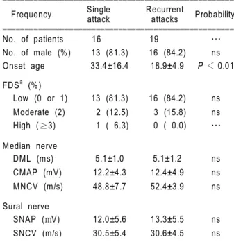
Table 3. Comparison of the clinical and electrophysiological aspects
of HNPP patients who had experienced a single attack and those who had experienced recurrent attacks. Values are mean±SD, and percentages are in parentheses. FDSa, functional disability scale; DML, distal motor latency; CMAP, compound muscle action potential; MNCV, motor nerve conduction velocity; SNAP, sensory nerve action potential; SNCV, sensory nerve conduction velocity; ns, not significant. ꠚꠚꠚꠚꠚꠚꠚꠚꠚꠚꠚꠚꠚꠚꠚꠚꠚꠚꠚꠚꠚꠚꠚꠚꠚꠚꠚꠚꠚꠚꠚꠚꠚꠚꠚꠚꠚꠚꠚꠚꠚꠚꠚꠚꠚꠚꠚꠚꠚꠚꠚꠚ
Single Recurrent
Frequency attack attacks Probability ꠏꠏꠏꠏꠏꠏꠏꠏꠏꠏꠏꠏꠏꠏꠏꠏꠏꠏꠏꠏꠏꠏꠏꠏꠏꠏꠏꠏꠏꠏꠏꠏꠏꠏꠏꠏꠏꠏꠏꠏꠏꠏꠏꠏꠏꠏꠏꠏꠏꠏꠏꠏ No. of patients 16 19 … No. of male (%) 13 (81.3) 16 (84.2) ns Onset age 33.4±16.4 18.9±4.9 P < 0.01 FDSa (%) Low (0 or 1) 13 (81.3) 16 (84.2) ns Moderate (2) 2 (12.5) 3 (15.8) ns High (≥3) 1 ( 6.3) 0 ( 0.0) … Median nerve DML (ms) 5.1±1.0 5.1±1.2 ns CMAP (mV) 12.2±4.3 12.4±4.9 ns MNCV (m/s) 48.8±7.7 52.4±3.9 ns Sural nerve SNAP (mV) 12.0±5.6 13.3±5.5 ns SNCV (m/s) 30.5±5.4 30.6±4.5 ns ꠏꠏꠏꠏꠏꠏꠏꠏꠏꠏꠏꠏꠏꠏꠏꠏꠏꠏꠏꠏꠏꠏꠏꠏꠏꠏꠏꠏꠏꠏꠏꠏꠏꠏꠏꠏꠏꠏꠏꠏꠏꠏꠏꠏꠏꠏꠏꠏꠏꠏꠏꠏ
enced recurrent attacks showed a significantly earlier onset age (P < 0.01). However, there were neither electrophysiological nor functional differences between the two sets of patients.
Electrophysiological and pathological findings
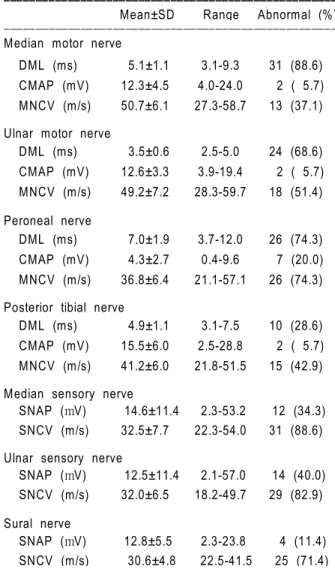
All thirty-five patients showed a marked slowing of motor and sensory nerve conduction (Table 4). The MNCVs in this study were frequently reduced, for example, in 74.3% of cases in terms of peroneal nerve conduction. Also, the distal motor latency was prolonged in the median nerve (88.6%). Neither a con-duction block nor a temporal dispersion of action potentials was exhibited in this study.
SNAP and SNCV were abnormal in many patients tested. Finger-wrist segments of the median and the ulnar nerves recorded abnormal results in more than 80% of HNPP deletion patients.
When we compared symptomatic and asymptomatic sites in patients showing a deletion in the 17p11.2-p12 region, nerve conduction studies demonstrated diffuse mild to moderate slowing of NCV that was noticeable worse over the common entrapment sites, regardless of the clinical manifestations.
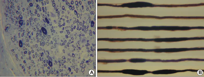
Sural nerve biopsy was performed in four patients with the deletion. Histopathologic examinations showed relatively preserved density of myelinated fibers. No onion bulb formation was found. Teased fiber analysis confirmed the presence of focally folded myelin (toma-cula) about 56.4%. The histopathologic findings were consistent with tomacula neuropathy in all biopsied patients (Figure 4).
D iscussion
HNPP with deletion is the reciprocal product of an unequal crossing-over event within the chromosome 17p11.2-p12 region. By mutational screening of HNPP families, the deletion was found in 35 patients from 19 families (79%) among 24 HNPP diagnosed families. This frequency of HNPP with the deletion in Korean patients is consistent with the range of 72 to 86% reported in other populations (Mariman et al., 1994; Nelis et al., 1996; Timmerman et al., 1997). HNPP is known as a genetically homogeneous disease com-pared with CMT, though frame-shift mutations in the PMP22 gene lead to HNPP in rare cases (Young et al., 1997). Thus, causative mutations in PMP22 are expected in the 21% of HNPP patients with no dele-tion. In addition, it is possible that unknown genes are related to HNPP patients in Koreans. The detection of HNPP deletion in this study would be helpful for the further analysis of the genetic and pathophysio-logic causes of peripheral neuropathies.
We investigated the clinical differences between 35 HNPP patients with chromosome 17p11.2-p12 dele-tion and 34 CMT1A patients with these duplicadele-tion. The clinical assessments of HNPP patients were gen-erally less severe than those of CMT1A patients. HNPP usually develops as a painless neuropathy after minor trauma or compression (Gouider et al., 1994; Parey-son et al., 1996). The mean onset ages between HNPP and CMT1A were not significantly different. However, onset in the pre-teens was found to be seven times more frequent in CMT1A than in HNPP. Several clinical symptoms, such as disease duration, muscular atrophy, and foot deformity indicated that CMT1A patients were more severely affected than HNPP patients. FDS values also implied the greater
Table 4. Electrophysiological testing abnormalities of HNPP patients
with chromosome 17p11.2-p12 deletion. Values are mean±SD, and percentages are in parentheses. DML, distal motor latency; CMAP, compound muscle action potential; MNCV, motor nerve conduction velocity; SNAP, sensory nerve action potential; SNCV, sensory nerve conduction velocity.
ꠚꠚꠚꠚꠚꠚꠚꠚꠚꠚꠚꠚꠚꠚꠚꠚꠚꠚꠚꠚꠚꠚꠚꠚꠚꠚꠚꠚꠚꠚꠚꠚꠚꠚꠚꠚꠚꠚꠚꠚꠚꠚꠚꠚꠚꠚꠚꠚꠚꠚꠚꠚ Mean±SD Range Abnormal (%) ꠏꠏꠏꠏꠏꠏꠏꠏꠏꠏꠏꠏꠏꠏꠏꠏꠏꠏꠏꠏꠏꠏꠏꠏꠏꠏꠏꠏꠏꠏꠏꠏꠏꠏꠏꠏꠏꠏꠏꠏꠏꠏꠏꠏꠏꠏꠏꠏꠏꠏꠏꠏ Median motor nerve
DML (ms) 5.1±1.1 3.1-9.3 31 (88.6) CMAP (mV) 12.3±4.5 4.0-24.0 2 ( 5.7) MNCV (m/s) 50.7±6.1 27.3-58.7 13 (37.1) Ulnar motor nerve
DML (ms) 3.5±0.6 2.5-5.0 24 (68.6) CMAP (mV) 12.6±3.3 3.9-19.4 2 ( 5.7) MNCV (m/s) 49.2±7.2 28.3-59.7 18 (51.4) Peroneal nerve DML (ms) 7.0±1.9 3.7-12.0 26 (74.3) CMAP (mV) 4.3±2.7 0.4-9.6 7 (20.0) MNCV (m/s) 36.8±6.4 21.1-57.1 26 (74.3) Posterior tibial nerve
DML (ms) 4.9±1.1 3.1-7.5 10 (28.6) CMAP (mV) 15.5±6.0 2.5-28.8 2 ( 5.7) MNCV (m/s) 41.2±6.0 21.8-51.5 15 (42.9) Median sensory nerve
SNAP (mV) 14.6±11.4 2.3-53.2 12 (34.3) SNCV (m/s) 32.5±7.7 22.3-54.0 31 (88.6) Ulnar sensory nerve
SNAP (mV) 12.5±11.4 2.1-57.0 14 (40.0) SNCV (m/s) 32.0±6.5 18.2-49.7 29 (82.9) Sural nerve SNAP (mV) 12.8±5.5 2.3-23.8 4 (11.4) SNCV (m/s) 30.6±4.8 22.5-41.5 25 (71.4) ꠏꠏꠏꠏꠏꠏꠏꠏꠏꠏꠏꠏꠏꠏꠏꠏꠏꠏꠏꠏꠏꠏꠏꠏꠏꠏꠏꠏꠏꠏꠏꠏꠏꠏꠏꠏꠏꠏꠏꠏꠏꠏꠏꠏꠏꠏꠏꠏꠏꠏꠏꠏ
severity of CMT1A. The frequency of patients with a moderate or high level of FDS was 0.17 and 0.62 in each HNPP and CMT1A. We also found that the FDS was related to the onset age in CMT1A, but not in HNPP (Figure 3). Thus, it seems that the FDS is a more useful scale for CMT1A.
Clinical symptoms of HNPP appeared at less than thirty years of age in more than 88% of cases. In addition, the percentage of male patients in HNPP was found to be more than 80%. In Korea, most men conscripted to the military for 3 years in their third decade. Military service might explain why most HNPP patients in Korea develop pressure palsies during this period. Perhaps nerve compression due to military training or greater physical activity is directly related to disease onset.
Eight (22.9%) of HNPP patients with the deletion were asymptomatic and diagnosed only by genetic analysis, which agrees with the results of previous studies (Mariman et al., 1994; Pareyson et al., 1996). So, it would be necessary to perform nerve conduc-tion studies and genetic analyses in asymptomatic family members.
In most patients, prolonged distal motor latency and a slow MNCV in the carpal tunnel area of the median nerve showed that those sites are liable to suffer myelin damage by nerve compression. In other words, HNPP patients, with congenital abnormalities for compact myelin, easily damage the peripheral ner-vous system by external compression. Those pheno-mena differ from both CMT1A and acquired demyelina-ting neuropathy (Airaksinen et al., 1985; Maritinelli et al., 1989; Uncini et al., 1995).
It is well known that both conduction block and temporal dispersion are the electrophysiological
charac-teristics of demyelinating neuropathy (Behse et al., 1972; Chance et al., 1994; Uncini et al., 1995). However, all HNPP patients in this study showed a long duration of compound muscle action potentials without a conduction block or a temporal dispersion. These findings differentiate HNPP from both CMT1A and acquired demyelinating neuropathy. In addition, nerve conduction studies demonstrated diffuse slow-ing of NCV, which was notably worse over common entrapment sites, regardless of the clinical manifesta-tions. Moreover, the electrophysiological findings were frequently more severe in the asymptomatic side than in the symptomatic state. Therefore, in HNPP patients, it was relatively common that clinical symptoms were not in accord with electrophysiological abnormalities. These findings show that the clinical manifestations of HNPP are more related to nerve compression than congenital demyelination.
When we compared HNPP patients, who had ex-perienced a single attack or recurrent attacks, we found that the latter had a lower age of onset. Inter-estingly, these findings indicate the possibility that an indivisual with early symptom onset has a higher likeli-hood of recurrence. These findings have not been re-ported previously. The identification of prognostic indi-cators and a correct diagnosis might help to prevent recurrent attacks. Though these hypotheses require fur-ther prospective studies, these results indicate that who experience the clinical symptoms of HNPP at an early age must take precautions to nerve compression. Even though tomacula is not specific, it is the most pathognomonic finding of HNPP in a nerve biopsy (Bradley et al., 1975; Jacobs and Gregory, 1991). In this study, we found that tomacula is present in about 56.4% of cases. The reason for the presence of
to-Figure 4. Transverse semithin section of sural nerve. Thickening of myelin sheath (tomacula) are occasionally seen (A, Toluidine blue, ×400).
Consecutive lengths along one teased myelinated fiber from the sural nerve of a patient with HNPP demonstrates multiple characteristic tomacula (B, Osmium tetroxide, ×400).
macula is not known, however, the observation of tomacula is useful in the diagnosis of HNPP. We report upon the clinical, electrophysiological and morphological aspects of the Korean HNPP patients with deletion. In addition, our findings suggest the possibility that HNPP patients with an earlier sym-ptom onset face an increased likelihood of recurrent attacks.
Acknow ledgm ent
This study was supported by the research fund (#R05- 2003-000-11496-0) from KOSEF, and Kongju National University (2003).
R eferences
Airaksinen EM, Livanainen M, Karil P, Sainio K, Haltia M. Hereditary recurrent brachial plexus neuropathy with dysmor-phic features. Acta Neurol Scand 1985;71:309-16
Bassam SJ, Caetano-Anolles G, Gresshoff PM. Fast and sensitive silver staining of DNA in polyacrylamide gels. Anal Biochem 1991;196:80-3
Behse F, Buchthal F, Carlsen F, Knappeis GG. Hereditary neu-ropathy with liability to pressure palsies. Brain 1972;95:777-94 Bradley WG, Madrid R, Thrush DC, Campbell MJ. Recurrent brachial plexus neuropathy. Brain 1975;98:381-98
Chance PF, Alderson MK, Leppig KA, Lensch MW, Matsuna-mi N, SMatsuna-mith B, Swanson PD, Odelberg SJ, Distsche CM, Bird TD. DNA deletion associated with hereditary neuropathy with liability to pressure palsies. Cell 1993;72:143-51 Chance PF, Lensch MW, Lipe H, Brown RH Sr, Brown RH Jr, Phil D, Bird TD. Hereditary neuralgic amyotrophy and hereditary neuropathy with liability to pressure palsies: two distinct genetic disorders. Neurology 1994;44:2253-7 Georgiou DM, Zidar J, Korosec M, Middleton LT, Kyriakides T, Christodoulou K. A novel NF-L mutation Pro22Ser is asso-ciated with CMT2 in a large Slovenian family. Neurogenetics 2002;4:93-6
Gouider R, LeGuern E, Emile J, Tardieu S, Cabon F, Samid M, Weissenbach J, Agid Y, Bouche P, Brice A. Hereditary neuralgic amyotrophy and hereditary neuropathy with liability to pressure palsies: two distinct clinical, electrophysiologic, and genetic entities. Neurology 1994;44:2250-2
Hattori N, Yamamoto M, Yoshihara T, Koike H, Nakagawa M, Yoshikawa H, Ohnishi A, Hayasaka K, Onodera O, Baba M, Yasuda H, Saito T, Nakashima K, Kira J, Kaji R, Oka N, Sobue G; Study Group for Hereditary Neuropathy in Ja-pan. Demyelinating and axonal features of Charcot-Marie- Tooth disease with mutations of myelin-related proteins (PMP22, MPZ and Cx32): a clinicopathological study of 205 Japanese patients. Brain 2003;126:134-51
Jacobs JM, Gregory R. Uncompacted lamellae as a feature of tomaculous neuropathy. Acta Neuropathol 1991;83:87-91 Le Guern E, Sturtz F, Gugenheim M, Gouider R, Bonne-bouche C, Ravise N, Gonnaud PM, Tardieu S, Bouche P,
Chazot G. Detection of deletion within 17p11.2 in 7 French families with hereditary neuropathy with liability to pressure palsies (HNPP). Cytogenet Cell Genet 1994;65:261-4 Lobsiger CS, Taylor V, Suter U. The early life of a Schwann cell. Biol Chem 2002;383:245-53
Mariman EC, Gabreels-Festen AA, van Beersum SE, Valen-tijn LJ, Baas F, Bolhuis PA, Jongen PJ, Ropers HH, Ga-breels FJ. Prevalence of the 1.5-Mb 17p deletion in families with hereditary neuropathy with liability to pressure palsies. Ann Neurol 1994;36:650-5
Maritinelli P, Fabbri R, Moretto G, Gabellini AS, D'Alessandro R, Rizzuto N. Recurrent familial brachial plexus palsies as the only clinical expression of 'tomaculous' neuropathy. Eur Neurol 1989;29:61-6
Martini R. The effect of myelinating Schwann cells on axons. Muscle Nerve 2001;24:456-66
Mersiyanova IV, Ismailov SM, Polyakov AV, Dadali EL, Fedotov VP, Nelis E, Lofgren A, Timmerman V, van Broeck-hoven C, Evgrafov OV. Screening for in the peripheral myelin genes PMP22, MPZ and Cx32 (GJB1) in Russian Charcot- Marie-Tooth Neuropathy patients. Hum Mutat 2000;15:340-7 Nelis E, Van Broeckhoven C, De Jonghe P, Lofgren A, Van-denberghe A, Latour P, Le Guern E, Brice A, Mostacciuolo ML, Schiavon F, Palau F, Bort S, Upadhyaya M, Rocchi M, Archidiacono N, Mandich P, Bellone E, Silander K, Savon-taus ML, Navon R, Goldberg-Stern H, Estivill X, Volpini V, Friedl W, Gal A. Estimation of the mutation frequencies in Charcot-Marie-Tooth disease type 1 and hereditary neuropa-thy with liability to pressure palsies: a European collaborative study. Eur J Hum Genet 1996;4:25-33
Pareyson D, Scaioli V, Taroni F, Botti S, Lorenzetti D, Solari A, Ciano C, Sghirlanzoni A. Phenotypic heterogeneity in heredi-tary neuropathy with liability to pressure palsies associated with chromosome 17p11.2-12 deletion. Neurology 1996; 46:1133-7 Timmerman V, Rautenstrauss B, Reiter LT, Koeuth T, Lof-gren A, Liehr T, Nelis E, Bathke KD, De Jonghe P, Grehl H, Martin JJ, Lupski JR, Van Broeckhoven C. Detection of the CMT1A/HNPP recombination hotspot in unrelated pa-tients of European descent. J Med Genet 1997;34:43-9 Uncini A, Di Guglielmo G, Di Muzio A, Gambi D, Sabatelli M, Mignogna T, Tonali P, Marzella R, Finelli P, Archidiacono N. Differential electrophysiological features of neuropathies associated with 17p11.2 deletion and duplication. Muscle Nerve 1995;18:628-35
Verhagen WI, Gabreels-Festen AA, van Wensen PJ, Joosten EM, Vingerhoets HM, Gabreels FJ, de Graaf R. Hereditary neuropathy with liability to pressure palsies: a clinical, elec-troneurophysiological and morphological study. J Neurol Sci 1993;116:176-84
Yoshihara T, Yamamoto M, Hattori N, Misu K, Mori K, Koike H, Sobue G. Identification of novel sequence variants in the neurofilament-light gene in a Japanese population: analysis of Charcot-Marie-Tooth disease patients and normal indivi-duals. J Peripher Nerv Syst 2002;7:221-4
Young P, Wiebusch H, Stogbauer F, Ringelstein B, Assmann G, Funke H. A novel frameshift mutation in PMP22 accounts for hereditary neuropathy with liability to pressure palsies. Neurology 1997;48:450-2