저작자표시-비영리-변경금지 2.0 대한민국 이용자는 아래의 조건을 따르는 경우에 한하여 자유롭게 l 이 저작물을 복제, 배포, 전송, 전시, 공연 및 방송할 수 있습니다. 다음과 같은 조건을 따라야 합니다: l 귀하는, 이 저작물의 재이용이나 배포의 경우, 이 저작물에 적용된 이용허락조건 을 명확하게 나타내어야 합니다. l 저작권자로부터 별도의 허가를 받으면 이러한 조건들은 적용되지 않습니다. 저작권법에 따른 이용자의 권리는 위의 내용에 의하여 영향을 받지 않습니다. 이것은 이용허락규약(Legal Code)을 이해하기 쉽게 요약한 것입니다. Disclaimer 저작자표시. 귀하는 원저작자를 표시하여야 합니다. 비영리. 귀하는 이 저작물을 영리 목적으로 이용할 수 없습니다. 변경금지. 귀하는 이 저작물을 개작, 변형 또는 가공할 수 없습니다.
A Doctoral Dissertation
A study on the neuroprotective mechanism of
nobiletin through regulation of mitochondrial
membrane potential in primary cortical neurons and
isolated brain mitochondria
Ji Hyung Lee
Department of Medicine
Graduated Schcool
Jeju National University
CONTENTS
LIST OF ABBREVIATIONS………...………...……….1 LIST OF FIGURES………..………3 LIST OF TABLES……….…...5 ABSTRACT……….….6 INTRODUCTION….………..……….8MATERIALS AND METHODS………..………….10
RESULTS…...………...18
DISCUSSION……….…53
REFERENCES………..….……57
ABSTRACT IN KOREAN……….……...61
1
LIST OF ABBREVIATIONS
5-HD 5-Hydroxydecanoate
AM Acetoxymethyl ester
ATP Adenosine triphosphate
mitoKATP Mitochondrial ATP-sensitive K+ channels
Ara-C Arabinofuranosyl cytidine
Ca2+ Calcium ion
CCCP Carbonyl cyanide m-chlorophenylhydrazone
FCCP Carbonyl cyanide p-(trifluoromethoxy) phenylhydrazone
CPE Citrus polymethoxylated flavone
CCD Cooled-charged device
DIV Days in vitro
DPPH 1,1-diphenyl-2-picrylhydrazyl
EM Electron microscopy
ETC Electron transport chain
FBS Fetal bovine serum
Fura-2 AM Fura-2 acetoxymethyl ester
HT-22 Immortalized clonal mouse hippocampal cells
IPC Ischemic pre-conditioning
MEM Minimal essential medium
2
MCU Mitochondrial calcium uniporter
mitoBKCa Mitochondrial large-conductance Ca2+-activated K+ channels
MTT 3-(4,5-dimethylthiazol-2-yl)-2,5-diphenyl tetrazolium bromide
NAC N-acetyl cysteine
OCR Dxygen consumption rate
PC12 Rat pheochromocytoma cells
ROS Reactive oxygen species
Rhod-2 AM Rhod-2 acetoxymethyl ester
SD Sprague-Dawley
3
LIST OF FIGURES
Figure 1. The effects of Citrus sunki Hort. ex Tanaka (CPE) on basal ΔΨm and cell viability
against glutamate toxicity in primary cortical neurons ……….…..20
Figure 2. The effects of nobiletin on basal ΔΨm and cell viability against glutamate toxicity in
primary cortical neurons………...…22
Figure 3. Dose determination of nobiletin treatment.…………...……...………...……24
Figure 4. The effects of nobiletin on glutamate-induced overload of cytosol calcium in
primary cortical neurons………...…26
Figure 5. The effects of nobiletin on glutamate-induced overload of mitochondrial calcium
overload in primary cortical neurons……….………....27
Figure 6. Identification of physiological activity of isolated brain mitochondria...30
Figure 7. The effects of nobiletin on mitochondrial ROS generation in glutamate-stimulated
4
Figure 8. The effects of nobiletin on mitochondrial ROS generation in isolated brain
mitochondria exposed to high concentration of Ca2+………...…….………35
Figure 9. The comparison of ROS scavenging activities of Citrus compounds in cell-free system....……...………..……….……...36
Figure 10. The effects of K+ influx on nobiletin-induced partial mitochondrial depolarization in isolated brain mitochondria………..….….40
Figure 11. The effects of mitochondrial K+ channel inhibitors on nobiletin- induced ΔΨm
changes in primary cortical neurons……….……….…….42
Figure 12. The effects of rotenone, a complex Ⅰ inhibitor, on mitochondrial membrane potential………..46
Figure 13. Rotenone inhibits the action of nobiletin, which attenuate mitochondrial calcium
overload in the glutamate toxicity model……….….………..47
Figure 14. The effects of nobiletin on rotenone-induced mitochondrial ROS in primary cortical neurons………..………..…..………..….……50
Figure 15. Schematic neuroprotective mechanism of nobiletin through mitochondrial
5
LIST OF TABLES
Table 1. Detailed informations of equipment and measuring protocols used for real-time imaging-based fluorometry analysis in living cells……….………….17
Table 2. Detailed informations of equipment and measuring protocols for ∆Ψm and ROS in
6
ABSTRACT
Mitochondrial calcium overload is an important event at the cellular level in determining the fate of neuronal survival and death and is regulated by a variety of channels, antiporters and pumps, as well as an electron transport chains (ETC) in the mitochondrial inner membrane. There are two motivating forces for calcium influx into the mitochondria, one of which is the calcium ion (Ca2+) concentration gradient, and the other is mitochondrial membrane potential
(∆Ψm). ∆Ψm is regulated within the range of -120 to -180 mV under physiological conditions.
The negative charge of ∆Ψm has a significant effect on Ca2+ influx. Therefore, pharmacological
manipulation of ∆Ψm could be a promising strategy to prevent neuronal cell death against brain
insults by reducing calcium influx into the mitochondria.
Based on this hypothesis, it was investigated here whether nobiletin, a Citrus polymethoxylated flavone (CPE), prevents neuronal cell death by reducing mitochondrial calcium overload and oxidative stress via regulating ∆Ψm against neuronal insult in primary
cortical neurons and brain mitochondria isolated from rat cortices. Results demonstrated that nobiletin (100 μM) treatment significantly increased cell viability against glutamate toxicity (100 μM, 20 min) in primary cortical neurons. Real-time imaging based fluorometry data proved that nobiletin evoked partial mitochondrial depolarization in these neurons. Nobiletin-induced partial mitochondrial depolarization in intact neurons was also confirmed in the isolated mitochondrial model using a fluorescence microplate reader. Nobiletin significantly reduced mitochondrial calcium overload and reactive oxygen species (ROS) generation in glutamate (100 μM)-stimulated cortical neurons. In the isolated mitochondrial model, nobiletin decreased ROS production when treated with high concentrations of Ca2+ (5 μM) to mimic glutamate toxicity. In experiments to determine whether nobiletin has its own ROS scavenging
7
ability, CPE and hesperidin exhibited considerable ROS scavenging activity, but nobiletin did not exhibit such an effect. Furthermore, nobiletin effects on basal ΔΨm were completely
abolished in isolated brain mitochondria maintained in K+-free medium. To elucidate the molecular target of nobiletin, iberiotoxin 10 nM and 5-Hydroxydecanoate (5-HD) 500 μM were used to inhibit mitochondrial large-conductance Ca2+-activated K+ channels (mitoBKCa) and
mitochondrial ATP-sensitive K+ channels (mitoKATP), respectively. The results suggested that
K+ influx into the mitochondrial matrix by nobiletin is likely mediated by mitoBKCa and mitoKATP.
In addition, nobiletin markedly reduced rotenone-induced mitochondrial ROS generation, suggesting the possibility that complex I of ETC might be another mitochondrial target of nobiletin. However, more detailed studies should be conducted to determine the exact molecular targets of nobiletin in mitochondria. Taken together, results demonstrate that nobiletin-induced partial depolarization of ∆Ψm decreased mitochondrial Ca2+ overload and
ROS generation, which are the important parameters of neuronal cell death. The K+ influx into the mitochondria matrix is critically engaged in the partial mitochondrial depolarization-related neuroprotective effect of nobiletin. Nobiletin-induced mitochondrial K+ influx is likely mediated, at least in part, by the activation of mitochondrial K+ channels.
Keywords: nobiletin; calcium; mitochondrial calcium; mitochondrial K+ channels; mitochondrial membrane potential
8
1. Introduction
The main function of mitochondria is to produce adenosine triphosphate (ATP). However, since the 20th century, many studies have focused on the regulatory function of mitochondria in determining the fate of cellular survival and death. Electron transport chains of mitochondria generate an H+ electrochemical gradient as electrons pass through complex I, III, and IV,
activating ATP synthase. The complete process is called oxidative phosphorylation. In healthy cells, the charge imbalance due to the generation of an electrochemical gradient through oxidative phosphorylation forms the basis of ∆Ψm. Although a transient depolarization of the
∆Ψm, through the “flickering” of one or several pores of the mitochondrial inner membrane,
may occur under physiological conditions (Kroemer et al., 2007), a long-lasting dissipation of ∆Ψm is often associated with cell death. ∆Ψm dissipation is induced by proapoptotic stimuli
such as Bax, Bak, tBid, Ca2+, and cytosolic metabolites. In particular, mitochondrial calcium plays a key role in the regulatory mechanism of cell survival and death. Mitochondrial Ca2+ overload and oxidative stress can facilitate the opening of a high-conductance pore in the inner mitochondrial membrane. Pore opening is followed by osmotic swelling and rupture of the mitochondrial membrane that leads to the release of mitochondrial proteins including cytochrome c, which induces apoptosis, into the cytosol (Orrenius et al. 2015). Overload of mitochondrial calcium during brain insults results in oxidative stress, impaired mitochondrial function, neuronal cell death, and neuroinflammation that are implicated in the pathogenesis of many neurodegenerative diseases (Kroemer et al., 2007, Duchen et al., 2012).
Gunter (1990) described the driving forces of Ca2+ into mitochondria by an equation: ΔμCa = RT ln ([Ca2+]out/[Ca2+]in) + 2F(Ψout – Ψin). According to this equation, there are two different
9
mitochondrial calcium ([Ca2+]m), ⅱ) ∆Ψm gradient. Based on the correlation between [Ca2+]m
and ∆Ψm, it has been proposed that pharmacological manipulation of ΔΨm can be a key strategy
to prevent neurotoxic [Ca2+]m overload and neuronal cell death by reducing the driving forces
of Ca2+ into the mitochondrial gradient (Ishida et al., 2001). Regarding these ideas, it has been
reported that mild mitochondrial depolarization reduces mitochondrial calcium overload in different cell types (Sanz-Blasco et al., 2008, Valero et al., 2008).
In Wu’s study (2013), the authors demonstrated that ethanolic peel extract of CPE induces mild mitochondrial depolarization intrinsically. Based on this property, CPE significantly attenuated mitochondrial calcium overload and cell death against H2O2 toxicity in immortalized
clonal mouse hippocampal (HT-22) cells. Various flavonoid compounds in CPE (i.e., rutin, hesperidin, sinensetin, tangeretin, and nobiletin) belong to the polymethoxylated flavone (PMF) group (Wu et al., 2013). Nobiletin exerts several beneficial effects on improving cognitive function or motor deficits in several animal models such as cerebral ischemia (Yamamoto et al., 2009), Parkinson’s and Alzheimer’s diseases (Yabuki et al., 2014, Onozuka et al., 2008). In addition, nobiletin induced neurites outgrowth in rat pheochromocytoma (PC12) cells (Nagase et al., 2005) and significantly suppressed microglial activation and neuroinflammation (Cui et al., 2010). These results suggest that nobiletin is a promising candidate as a neuroprotective agent and warrants a thorough examination.
Based on this history, I investigated here whether nobiletin prevents glutamate toxicity induced by neurotoxic mitochondrial calcium overload and neuronal cell death through K+ influx and ∆Ψm across the mitochondrial inner membrane in primary cerebrocortical neurons
or isolated brain mitochondria. I also examined whether complex Ⅰ is directly involved in the regulation of ∆Ψm by nobiletin.
10
2. Materials and Methods
2.1. Materials
Tetramethylrhodamine ethyl ester (TMRE), rhod-2 acetoxymethyl ester (Rhod-2 AM), MitoSOX Red, Fura-2 acetoxymethyl ester (Fura-2 AM) were purchased from Invitrogen (Carlsbad, CA, USA). Minimal essential medium (MEM), Neurobasal medium, fetal bovine serum (FBS), B-27 serum-free supplement, glutamine and penicillin/streptomycin were purchased from Gibco BRL (Gland Island, NY, USA). Nobiletin was isolated and purified from the peel of Citrus sunki Hort. ex Tanaka in Jeju National University, Department of Biology as described previously (Choi et al., 2007). All other reagents were obtained from Sigma-Aldrich (St Louis, MO, USA) unless otherwise indicated.
2.2 Primary culture of cortical neurons
Primary cortical neurons were prepared from cerebral cortices of postnatal 1-day-old Sprague- Dawley (SD) rats. The neonatal brain were dissected and the cortices were transferred to plating medium (containing MEM supplemented with 10% FBS, 25 mM glucose, 1 mM sodium pyruvate, 25 mM glutamine and 1% penicillin/streptomycin) and dissociated by trituration using fire-polished glass Pasteur pipettes. Then, cells were plated onto poly-L-lysine-coated round glass coverslips placed in 12-well plates at a density of 1.3 × 10⁵/well. After 6 h, plating medium was replaced to Neurobasal media supplemented with 2% B-27, 50 mM glutamine and 1% penicillin/streptomycin, and a half of culture medium was replaced every four days. Cultured neurons were incubated at 37°C in a humidified 5% CO2/95% air atmosphere. The
11
cells were used after 7 days in vitro (DIV). The study was approved by Animal Care and Use Committee of Jeju National University and all experiments were performed in accordance with the guidelines.
2.3. Dual real-time imaging-based fluorometry of both cytosolic and mitochondrial calcium levels in the same cortical neurons
Fura-2 and Rhod-2 were used to measure cytosolic and mitochondrial calcium levels. Cell-permeable acetoxymethyl ester (AM) forms were used for these two probes. Fura-2 is a ratio-metric fluorescent indicator to measure [Ca2+]c using the ratio of emitted fluorescence intensity
at excitation wavelengths of 340 and 380 nm (Eun et al., 2001). Another calcium indicator Rhod-2, used for a selective [Ca2+]m probe, exhibits charge-driven uptake into the mitochondria
and evokes increase of fluorescence upon Ca2+ binding (Wu et al., 2013). Fura-2 and Rhod-2
were dissolved in DMSO at a concentration of 5 mM and 2 mM respectively and there are kept frozen in aliquot of 10 μL. The cortical neurons cultured on a coverslip were loaded with 10 μM Fura-2 AM and 0.1% Pluronic F127 for 45 min at 37°C. And then, cells were loaded with 2 μM Rhod-2 AM, transferred to 35 mm petri dish with media and incubated for 30 min at 4°C. After washing three times with normal Tyrode solution, a coverslip was transferred to the recording chamber. Cells were continuously superfused with normal Tyrode solution (in mM): NaCl 145, KCl 5, CaCl2 2, MgCl2 1.3, HEPES 10, glucose 10, pH adjusted to 7.4 with NaOH,
the osmolarity was adjusted to approximately 310 mOsm. Live-cell recording was performed with preheated normal Tyrode solution and stable perfusion at 1ml/min and the solution in the recording chamber was maintained at about 37℃. Neurons were illuminated at 340 and 380 nm with a monochromator through a T400lp dichroic mirror and an emission was monitored
12
through a 510/80 m filter for Fura-2. Rhod-2 fluorescence was measured using a 540/25 nm excitation filter, an AT565DC dichroic mirror and a 605/55 m emission filter (Jousset et al., 2008). High-speed filter switching device (Sutter Instrument, Lambda, DG-4) with a 175W xenon arc lamp (ILC technology, Sunnyvale, CA) as a light source was used for dual recording of Fura-2 and Rhod-2, the light source was switched at a speed of less than 2 ms to generate respective excitation wavelengths. The fluorescence images were acquired at 6 s intervals using an inverted microscope Olympus IX71 (Olympus) and a cooled-charged device (CCD) camera (Cascade, Roper Scientific). The data were analyzed using Metafluor software (Molecular Devices). The regions of selected from the initial image were converted to relative fluorescence intensity unit data. Also, the area for the background fluorescence was selected and subtracted from the measured value. All measurements were obtained after stabilization of the cell for 5 min and were displayed as a ratio of 380/340.
2.4. Real-time imaging-based fluorometry of ∆Ψm and mitochondrial ROS in intact
cortical neurons
The cationic fluorescent probe TMRE is sequestered by mitochondria in proportion to ΔΨm
(Scaduto and Grotyohann., 1999). The cationic MitoSOX Red is also selectively targeted to the mitochondria. Once MitoSOX Red is oxidized by superoxide anions which are the predominant ROS in mitochondria, it exhibits red fluorescence. The cortical neurons were loaded for 15 min at 37°C with 25 nM TMRE and 5 μM MitoSOX Red for ∆Ψm and mitochondrial superoxide,
respectively. After then, the neurons on the cover glass were washed three times with normal Tyrode solution. The cover glass was transferred to the recording chamber which was continuously superfused with normal Tyrode solution. TMRE and MitoSOx Red fluorescence
13
were measured using a 540/25 nm excitation filter, an AT565DC dichroic mirror and a 605/55 m emission filter. After stabilization of cells in the chamber for 5 min, the cells were treated with the reagents and fluorescence value were obtained at a single wavelength value which was subtracted background intensity. Digitized fluorescence images were acquired at 30 s intervals using an inverted microscope Olympus IX71 (Olympus) with a CCD camera (Cascade), and analyzed in a personal computer using Metafluor software (Molecular Devices). The equipment and protocols used for real-time image-based fluorescence measurements are listed in Table 1.
2.5. Preparation of pure mitochondria isolated from rat brain cortices
The isolated mitochondria were obtained from rat brain cortices, as previously described (Iglesias-González et al., 2013).In short, the cortices were removed from the brains of 14-16 day-old SD rats. The fragmented cortices were placed in EGTA-containing isolation buffer (IB) and homogenized in the Dounce-type tissue grinders (Kimble chase). Pestle A and B are used sequentially. The clearance of pestle A and B are 0.0035-0.0065 mm and 0.0010-0.0030 mm respectively. The IB contained (in mM): Mannitol 225, Sucrose 75, HEPES 5, ECTA 3, BSA 0.1%, titrated with KOH to pH 7.4. The homogenates were centrifuged at 600 × g for 10 min. The supernatant was transferred to a new tube and then centrifuged again at 600 × g for 10 min. Then, supernatants were centrifuged at 12,000 × g for 10 min. The pellets containing mitochondria were resuspended in IB buffer without EGTA and homogenized using Dounce-type tissue grinders. The clearance of pestle A and B used in this stage were 0.0028-0.0047 mm and 0.0008-0.0022 mm respectively. The homogenates were centrifuged at 12,000 × g for 10 min. All the above procedures were carried out at 4°C. The isolated mitochondrial proteins
14
were quantified using Bio-Rad protein assay dye. Electron microscopy (EM) and oxygen consumption rate (OCR) measurement were conducted to evaluate morphology and metabolic activity of isolated brain mitochondria.
2.6. Measurement of ∆Ψm and ROS in isolated brain mitochondria using a microplate
reader
After preparation of pure mitochondria isolated from rat brain cortices, an isolated brain mitochondrial model was set up. The mitochondria suspension (500 μg of protein/ml) was incubated in recording buffer for 10 min at 37°C with 25 nM TMRE and 50 μM 2',7'-dichlorodihydrofluorescein diacetate (DCF-DA) for ΔΨm and mitochondrial ROS, respectively
(Scaduto and Grotyohann, 1999, Blattner et al., 2001, Wu et al., 2013). The recording buffer contained (in mM): KCl 100, HEPES 20, Tris 20, NaCl 10, succinate 5, KH2PO4 1, EGTA 0.02,
rotenone 0.002, oligomycin 0.001 and CaCl2 0.0001. The loaded mitochondria in suspension were treated with nobiletin or other reagents in 96 well plates. The mitochondrial suspension was divided into 200 μL/well, thus each well contained 100 μg of protein/ml. And then, fluorescence intensities were measured using a fluorescence microplate reader (SPECTRA FluoR, Tecan and SpectraMax i3, Molecular devices). As shown in Table 2, when measuring the ∆Ψm and mitochondrial ROS using SPECTRA FlioR, fluorescence was measured with
excitation/emission wavelength of 485 nm/595 nm and 485 nm/535 nm for TMRE and DCF-DA, respectively. Fluorescence was measured with excitation/emission wavelength of 540 nm/595 nm and 493 nm/520 nm for TMRE and DCF-DA, respectively.
15
2.7. Measurement of oxygen consumption rate (OCR)
The OCR of isolated mitochondria was measured using a Seahorse XF-24 extracellular flux analyzer from Seahorse Bioscience (Santa Clara, CA, USA) following the manufacturer’s protocol. Briefly, 5 μg of isolated mitochondria were suspended in 50 μL of assay medium and transferred to each well for OCR measurement. Mitochondrial assay medium contained (in mM): mannitol 220, sucrose 70, KH2PO4 10, MgCl2 5, HEPES 2, EGTA 1 and fatty acid-free
BSA 0.2% (w/v), pH 7.2. One day before the experiment, sensor cartridge was placed into calibration buffer and incubated overnight in a non-CO2 condition at 37°C. The reagents listed
below were added sequentially according to the manufacturer’s protocol: ADP (2 mM), oligomycin (2 μg/ml) as an inhibitor of mitochondrial ATP synthase, 2 μM carbonyl cyanide p-(trifluoromethoxy) phenylhydrazone (FCCP) as an electron transport chain accelerator, rotenone/antimycin A (0.5 μM) as a complex I and III inhibitor. The OCR was recorded by sensor cartridge and analyzed using Seahorse XF-24 software.
2.8. DPPH free radical scavenging assay
DPPH (1,1-diphenyl-2-picrylhydrazyl) was used to investigate free radical scavenging activity of nobiletin in the solution state, as previously described (Cui et al., 2015). This assay is based on redox reaction. The odd electron of N (nitrogen atom) in DPPH is reduced by receiving an H (hydrogen atom) from antioxidants. Purple-color DPPH is changed to yellow-color when it is reduced by antioxidant sample. The mixture of DPPH and nobiletin was incubated in dark at room temperature for 1 h, and the absorbance was read at 517 nm using a microplate reader (Tecan, Sunrise, AT, USA).
16
2.9. Cell viability assay
MTT [3-(4,5-dimethylthiazol-2-yl)-2,5-diphenyl tetrazolium bromide] was used to investigate cytoprotective effect of nobiletin on cell viability, as previously described (Choi et al., 2016). Water-soluble MTT was converted into an insoluble purple formazan by mitochondria in living cell. Primary cortical neurons were plated at a density of 7.5 × 10⁴/well in cell culture plates. Arabinofuranosyl cytidine (Ara-C) 1 μM was treated in the cell culture well at DIV 4 to prevent growth of glial cell. Subsequently, the Ara-C concentration was kept constant at 1 μM in cell culture medium. After reagents treatment, MTT solution (2mg/ml) was added to cell culture medium for 1h at 37 ℃. The supernatant was discarded and the formazan dissolved in DMSO. Absorbance was subsequently read at 540 nm using a microplate reader (Model 550, Bio-Rad, USA).
2.10. Statistical analysis
Data were expressed as mean values ± standard error of the mean (SEM). The significance of data was analyzed using Student’s t-test using the sigma plot 8.0 software. The differences between groups were considered to be significant when p < 0.05.
17
Table 1. Equipment used for real-time imaging-based fluorometry analysis in living cells
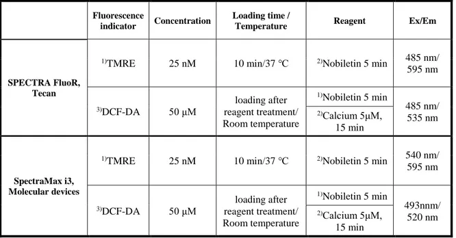
Table 2. Measuring protocols for ∆Ψm and ROS in isolated brain mitochondria
Fluorescence
indicator Concentration
Loading time /
Temperature Reagent Ex/Em
SPECTRA FluoR, Tecan
1)TMRE 25 nM 10 min/37 ℃ 2)Nobiletin 5 min 485 nm/
595 nm 3)DCF-DA 50 μM loading after reagent treatment/ Room temperature 1)Nobiletin 5 min 485 nm/ 535 nm 2)Calcium 5μM, 15 min SpectraMax i3, Molecular devices
1)TMRE 25 nM 10 min/37 ℃ 2)Nobiletin 5 min 540 nm/
595 nm 3)DCF-DA 50 μM loading after reagent treatment/ Room temperature 1)Nobiletin 5 min 493nnm/ 520 nm 2)Calcium 5μM, 15 min 1), 2), 3) indicate the processing sequence
Real-time fluorescence analysis
Indicator Measurement Excitation
filter
Emission
filter Dichroic mirror Loading protocol
Fura-2 Cytosolic calcium ET340x/
ET380x ET510/80m T400lp
10μM, For 45 min at 37 ℃
Rhod-2 Mitochondrial
calcium AT540/25x AT605/55m AT565DC
2μM, For 45 min at 4 ℃
TMRE Mitochondrial
membrane potential AT540/25x AT605/55m AT565DC
25nM, For 15 min at 37 ℃
MitoSOX Red
Mitochondrial
ROS AT540/25x AT605/55m AT565DC
5μM, For 15 min at 37 ℃
18
3. Results
3.1. Nobiletin, the key compound of CPE, exhibits neuroprotective effects based on mild mitochondrial depolarization
It was previously reported that CPE evokes partial mitochondrial depolarization in HT-22 cell, while carbonyl cyanide m-chlorophenylhydrazone (CCCP, 10 μM), a well-known ∆Ψm
dissipation-inducer, evokes complete mitochondrial depolarization (Wu et al., 2013). Driving force of mitochondrial calcium overload can be reduced by inducing mitochondrial depolarization that blocks apoptosis. Based on this study, I investigated here whether nobiletin, an active compound of CPE, induces partial depolarization of ∆Ψm in resting state and reveals
neuroprotective activity against glutamate toxicity in primary cortical neurons.
First, to determine whether the effect of CPE was maintained in primary cortical neurons, ∆Ψm and cell viability were measured in cortical neurons from embryonic 20-day SD rats. Fig.
1 shows the effect of CPE on ∆Ψm and cell viability. CPE induced mitochondrial depolarization
in a dose-dependent manner (Fig.1A, B). The TMRE fluorescence values in 500 μg/ml group and 1000 μg/ml group at 13 minutes were 72.44 5.67, and 68.42 15.28 %, respectively (Fig.1B). 500 μg/ml CPE significantly enhanced cell viability against glutamate toxicity (100 μM, 20 min) in primary cortical neuron (Fig. 1C).
Various concentrations of nobiletin (30, 50, 100 and 200 µM) were superfused over TMRE-loaded cortical neurons on a cover slip in a recording chamber. ∆Ψm values were recorded and
analyzed using real-time imaging-based fluorometry (See ‘Materials and Methods’ for more detailed description). TMRE fluorescence values from individual cells were normalized to values before drug treatment in Fig. 2A. Recording traces reveal the averaged recordings of
19
TMRE intensities obtained from individual cells. Results demonstrated that nobiletin treatment significantly evoked mild mitochondrial depolarization in dose-dependent manner. Normalized TMRE values at 13-min points were 94.71 5.47%, 73.06 4.79%, 61.47 6.06% and 54.26 10.37% in nobiletin (30, 50, 100 and 200 M)-treated groups, respectively (Fig. 2B), compared to the control group.
It is well established that glutamate toxicity results in massive and global calcium influx into the cytosol and subsequent mitochondrial calcium overload and evokes cell death (Duchen et al., 2012). The neuroprotective effect of nobiletin against glutamate toxicity was examined in primary cortical neurons using MTT assay. Nobiletin (100 μM) was pretreated for 10 min and glutamate (100 μM) was added for 20 min in the presence of nobiletin. As shown in Fig. 2C, nobiletin significantly enhanced neuronal cell viability to 80.32 4.80% against glutamate toxicity (100 μM, 20 min). The working concentration of nobiletin was determined as 100 μM which shows the maximum depolarization effect without cytotoxicity (Fig.3). Therefore, subsequent experiments were conducted at 100 μM of nobiletin.
Results suggest that the effects of each active compound in the CPE, including nobiletin, can be reproduced in primary cortical neurons and nobietin-induces partial mitochondrial depolarization does not adversely affect to the physiological activity of neuronal cells at concentrations below 100 μM. The beneficial effects of CPE on ∆Ψm and neuronal cell viability
21
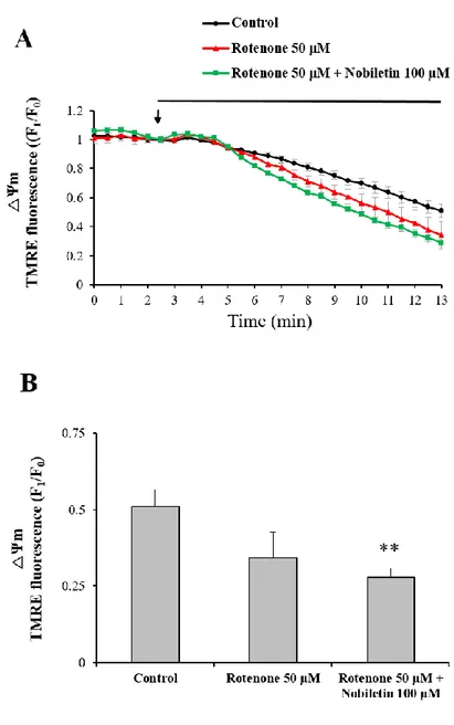
Figure 1. The effects of Citrus sunki Hort. ex Tanaka (CPE) on basal ΔΨm and cell viability
against glutamate toxicity in primary cortical neurons.
(A) Recording traces of ∆Ψm using real-time imaging-based fluorometry with TMRE. After
TMRE (25 nM) was loaded for 15 min, the base line was measured for 5 min. CPE (500, 1000 μg/ml) was superfused in a recording chamber from 5 min to the end of the measurement. TMRE fluorescence values from individual cells were normalized to the value at starting point of drug treatment. (B) Quantification of TMRE fluorescence at the end of the measurement, 13 min time point, was compared with among different group. (C) Neuroprotective effects of CPE on glutamate-induced neurotoxicity. Primary cortical neurons were pretreated with CPE for 10 minutes and then treated with glutamate (100 μM) for 40 minutes. The neuroprotective effects of CPE were analyzed by MTT assay. Values indicate the mean ± S.E.M. *p < 0.05, **p < 0.01, ***p < 0.001 as compared with control group, #p < 0.05 as compared with glutamate
22
23
Figure 2. The effects of nobiletin on basal ΔΨm and cell viability against glutamate toxicity
in primary cortical neurons.
(A) Recording traces of ∆Ψm using real-time imaging-based fluorometry with TMRE. Various
concentrations of nobiletin were superfused over primary cortical neurons on a cover slip in a recording chamber from the arrow point. TMRE fluorescence values from individual cells were normalized to values before drug treatment shown as an arrow. (B) Quantification of ∆Ψm at
the end of experiment. (C) Effects of nobiletin on cell viability against glutamate toxicity (100 μM, 20 min) were investigated using MTT assay. Values indicate the mean ± S.E.M. *p < 0.05, ***p < 0.001 as compared with the control group and #p < 0.05 as compared with glutamate alone-treated group.
24
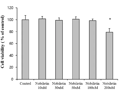
Figure 3. Dose determination of nobiletin treatment.
The toxicity of nobiletin on cortical neurons. Various concentrations of nobiletin (10, 30, 50, 100 and 200 µM) were treated for 30min. After 3 times wash out, neurobasal medium was added to cells. The cell viability was measured after 24 hours. *p < 0.05 as compared with untreated controls.
25
3.2. Nobiletin reduces glutamate-induced mitochondrial calcium overload
High concentration of glutamate induces excess Ca2+ influx from extracellular space to
cytosol via glutamate receptor/channels and subsequently excess cytosol Ca2+ is moved into mitochondria (Duchen et al., 2012). Mild mitochondrial depolarization attenuates mitochondrial calcium overload by reducing driving force of Ca2+ uptake into mitochondria (Ishida et al., 2001, Sanz-Blasco et al., 2008, Valero et al., 2008). Therefore, I investigated here whether nobiletin attenuates mitochondrial Ca2+ overload against glutamate toxicity.
Cytosol and mitochondrial Ca2+ levels were simultaneously recorded and analyzed in both regions, cytoplasm and mitochondria, using dual real-time imaging-based fluorometry with Fura-2 AM and Rhod-2 AM (Fig. 4). Glutamate (100 μM) in the presence or absence of nobiletin (100 μM) was superfused over Fura-2 AM and Rhod-2 AM-loaded cortical neurons on a cover slip in a recording chamber. Cytosol and mitochondrial Ca2+ levels from individual
cells, indicated as Fura-2 AM and Rhod-2 AM fluorescence values, were normalized to values before drug treatment. The cytosolic Ca2+ levels were increased by 2.02 0.07 in glutamate group. Nobiletin markedly abolished glutamate-induced mitochondrial calcium overload in cortical neurons by 85.56 4.10 % (Fig. 5). However, nobiletin did not affect glutamate-induced increase of [Ca2+]c (Fig.4). Compared with the control group, nobiletin alone did not
affect the intracellular Ca2+ level. Taken together from Fig. 4 and 5, it was demonstrated that nobiletin capable of evoking mild mitochondrial depolarization, potently blocked glutamate-induced mitochondrial Ca2+ overload in primary cortical neurons.
26
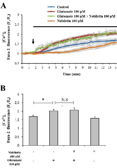
Figure 4. The effects of nobiletin on glutamate-induced overload of cytosol calcium in primary cortical neurons.
Dual real-time imaging-based fluorometry of [Ca2+]c and [Ca2+]m were simultaneously
conducted in the same neurons. (A) Fura-2 fluorescence values from individual cells were normalized to values before drug treatment. (B) Quantification of Fura-2 fluorescence values at the end of experiment. Values indicate the mean ± S.E.M. *p < 0.05 as compared with the control group compared. N.S., not statistically significant.
27
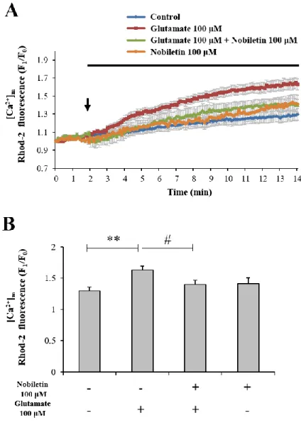
Figure 5. The effects of nobiletin on glutamate-induced overload of mitochondrial calcium overload in primary cortical neurons.
(A) Rohd-2 fluorescence values from individual cells were normalized to values before drug treatment. (B) Quantification of Rohd-2 fluorescence values at the end of experiment. Values indicate the mean ± S.E.M. **p < 0.01 as compared with the control group and #p < 0.05 as compared with glutamate alone-treated group.
28
3.3. Establishment of isolated mitochondrial model
The concentration of K+ in the neuronal cytoplasm is about 140 mM, which is among the highest among intracellular ions of the resting neuron. It has been demonstrated that mitoBKCa
and mitoKATP channel exist in mitochondria (Szewczyk et al., 2009). For this reason, I
hypothesized that mitochondrial depolarization by nobiletin will be related to the influx of K+ into mitochondria. To make sure the relationship between K+ and nobiletin, isolated
mitochondrial model was adopted. First, an experiment was conducted to confirm the physiological function of the isolated mitochondria (Fig. 6). Figure 6.A is the schematic experimental procedure for mitochondria isolation. The fragmented cortex was placed in EGTA-containing isolation buffer (IB) and homogenized. After centrifugation at 600 ⅹ g for 10 min, supernatant was transferred to a new micro-tube. This process was repeated twice. The supernatant was re-centrifuged at 12,000 ⅹ g for 10 min. The settled pellets were suspended in EGTA-free IB buffer and homogenized with dounce-type glass homogenizer.
The finally obtained mitochondria are suspended in the recording buffer. In the EM results, the black arrow indicates the mitochondria and the cristae structure in isolated mitochondria is well maintained (Fig. 6B)
Afterwards, the oxygen consumption rate (OCR) was measured to test the physiological activity of isolated mitochondria (Fig. 6C). Treatment of oligomycin, an inhibitor of mitochondrial ATP synthase, reduced OCR. FCCP is generally used as protonophore and uncoupler of oxidative phosphorylation. When treated with FCCP, the OCR was increased to compensate for the electrochemical gradient. It is speculated that OCP was not elevated when treated with FCCP compared to ADP treatment because the biochemical activity of isolated mitochondria was decreased because the time required for measurement was set too long.
29
Rotenone and antimycin A are inhibitors of complex Ⅰ and Ⅲ, respectively. These reagents completely reduced the OCR due to the shutting down of the electron transport chains.
These results suggest that isolated mitochondria maintain the function of the electron transport system at least during the experiment and that the basal ∆Ψm is also normally formed.
So isolated mitochondrial model according to the described method is appropriate for physiological experiment.
31
Figure 6. Identification of physiological activity of isolated brain mitochondria.
(A) Schematic experimental procedure for mitochondria isolation from the cortices of SD rats. The fragmented cortex was homogenized using 15ml dounce-type glass tissue grinder (clearance of pestle; A is 0.0035-0.0065; B is 0.0035-0.0065). The mitochondria pellets obtained after centrifugation at 12000 × g was homogenized using 7ml dounce-type glass homogenizer (clearance of pestle; A is 0.0028-0.0047; B is 0.0008-0.0022). (B) The oxygen consumption rate (OCR) of isolated mitochondria. OCR was measured in the absence of ADP as in the other experimental conditions. Final concentrations of treated reagents were oligomycin 2 µg/ml, FCCP 2 µM, rotenone 2 µM and antimycin A 0.5 µM.
32
3.4. Nobiletin attenuates glutamate-induced mitochondrial ROS generation
Mitochondrial Ca2+ overload can lead to ROS generation and oxidative stress although Ca2+ has no direct effect in electron transport chains or oxidation/reduction in mitochondria (Peng et al., 2010). In this regard, I investigated whether nobiletin inhibits glutamate toxicity-induced mitochondrial ROS generation in intact primary cortical neurons. In addition, this issue was explored in isolated brain mitochondria model.
In intact cortical neurons, mitochondrial superoxide levels were recorded and analyzed using real-time imaging-based fluorometry with MitoSOX Red. Glutamate (100 μM) in the presence or absence of nobiletin (100 μM) was superfused over MitoSOX Red-loaded cortical neurons on a cover slip in a recording chamber. Mitochondrial ROS levels from individual cells were normalized to values before drug treatment. Nobiletin almost blocked glutamate-induced mitochondrial ROS in intact cortical neurons (Fig. 7A and B).
High concentration of CaCl2 (5 μM) was treated to isolated mitochondria to mimic glutamate
toxicity model in intact cortical neurons. Glutamate treatment did not work in isolated mitochondria since glutamate receptors were expressed in plasma membrane, not in mitochondria membrane. In isolated mitochondrial model as revealed in Fig. 8, high concentration of Ca2+ was added in the medium instead of glutamate. Using a fluorescence microplate reader, mitochondrial ROS was measured using DCF-DA. Nobiletin significantly attenuated ROS production in isolated brain mitochondria exposed to high concentration of Ca2+ in a dose (30, 50 and 100 M)-dependent manner (Fig. 8), consistent to ROS data from
intact cortical neurons.
If nobiletin has free radical scavenging activity, it becomes difficult to interpret whether the ROS reduction effect is due to ∆Ψm regulation or by its own ROS elimination. Therefore, an
33
experiment was conducted to investigate the ROS scavenging activity of nobiletin. Data from DPPH free radical scavenging assay revealed that nobiletin did not exhibit the direct free radical-scavenging activity in cell-free and mitochondria-free tube system. N-acetyl cysteine (NAC), a well-known anti-oxidant, was used as a positive control in DPPH assay (Fig. 9A). A large part of the ROS scavenging activity of CPE is due to hesperidin, which is the most abundant compound in CPE. (Fig 9 B and C).
Taken together, results suggest that nobiletin markedly reduces mitochondrial ROS production against glutamate toxicity and mitochondrial calcium overload in intact primary cortical neurons (Fig. 7A and B) and isolated brain mitochondria (Fig. 8) although it does not have free radical scavenging activity (Fig. 9A). Fig.9 implicate that the ROS reduction effect of nobiletin is based on the modulating calcium-induced ROS generation through reducing Ca2+ overload into the mitochondria matrix.
34
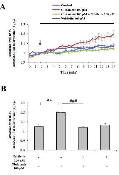
Figure 7. The effects of nobiletin on mitochondrial ROS generation in glutamate-stimulated cortical neurons.
(A) Recording traces of mitochondrial superoxide using real-time imaging-based fluorometry with MitoSOX Red (See ‘Materials and methods’ for the detailed description). MitoSOX Red fluorescence values from individual cells were normalized to values before drug treatment shown as an arrow. (B) Quantification of MitoSOX Red fluorescence values at the end of experiment for panel A. Values indicate the mean ± S.E.M. **P < 0.01 as compared with untreated controls and, ###p<0.001 as compared with glutamate treated group.
35
Figure 8. The effects of nobiletin on mitochondrial ROS generation in isolated brain mitochondria exposed to high concentration of Ca2+.
Effects of nobiletin on mitochondrial ROS generation were measured with DCF-DA indicator using a fluorescence microplate reader in an isolated brain mitochondrial model (See ‘Materials and methods’ for the detailed description). Values indicate the mean ± S.E.M. ***P < 0.001 as compared with untreated controls and, #p<0.05, ##p<0.01, ###p<0.001 as compared with CaCl2
37
Figure 9. The comparison of ROS scavenging activities of Citrus compounds in cell-free system.
(A) Antioxidant activity in various concentrations of nobiletin was measured using DPPH assay. (B), (C) Free radical scavenging activity of CPE and hesperidin in the cell-free system. Various concentrations of CPE and hesperidin were reacted with the DPPH solution for 1 hr (See ‘Materials and methods’ for the detailed description). Values indicate the mean ± S.E.M. *p<0.05, **P < 0.01, ***P < 0.001 as compared with untreated controls.
38
3.5. Nobiletin induces partial mitochondrial depolarization through K+ influx across
mitochondrial inner membrane
Influx of K+ from cytosol into the mitochondrial matrix induces mild uncoupling, resulting in dissipation of ∆Ψm (Ishida et al., 2001). K+ is dominant cations among intracellular ions in
resting neurons. Based on this background, I hypothesized that nobiletin-induced mild mitochondrial depolarization is mainly mediated by the influx of K+ into mitochondria. To
confirm the correlation between mitochondrial K+ influx and nobiletin effect on ∆Ψm, a isolated
brain mitochondrial model was used. The effect of FCCP on ∆Ψm was investigated as positive
control using a fluorescence microplate reader in isolated brain mitochondrial model. When depolarization was induced by FCCP, TMRE intensity increased in dose-dependent manner (Fig. 10A). TMRE, cationic fluorescent dye, is sequestered in mitochondria and it is released by mitochondrial depolarization (Scaduto and Grotyohann,1999). This result was due to the measurement of the total TMRE fluorescence emitted from the mitochondria into the recording buffer in a well. KCl (100 mM) was replaced with CsCl (100 mM) to remove K+ effects in the medium and thereby K+ dependence in nobiletin effect on ΔΨm was investigated. Fig. 10B
demonstrated that nobiletin effects on mild mitochondrial depolarization were not shown in the absence of K+ in the medium when KCl (100 mM) was replaced with CsCl (100 mM) in an isolated mitochondria model. On the other hand, mitochondrial depolarization was induced by nobiletin in a dose-dependent manner in the KCl (100 mM) condition. Fig. 10C shows the difference in the change of ΔΨm for 100uM nobiletin in environments with or without K+ in
time-dependent manner. In the absence of K+, depolarization of ΔΨm was not observed until 15
min.
39
investigate the molecular target of nobiletin, I examined whether the membrane depolarization disappeared by using K+ channel inhibitors. Iberiotoxin 10 nM and 5-HD 500 μM were used
to inhibit mitoBKCa and mitoKATP channels, respectively. As a result, Fig. 11A demonstrated that
mitochondrial depolarization by CPE was significantly inhibited by 500 μM 5-HD.In Fig. 11B, membrane depolarization by nobiletin was significantly inhibited in the group treated with 10 nM iberiotoxin in primary cortical neurons. In the group treated with 500 μM 5-HD, partial mitochondrial depolarization tended to disappear, but there was no significant difference. The
mitoBKCa channels may be closely involved in mitochondrial polarization by nobilrtin, but the
association mitoKATP channels can not be ruled out because the depolarization of CPE was
disappeared by 5-HD.Results suggest that nobiletin induces partial membrane depolarization by promoting influx of K+ across mitochondrial inner membrane. K+ influx into mitochondrial matrix by nobiletin is probably mediated by mitochondrial K+ channels.
40
41
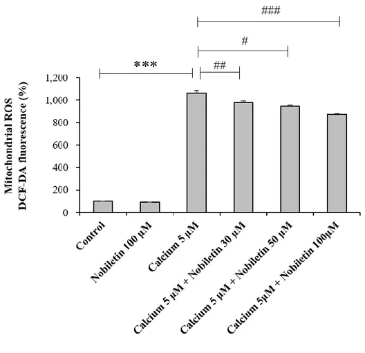
Figure 10. The effects of K+ influx on nobiletin-induced partial mitochondrial
depolarization in isolated brain mitochondria.
(A) The effect of FCCP on ∆Ψm was investigated with TMRE in an isolated brain mitochondrial
model (See ‘Materials and methods’ for the detailed description) using a fluorescence microplate reader, as a positive control. (B and C). The effect of nobiletin on ∆Ψm was
measured with TMRE in the presence or absence of K+ in the medium using a fluorescence microplate reader in an isolated brain mitochondrial model. To remove K+ in the medium, KCl
(100 mM) was replaced to CsCl (100 mM). Dose responses 10 min after nobiletin treatment (B) and time courses (C) were analyzed. Values indicate the mean ± S.E.M. *p < 0.05, **p < 0.01, ***p < 0.001 as compared with the control group.
42
Figure 11. The effects of mitochondrial K+ channel inhibitors on nobiletin- induced ΔΨ m
changes in primary cortical neurons.
(A) CPE Nobiletin was superfused with 500μM 5-HD in a recording chamber (B) Nobiletin was superfused with 10 nM iberiotoxin and 500μM 5-HD in a recording chamber, TMRE fluorescence values from individual cells were normalized to the value at starting point of drug
43
treatment. Quantification of TMRE fluorescence at the end of the measurement. Values indicate the mean ± S.E.M. *p < 0.05 as compared with untreated controls and, #p<0.05, as compared
44
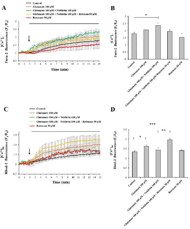
3.6. Rotenone attenuate the effect of nobiletin to reducing mitochondrial calcium in glutamate toxicity model
Some studies (Sherer et al., 2003) have used a rotenone-induced Parkinson's model which was involved in oxidative damage of rotenone. Nobiletin has beneficial effects in improving cognitive function or motor deficits in Parkinson’s diseases (Yabuki et al., 2014). The main source of intracellular ROS is the mitochondrial electron transport system. To investigate the possibility of nobiletin to interact with the mitochondrial electron transport system, I hypothesized that nobiletin stimulates the function of complex Ⅰ as an antagonist to rotenone. To determine the effect of rotenone on △Ψm, △Ψm was observed using real-timeimage based
recording when rotenone (50 μM) and nobiletin (100 μM) was added to neuronal cells in primary neuron culture system (Fig. 12 A and B). The mitochondrial depolarization was induced more significantly in rotenone and nobiletin co-treated group. Rotenone induced mitochondrial depolarization like nobiletin but showed no significance compared with the control group. The TMRE fluorescence values in rotenone 50 μM group and rotenone 50 μM + nobiletin 100 μM group at 15 minutes were 50.36 17.73, and 43.97 6.18 % respectively (Fig. 12 B).
[Ca2+]
c and [Ca2+]m were measured simultaneously in living individual neurons. Because
nobiletin reduces mitochondrial calcium overload induced by glutamate, the [Ca2+]c increases
and the [Ca2+]m decreases. However, these nobiletin effects were disappeared by rotenone, so,
[Ca2+]m increased and [Ca2+]c decreased (Fig. 13A and C). The Fura-2 fluorescence values at
the end of the measurement were 114.92 5.39 %, 132.29 9.64 %, 109.53 10.82 % and 86.30 14.70 % compared with control group respectively (Fig. 13C). The mitochondrial calcium reduced by nobiletin was increased to 146.31 3.12 % by rotenone (fig. 13D).
45
These results show that although nobiletin does not affect △Ψm induced by inhibition of
complex Ⅰ, nobiletin effects on [Ca2+]
46
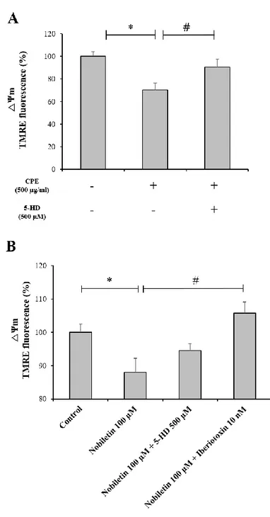
Figure 12. The effects of rotenone, a complex Ⅰ inhibitor, on mitochondrial membrane potential.
(A) Effects of rotenone and nobiletin on mitochondrial membrane potential. Rotenone 50 µM and nobiletin 100 µM were superfused in a recording chamber, TMRE fluorescence values from individual cells were normalized to the value at starting point of drug treatment. (B) Quantification of TMRE fluorescence at 15 min time point was compared with among different group. Values indicate the mean ± S.E.M. **p < 0.01 as compared with untreated controls.
47
Figure 13. Rotenone inhibits the action of nobiletin, which attenuate mitochondrial calcium overload in the glutamate toxicity model.
(A and C) Dual real-time imaging-based recording of [Ca2+]c and [Ca2+]m were simultaneously
48
were superfused in a recording chamber, Fura-2 and Rhod-2 fluorescence values from individual cells were normalized to the value at starting point of baseline measurement. (B and D) Quantification of fluorescence values of Fura-2 and Rhod-2 from individual cells at the end of the measurement (15 min). Values indicate the mean ± S.E.M. *p < 0.05, **p < 0.01, ***p < 0.001.
49
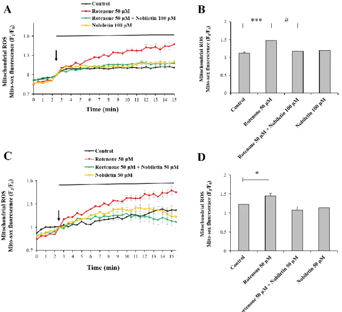
3.7. Rotenone-induced mitochondrial ROS was reduced by nobiletin
It is already demonstrated that when complex Ⅰ is inhibited by rotenone, ROS production is increased and apoptosis is induced. If nobiletin is antagonistic to rotenone, it may reduce ROS production caused by the inhibition of complex Ⅰ. Mitochondrial ROS was measured using MitoSOX Red, mitochondrial superoxide indicator, under a real-time imaging-based recording system. When treated with rotenone alone, ROS increased to 131.83 5.34 % compared to the control group. In the group treated with 50 μM rotenone and 100 μM nobiletin at the same time, ROS was decreased to 105.21 3.28 % similar to the control group (Fig. 14. A and B). When 50 μM nobiletin was co-treated with rotenone, the effect of ROS generation was attenuated but no significant difference was observed compared with the rotenone alone group (Fig. 14.C and D). The MitoSOX Red fluorescence values in 50 μM rotenone group, rotenone 50 μM + nobiletin 100 μM group and nobiletin 100 μM group at the end of the measurement were 118.97 3.36 %, 87.91 8.00 % and 93.23 7.46 % respectively. Taken together, there is a possibility that nobiletin inhibit rotenone action or promote the function of complex Ⅰ because nobiletin reduced ROS from the electron transport system when complex Ⅰ was inhibited. However, additional studies should be performed to determine the exact mechanism of action between nobiletin and complex Ⅰ.
50
Figure 14. The effects of nobiletin on rotenone-induced mitochondrial ROS in primary cortical neurons.
(A) After MitoSOX Red (5 μM) was loaded for 15 min, base line was measured for 2 min. Nobiletin (100 μM), and rotenone (50 μM ) were superfused in a recording chamber from 2min to the end of the measurement. MitoSOX Red fluorescence values from individual cells were normalized to the value at starting point of drug treatment. (B) Quantification of MitoSOX Red fluorescence at the end of the measurement, 15 min time point, was compared with among
51
different group. (C) Antioxidant activity of nobiletin (50 μM) in primary cortical neurons. (D) Quantification of MitoSOX Red fluorescence at the end of the measurement, 15 min time point. Nobiletin 50 μM also decreased ROS induced by rotenone, but no significant difference compared with rotenone alone treatment. Values indicate the mean ± S.E.M. *p<0.05, ***P < 0.001 as compared with untreated controls and, #p<0.05 as compared with rotenone 50 μM alone.
52
Figure 15. Schematic neuroprotective mechanism of nobiletin through mitochondrial depolarization.
Nobiletin induced mild mitochondrial membrane depolarization. This mild mitochondrial depolarization is based on regulation of K+ channels. The blockade of mitochondrial Ca2+
overload via depolarization of mitochondrial membrane plays a critical role in the mechanism underlying neuroprotective effects of nobiletin. Nobiletin markedly reduce electron transport chain complex I inhibitor rotenone-induced mitochondrial ROS generation.
53
4. Discussion
In this study, I demonstrated in primary cultured cortical neurons and brain mitochondria isolated from rat cortices that nobiletin prevents neurotoxic mitochondrial calcium overload, excess mitochondrial ROS, and neuronal cell death through ΔΨm regulation. Results
demonstrated that nobiletin evoked partial mitochondrial depolarization in intact cortical neurons (Fig. 2A and B) and isolated brain mitochondria (Fig. 10B and C). Nobiletin markedly attenuated mitochondrial calcium overload and ROS generation in glutamate (100 μM)-stimulated cortical neurons (Fig. 5 and Fig. 7) and isolated brain mitochondria exposed to a high concentration of Ca2+ (5 μM) (Fig. 8). In neuronal cells, NMDA and AMPA receptors in cell membranes are activated by excess glutamate. This glutamate receptor activation contributes to calcium overload into the cytosol and collapse of the mitochondrial membrane potential. In particular, the accumulation of calcium in the mitochondria is an important step in the progression to cell apoptosis, promoting the loss of ΔΨm, and the collapse of apoptotic
cell death (Abramov et al., 2008). Mitochondria take up Ca2+ electrophoretically from the cytosol through a mitochondrial calcium uniporter (MCU). Mitochondrial Ca2+ influx is rapidly induced by the mitochondrial membrane potential, whereas its release in exchange for protons or sodium is electroneutral. Therefore, treatment with high concentrations of glutamate results in the influx of calcium into the cytoplasm, as shown in Fig. 5, and this calcium is accumulated in the mitochondrial matrix. Fig. 2 and 5 suggest that nobiletin induces partial depolarization, which does not interfere with the normal physiological function of the cell, thereby reducing the electrophoretic force that influxes Ca2+ into the mitochondria..
Excess exposure to glutamate markedly evoked overload of both mitochondrial Ca2+ and ROS. Nobiletin treatment significantly reduced these two reciprocal parameters, as shown in Fig. 5 and 7. The exact mechanism by which mitochondrial Ca2+ stimulates ROS generation
54
inside mitochondria remains elusive. However, several plausible mechanisms have been proposed. Possible mechanisms include mitochondrial Ca2+-induced increase of the metabolic
rate, mitochondrial Ca2+-stimulated dissociation of cytochrome c, and mitochondrial Ca2+ -stimulated opening of the mitochondrial permeability transition pore with cytochrome c release (Peng and Jou, 2010). Mitochondrial ROS can inversely affect Ca2+ dynamics and modulate Ca2+ surge. The reciprocal crosstalk between mitochondrial Ca2+ and ROS may result in a feedforward, self-amplified loop evoking subsequent cellular damage and death (Feissner et al., 2009).
The main finding in this study may be the regulation of mitochondrial K+ influx by nobiletin. I demonstrated that the neuroprotective effect of nobiletin via mild mitochondrial depolarization is largely mediated by the influx of K+ into mitochondria (Fig. 10B and C). Nobiletin effects on basal ΔΨm were completely abolished in brain mitochondria isolated from
rat cortices that were maintained in K+-free medium. Results suggest that K+ influx into the mitochondrial matrix is critically involved in the nobiletin effect on ΔΨm. The mitochondrial
K+ influx is likely mediated, at least in part, by the activation of mitochondrial K+ channels. However, further detailed studies should be conducted to determine the exact molecular targets of nobiletin in mitochondria.
There are several ion channels/transporters and electron transport chains on the inner membrane of mitochondria, which are proposed as potential molecular targets of nobiletin in mitochondria. It does not seem that MCU, the major route for mitochondrial Ca2+ uptake, is involved in nobiletin-induced ∆Ψm depolarization. If nobiletin might attenuate
glutamate-induced mitochondrial Ca2+ overload through inhibition of MCU, it should hyperpolarize the
mitochondrial membrane rather than depolarize it. Several studies (Szewczyk et al., 2009) have revealed that K+ channels/transporters are present in the mitochondrial inner membrane: mitoKATP, mitoBKCa, voltage-gated potassium channel Kv1.3, twin-pore domain TASK-3
55
potassium channels, and K+/H+ exchangers. The mitoBKCa and mitoKATP channels are major
mitochondrial K+ channels. It was demonstrated using brain mitochondria isolated from rat
cortices that the neuroprotective effect of nobiletin via mild mitochondrial depolarization is largely mediated by the influx of K+ into mitochondria (Fig. 10B and C). These results suggest
that nobiletin-induced mitochondrial K+ influx is likely mediated, at least in part, by the activation of mitochondrial K+ channels. Based on this idea, I further explored mitochondrial targets of nobiletin. Some preliminary findings related to mitoBKCa and mitoKATP are shown in
Fig. 11. I investigated whether nobiletin-induced ∆Ψm depolarization is blocked by iberiotoxin
and 5-HD as they are well-known inhibitors of mitoBKCa and mitoKATP channels, respectively. As
shown in Fig. 11, preliminary findings indicated that nobiletin-induced ∆Ψm depolarization was
significantly inhibited in the group treated with iberiotoxin (10 nM). These results suggest that nobiletin may induce the neuroprotective ∆Ψm depolarization by promoting the influx of K+
into mitochondria through the activation of mitoBKCa. However, further detailed studies should
be conducted to determine the exact molecular targets of nobiletin in mitochondria.
Among flavonoids, naringenin is one of the flavanones abundant in the genus Citrus, such as in grapefruit and orange; it has been widely studied recently.The activation of mitoBKCa is
involved in a cardioprotective mechanism of naringenin against myocardial ischemia/reperfusion (Testai et al., 2013). Naringenin is proposed as one of the BKCa channel
openers in vascular smooth muscle cells (Saponara et al., 2006). An electrophysiological study recently revealed that channel activities of mitoKATP and mitoBKCa were enhanced after treatment
with 10 μM of naringenin in a single channel study using mitoplasts isolated from primary human dermal fibroblast cells (Bednarczyk et al., 2017). These recent reports focus special attention on the intracellular pathways mediated by Citrus flavonoids regarding their beneficial effects on several physiological and pathophysiological conditions.
56
In Figures 12, 13, and 14, I demonstrated the relationship between nobiletin and electron transport chain complex I, which was inhibited by rotenone. These results suggest an antagonistic relationship between nobiletin and rotenone in the regulation of [Ca2+]m andROS,
but their relationship does not seem to be directly related to the regulation of ∆Ψm. Further
studies should be conducted to obtain more precise information on the interaction between nobiletin and complex I.
In conclusion, I reveal here that nobiletin evokes partial mitochondrial depolarization in intact cortical neurons as well as isolated brain mitochondria, and thereby prevents neurotoxic mitochondrial calcium overload, excess mitochondrial ROS, and neuronal cell death. Furthermore, K+ influx into the mitochondrial matrix is critically involved in the nobiletin effect on ∆Ψm. In addition, results suggest that the mitochondrial K+ influx is likely mediated,
at least in part, by the activation of mitochondrial K+ channels. All these findings reveal a
beneficial role for nobiletin-induced partial mitochondrial depolarization in neuroprotection, which is similar to the ischemic pre-conditioning (IPC) in the endogenous mechanism of cardioprotection. Therefore, pharmacological manipulation of ∆Ψm through novel substances
57
References
1. Abramov AY, Duchen MR. 2008. Mechanisms underlying the loss of mitochondrial membrane
potential in glutamate excitotoxicity. Biochim. Biophys. Acta. 1777 : 953–964
2. Bednarczyk P, Kicinska A, Jarmuszkiewicz W, Debowska R, Szewczyk A. 2017. Flavonoids as
natural modulators of mitochondrial potassium channel. Biophys. J. 112:405a-406a.
3. Blattner JR, He L, Lemasters JJ. 2001. Screening assays for the mitochondrial permeability
transition using a fluorescence multiwell plate reader, Anal. Biochem. 295:220-226.
4. Choi SY, Kim JY, Jo Y, Kim HD, Song M, Moon C, Kim CH, Kim K, Sesaki H, Rhyu IJ, Kim H, Sun W. 2016. Constriction of the mitochondrial inner compartment is a priming event for
mitochondrial division. Nat. Comm. 8:15754-15770.
5. Choi SY, Hwang JH, Ko HC, Park JG, Kim SJ. 2007. Nobiletin from citrus fruit peel inhibits
the DNA-binding activity of NF-kappaB and ROS production in LPS-activated RAW 264.7 cells. J. Ethnopharmacol. 113:149-155.
6. Cui Y, Wu J, Jung SC, Park DB, Maeng YH, Hong JY, Kim SJ, Lee SR, Kim SJ, Kim SJ, Eun SY. 2010. Anti-neuroinflammatory activity of nobiletin on suppression of microglial activation.
Biol. Pharm. Bull. 33:1814-1821.
7. Cui Y, Park JY, Wu J, Lee JH, Yang YS, Kang MS, Jung SC, Park JM, Yoo ES, Kim SH, Ahn JS, Suk K, Eun SY. 2015. Dieckol Attenuates Microglia-mediated Neuronal Cell Death via ERK,
58
8. Duchen MR. 2012. Mitochondria, calcium-dependent neuronal death and neurodegenerative
disease. Pflugers. Arch. 464:111–121.
9. Eun SY, Jung SJ, Park YK, Kwak J, Kim SJ, Kim J. 2001. Effects of capsaicin on Ca2+ release from the intracellular Ca2+ stores in the dorsal root ganglion cells of adult rats. Biochem. Biophy. Res. Commun. 285:1114-1120.
10. Feissner RF, Skalska J, Gaum WE, Sheu SS. 2009. Crosstalk signaling between mitochondrial
Ca2+ and ROS. Front. Biosci. 14:1197-1218.
11. Gunter TE, Pfeiffer DR. 1990. Mechanisms by which mitochondria transport calcium. Am. J.
Physiol. Cell. Physiol. 258:C755-C786.
12. Iglesias-González J, Sánchez-Iglesias S, Beiras-Iglesias A, Soto-Otero R, Méndez-Á lvarez E. 2013. A simple method for isolating rat brain mitochondria with high metabolic activity: Effects
of EDTA and EGTA. J. Neurosci. Methods. 213: 39– 42.
13. Ishida H, Hirota Y, Genka C, Nakazawa H, Nakaya H, Sato T. 2001. Opening of
mitochondrial KATP channels attenuates the ouabain-induced calcium overload in mitochondria. Circ. Res. 89:856-858.
14. Jousset H, Malli R, Girardin N, Graier W.F, Demaurex N, Frieden M. 2008. Evidence for a
receptor-activated Ca2+ entry pathway independent from Ca2+ store depletion in endothelial cells. Cell Calcium. 43:83–94.
15. Kroemer G, Galluzzi L, Brenner C. 2007. Mitochondrial membrane permeabilization in cell
59
16. Lee JH, Amarsanaa1 K, Wu JJ, Sang-Chan Jeon SC, Cui YJ, Jung SC6, Park BD, Kim SJ, Han SH, Kim HW, Rhyu IJ, Eun SY. 2018. Nobiletin attenuates neurotoxic mitochondrial
calcium overload through K+ influx and ∆Ψm across mitochondrial inner membrane. Korean J. Physiol. Pharmacol. 22(3):311-319.
17. Nagase H, Omae N, Omori A, Nakagawasai O, Tadano T, Yokosuka A, Sashida Y, Mimaki Y, Yamakuni T, Ohizumi Y. 2005. Nobiletin and its related flavonoids with CRE-dependent
transcription-stimulating and neuritegenic activities, Biochem. Biophy. Res. Commun. 337:1330-1336.
18. Onozuka H, Nakajima A, Matsuzaki K, Shin RW, Ogino K, Saigusa D, Tetsu N, Yokosuka A, Sashida Y, Mimaki Y, Yamakuni T, Ohizumi Y. 2008. Nobiletin, a citrus flavonoid, improves
memory impairment and Aβ pathology in a transgenic mouse model of Alzheimer's disease. J. Pharmacol. Exp. Ther. 326:739-744.
19. Peng TI, Jou MJ. 2010. Oxidative stress caused by mitochondrial calcium overload. Ann. N. Y.
Acad. Sci. 1201:183–188.
20. Sanz-Blasco S, Valero RA, Rodríguez-Crespo I, Villalobos C, Núñez L. 2008. Mitochondrial
Ca2+ overload underlies Aβ oligomers neurotoxicity providing an unexpected mechanism of neuroprotection by NSAIDs. PLoS One. 3:e2718.
21. Saponara S, Testai L, Iozzi D, Martinotti E, Martelli A, Chericoni S, Sgaragli G, Fusi F, Calderone V. 2006. (+/-)-Naringenin as large conductance Ca2+-activated K+ (BKca) channel opener in vascular smooth muscle cells. Br. J. Pharmacol. 149:1013-1021.
22. Scaduto RC Jr, Grotyohann LW. 1999. Measurement of mitochondrial membrane potential