C6 Glioma Cell Insoluble Matrix Components Enhance
Interferon-
␥-stimulated Inducible Nitric-oxide
Synthase/Nitric Oxide Production in BV2 Microglial Cells
*
Received for publication, March 20, 2007, and in revised form, October 31, 2007Published, JBC Papers in Press, November 2, 2007, DOI 10.1074/jbc.M610219200 Yoon-Jung Kim‡1, So-Young Hwang‡1, Ji-Sun Hwang‡, Jung-Weon Lee§, Eok-Soo Oh¶, and Inn-Oc Han‡2
From the‡Department of Physiology and Biophysics, Inha University, College of Medicine, Incheon 402-751, Korea, the §
Department of Tumor Biology, Cancer Research Institute, College of Medicine, Seoul National University, Seoul 110-799, Korea, and the¶Department of Life Sciences, Division of Molecular Life Sciences and Center for Cell Signaling Research, Ewha Womans
University, Seoul 120-750, Korea
Microglia are the primary central nervous system immune effector cells. Microglial activation is linked to interactions with extracellular cytokines and the extracellular matrix (ECM). Astrocytomas are characterized by their diffuse nature, which is regulated by insoluble ECM components produced by the tumor cells that are largely absent from normal central nervous system tissue. The present study examined the influence of astrocytoma (C6 rat glioma) insoluble matrix components on interferon-␥ (IFN-␥)-mediated inducible nitric-oxide synthase (iNOS) induction in microglial cells. We found that IFN-␥-stimulated iNOS induction and nitric oxide release was greater in microglia cultured on C6 glioma cell-derived matrices compared with microglia cultured on primary rat astrocyte-derived matrices. Culture of microglia on C6 glioma cell-derived matrices also led to activation of STAT1, augmentation of IFN-␥-induced STAT-3 activation, and an increase in IFN-␥-activated site (GAS)-luciferase reporter activity. In addition, culture of microglia on C6 glioma cell-derived matrices activated NF-B DNA binding activity and transcriptional activity. The results suggest that insol-uble matrix components derived from malignant glioma cells can regulate microglia activation. These factors may include ECM components, such as fibronectin, collagen, laminin, vitronectin, and other nondiffusible compounds, and laminin seems to a criti-cal regulator of this process. Microglia activation and subsequent brain inflammation may influence tumor growth, treatment, and metastasis. Better understanding of the regulation of microglial activation by astrocytoma-derived insoluble matrix components may be important in the development of immune-based treatment strategies against malignant brain tumors.
Microglia constitute 10 –15% of cells within the CNS3and are considered the primary immune effector cells resident
within this tissue (1). Not only do microglia play important protective roles in the CNS, such as removing harmful patho-gens and promoting tissue regeneration after injury; they are also linked to the activation of neuropathological conditions, including multiple sclerosis (2), Alzheimer disease, and human immunodeficiency virus infection-associated dementia (3). Microglia have also been identified in brain neoplasm. In human gliomas, intratumoral microglia density is higher than in peritumoral and normal brain tissue, and the number of microglia increases with the grade of malignancy (4, 5). Although the presence of microglia may represent a CNS anti-tumor response, it is also possible that gliomas attract microglia in order to promote tumor growth via microglia-derived immu-nomodulatory cytokines.
Although the effect of microglia activity on glioma develop-ment has been investigated in a number of studies, the effect of gliomas on microglia behavior has been relatively ignored. Gli-oma factors that may potentially affect microglia behavior include insoluble components of extracellular matrix (ECM) produced by brain tumors that are largely absent from normal CNS tissue. These components are of particular interest, since adhesive interactions between microglia and the glioma ECM are a crucial element of glioma-microglia interactions.
Cell adhesion to ECM or neighboring cells is necessary for cell survival, migration, proliferation, and differentiation (6 – 8). Regulation of adhesion is important in cells involved in mounting responses to infection or injury due to the necessity to attach and then detach from distinct biological substrates at different stages of the inflammatory process (9, 10). The recruitment of microglia and other inflammatory cells to a lesion requires the ability to migrate. Adhesive interactions with the tissue to which they are responding are critical deter-minants of migratory behavior (11, 12). In the CNS, astrocytes and their associated ECM form a large part of the potential substrate with which microglia might engage during migration. Pathological conditions, such as brain injury, induce astrocyto-sis and increase expression of many ECM molecules, including fibronectin, laminin, vitronectin, and several proteoglycans *This work was supported by Korean Government Korea Research
Founda-tion Grant (MOEHRD) KRF-2006-311-C00124. The costs of publicaFounda-tion of this article were defrayed in part by the payment of page charges. This article must therefore be hereby marked “advertisement” in accordance with 18 U.S.C. Section 1734 solely to indicate this fact.
1Both authors contributed equally to this work.
2To whom correspondence should be addressed: Dept. of Physiology and
Biophysics, College of Medicine, Inha University, 253 Yonghyun-Dong, Nam-Ku, Incheon, 402-751, Republic of Korea. Tel.: 82-32-890-0924; Fax: 82-32-890-0647; E-mail: iohan@inha.ac.kr.
3The abbreviations used are: CNS, central nervous system; ECM, extracellular
matrix; IFN, interferon; MTT,
3-(4,5-dimethylthiazol-2-yl)-2,5-diphenyltet-razolium bromide; mAb, monoclonal antibody; PBS, phosphate-buffered saline; PDL, poly-D-lysine; BSA, bovine serum albumin; IL, interleukin; FAK, focal adhesion kinase; NO, nitric oxide; iNOS, inducible nitric-oxide syn-thase; STAT, signal transducers and activators of transcription; TNF, tumor necrosis factor; GAS, IFN-␥-activated site.
at Ewha Medical Library on March 22, 2017
http://www.jbc.org/
(13–18). These molecules can modulate different aspects of glial behavior, including cell adhesion, migration, and microglia inflammatory responses. Studies focusing on the nature of microglia and matrix interactions are relatively recent and have shown that microglia behavior is greatly affected by binding to ECM proteins (19, 20). Those observations may be especially important in inflammatory responses where microglia are pri-mary mediators. Glioma cells produce ECM-modulating mol-ecules (21), and microglia inflammatory responses in a glioma-containing environment are likely to be profoundly affected by the ECM proteins encountered by the microglia. Microglia express several integrin types, the expression of which can be altered under various pathological conditions and by injection of lipopolysaccharide (22). However, it is not known which combinations of ECM components in disease states exert the major influence on microglial activation.
The present study examined the effect of glioma cell-derived insoluble matrix components on microglia activation. Specifi-cally, the study compared in vitro microglia responses to insol-uble matrix components derived from astrocytes and glioma cells. We found that IFN-␥-mediated iNOS induction was greatly increased when microglia were cultured on C6 glioma cell-derived matrices but not on normal astrocyte-derived mat-rices. Furthermore, microglial cells cultured on C6 glioma cell-derived matrices showed greater levels of STAT1 and NF-B activation and increased IFN-␥-mediated STAT3 activation compared with cells cultured on astrocyte-derived matrices or normal tissue culture plates. The current findings collectively suggest that glioma insoluble matrix components may be important regulators of microglia activity. In addition, they imply that these glioma-induced alterations to the ECM com-position, which affect microglia behavior, may be an important therapeutic target in the treatment of brain tumors.
EXPERIMENTAL PROCEDURES
Reagents—Recombinant mouse IFN-␥ was purchased from
Invitrogen. MTT was obtained from Sigma. Function-blocking monoclonal antibodies (mAbs) to human integrin subunits␣1 (mAb 1973Z),␣2(mAb 1950Z), and integrin␣6(GoH3) were obtained from Chemicon (Temecula, CA).
Cell Culture—The murine BV2 microglia cell line that exhib-its phenotypic and functional properties of reactive microglial cells has been described previously (23). BV2 and C6 rat astro-cytoma (glioma) cells were cultured in Dulbecco’s modified Eagle’s medium supplemented with 5% heat-inactivated endo-toxin-free fetal bovine serum (Hyclone, Logan, UT), 2 mM glu-tamine, 100g/ml streptomycin, and 100 units/ml penicillin under a humidified 5% CO2atmosphere at 37 °C.
Preparation of Primary Astrocytes and Microglia—Primary astrocytes and microglia were prepared from 1-day-old Spra-gue-Dawley rats as previously described (23) with some modi-fications. In brief, the cortices were triturated into single cells in minimal essential medium containing 10% fetal bovine serum and then plated into 75-cm2T-flasks for 10 –14 days. To pre-pare pure astrocytes, microglia were removed from the T-flasks by mild shaking. We subcultured the astrocyte 2 times before we obtained pure astrocyte for experiments. The astrocyte was confirmed by staining for astrocyte-specific glial fibrillary
acidic protein, resulting usually in ⬎99% purity. Detached microglia cells were used for the co-culture experiment. The enriched microglia were nearly pure (⬎95%) as judged by immunocytochemical staining for a microglia-specific marker, the CR3 complement receptor, detected by the antibody OX-42 (Roche Applied Science). The astrocyte cells remaining in the flasks after removal of the microglia were harvested with 0.1% trypsin and plated into dishes or plates.
Co-culture and Preparation of Conditioned Media—Cells of the BV2 murine microglial cell line were co-cultured with either primary astrocytes or C6 astrocytoma cell line cells as previously described, with some modifications (24). BV2 cells were seeded on top of subconfluent monolayers of astrocytes or C6 cells. Conditioned medium was collected from astrocytes or C6 glioma cells that had been cultured for 2 days. The medium was centrifuged at 13,000⫻ g, and supernatants were filtered to remove cellular material and stored sterile at⫺20 °C before being applied to BV2 cells.
Coating of Cell Culture Dishes with Astrocyte- or Glioma Cell-derived Matrices and with Various Purified ECM Components— C6 cells or astrocytes were cultured in cell culture dishes or plates (Nunc, Naperville, IL) as described above and maintained in Dul-becco’s modified Eagle’s medium containing 5% fetal bovine serum or in minimal essential medium containing 10% fetal bovine serum, respectively. Once confluent, cells were lysed in water for 30 min at 37 °C. Cell debris and soluble components were removed by washing the wells 10 –15 times in PBS, and the remaining adhered insoluble matrix components were then incubated in serum-free Dulbecco’s modified Eagle’s medium at 4 °C for over-night before microglial cells were seeded. In order to coat cell cul-ture dishes, fibronectin-, poly-D-lysine (PDL)- or laminin were added to plastic culture dishes or multiwell plates (final-coating concentration, 5g/cm2) and incubated for 1 h at room tem-perature. Dishes or plates were washed with PBS and incubated with 1% BSA in PBS for 1 h at 37 °C and then washed three times with PBS. For collagen-coated dishes, collagen was added to plastic culture dishes (final coating concentration, 5g/cm2) and allowed to air-dry for 1 h at room temperature. After wash-ing with PBS, plates were incubated with 1% BSA in PBS for 1 h at 37 °C and then washed three times with PBS.
Nitrite Assay—Nitric oxide (NO) production from activated microglia was determined by measuring the amount of nitrite, a relatively stable oxidation product of NO, as described previously (23). In brief, an aliquot of the conditioned medium was mixed with an equal volume of 1% sulfanilamide in water and 0.1% N-1-naphthylethylenediamine dihydrochloride in 5% phosphoric acid. The absorbance was determined at 540 nm. Sodium nitrite, diluted in culture medium to various concentrations ranging from 10 to 100Mwas used to generate a standard curve.
Cell Viability (MTT)—Primary cultured rat microglia or BV2 cells were grown in 24-well plates at a concentration of 2.5⫻ 104or 1.5⫻ 104cells/well followed by proper treatment. To measure the cell viability, 50l of 5 mg/ml MTT in growth medium was added to each well. After incubation for 1 h at 37 °C with MTT, cell medium was removed. The precipitated formazan, a product of the MTT tetrazolium ring by the action of mitochondrial dehydrogenases, was solubilized with Me2SO and quantified spectrophotometrically at 540 nm. The MTT
at Ewha Medical Library on March 22, 2017
http://www.jbc.org/
assay reflects the metabolic activity of cells and serves as a help-ful indicator of cell viability.
Reverse Transcription-PCR—Total cellular RNA was extracted with TRIzolTM(Invitrogen) according to the manu-facturer’s protocol. Total RNA (1g) was reverse transcribed for 1 h at 37 °C in a reaction mixture containing 5 units of RNase, 0.5 mMdNTP, 1⫻ reverse transcriptase buffer, and 5 units of reverse transcriptase (Qiagen). PCR was performed using primers for iNOS, IL-6, IFN response factor (IRF-1), TNF-␣, cyclooxygenase-2, and glyceraldehyde-3-phosphate dehydrogenase as follows: cyclooxygenase-2 (forward, GCTG-TACAAGCAGTGGCAAA; reverse, GTCTGGAGTGGGAG-GCACT); IL-6 (forward, CCGGAGAGGAGACTTCACAG; reverse, TGGTCTTGGTCCTTAGCCAC); TNF-␣ (forward, GACCCTCACACTCAGATCAT; reverse, TTGAAGAGAA-CCTGGGAGTA); iNOS (forward, ACTTCCGAGTGTG-GAACTCG; reverse, TGGCTACTTCCTCCAGGATG); IRF-1 (forward, ACCTCGGCTAGAGATGCAGA; reverse, CAGCT-TCCTCTTGGTTTTGC); and glyceraldehyde-3-phosphate dehydrogenase (forward, TCATTGACCTCAACTACAT-GGT; reverse, CTAAGCAGTTGGTGGTGCAG). Analysis of the resulting PCR products on 1% agarose gels showed single-band amplification products with the expected sizes.
Immunoblotting—Whole cell protein lysates of BV2 cells were prepared in lysis buffer (150 mMNaCl, 50 mMTris, 0.5% Nonidet P-40, and protease inhibitors, pH 8.0) and cleared from cellular debris by centrifugation. The supernatants were ali-quoted and stored at ⫺70 °C for further use. Samples were assayed for protein concentration using the Bradford (Bio-Rad) assay. Protein samples (20 – 40g for each) were separated by 10% SDS-PAGE and transferred to polyvinylidene difluoride membranes. The membrane was blocked with 5% BSA in TBST solution. The blots were incubated with antibodies of anti-iNOS (BD Transduction Laboratories), anti-phosphoextracel-lular signal-regulated kinase (New England Biolabs, Beverly, MA), anti-phospho-p38 mitogen-activated protein kinase (New England Biolabs), anti-phospho-Jun N-terminal kinase (New England Biolabs), anti-extracellular signal-regulated kinase (Santa Cruz Biotechnology, Inc., Santa Cruz, CA), anti-p38 mitogen-activated protein kinase (Santa Cruz Biotechnol-ogy), anti-Jun N-terminal kinase (Santa Cruz BiotechnolBiotechnol-ogy), polyclonal STAT1 antibody (Santa Cruz Biotechnology), phos-pho-STAT1 (Santa Cruz Biotechnology), phospho-STAT3 (Cell signaling Technology), phospho-Tyr416-Src (Cell Signal-ing Technology, Beverly, MA), c-Src (Santa Cruz Biotechnol-ogy), Rac1, FAK (BD Transduction Laboratories), phospho-Ser-3-cofilin, cofilin (Cell Signaling Technology, Danvers, MA) phospho-Tyr925-FAK, or phospho-Tyr397-FAK (BIOSOURCE International, Camarillo, CA) in blocking solution overnight at 4 °C according to the manufacturer’s directions for dilution. After extensive washing with TBST, horseradish peroxidase-conjugated secondary antibodies (1:3000 dilution in TBST; Cell Signaling Technology) were applied, and the blots were devel-oped by the ECL detection system (Amersham Biosciences). In some cases when nitrocellulose membrane was reprobed with another primary antibody, the membrane was stripped by incu-bation in a stripping buffer (62.5 mMTris, pH 6.8, 2% SDS, and 100 mM-mercaptoethanol) at 65 °C for 30 min, washed for 1 h
(three times for 20 min each) with TBST, reblocked with TBST containing 1% BSA plus 2% nonfat milk proteins, and then rep-robed with another primary antibody.
Electrophoretic Mobility Shift Assay—BV2 cells (2 ⫻ 107 cells) were treated with IFN-␥ for 30 min. Nuclear proteins were prepared according to the method previously used (23). Protein concentration was estimated using the Bradford assay (Bio-Rad) with BSA as the standard. The prepared nuclear extracts were stored in small aliquots at⫺80 °C until use. The double-stranded DNA oligonucleotide probe contained the consensus NF-B binding site (Promega, Madison, WI) labeled by polynucleotide kinase (New England Biolabs, Beverly, MA). Radiolabeled double-stranded oligonucleotides were purified through Sephadex G-25 spin columns. Probes were stored at ⫺20 °C until use. Aliquots of nuclear protein (4–6g) were incubated with labeled oligonucleotide in binding buffer (10 mM Tris-HCl (pH 7.5), 40 mM NaCl, 1 mM MgCl2, 0.5 mM EDTA, 5% EDTA, bromphenol blue, xylene cyanol, 1g of BSA, 1 mMdithiothreitol, 2 mg of poly(dI-dC), and probe) for 30 min on ice. The reaction samples were separated on 5% poly-acrylamide gel (poly-acrylamide/bispoly-acrylamide, 30:1) with 0.5⫻ Tris borate-EDTA buffer containing 2% glycerol, and the gel was electrophoresed at 200 V at room temperature for⬃1.5 h. The gel was then dried and exposed to x-ray film (Eastman Kodak Co.) with an intensifying screen at⫺80 °C.
Transient Transfection and Luciferase Assay—Transfection of the NF-B or GAS reporter genes into BV2 cells was per-formed using Lipofectamine plus transfection reagent as per the manufacturer’s instructions (Invitrogen). The NF-B reporter plasmid contained three copies of the B-binding sequence fused to firefly luciferase gene (Clontech), and the GAS-TA-luc contained two copies of the STAT1 enhancer ele-ment, located upstream of the minimal TA promoter, the TATA box from the herpes simplex virus thymidine kinase pro-moter (PTA). Located downstream of PTAis the firefly luciferase reporter gene (luc) (Clontech). After 6 h of treatment, cells were harvested, and a luciferase assay was performed as per the man-ufacturer’s description (BD Biosciences).
Rac1 in Vitro Pull-down Assay—Cellular Rac1 activity was measured with a pull-down assay using cell lysates (prepared as above) and the activation-specific probe GST-PAK1, as described previously (25).
Statistical Analysis—The data are expressed as the mean⫾ S.E. and analyzed for statistical significance using analysis of variance, followed by Scheffe’s test for multiple comparison. A
pvalue of⬍0.05 (shown by an asterisk in the figures) was con-sidered significant.
RESULTS
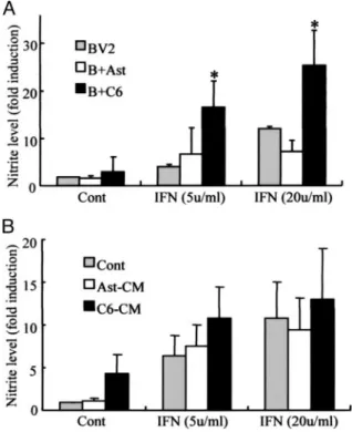
Co-culture with C6 Glioma Cells Enhances IFN-␥-induced NO Release by Microglia—The effect of astrocytes and C6 gli-oma cells on NO release by activated BV2 microglia was inves-tigated. BV2 cells were seeded on top of subconfluent layers of either astrocytes or C6 glioma cells and then stimulated with 5 or 20 units/ml IFN-␥ for 24 h. NO release was measured by determining nitrite concentrations in culture supernatants. We found that the addition of IFN-␥ resulted in NO release from microglia (Fig. 1A) but had little such effect when added to
at Ewha Medical Library on March 22, 2017
http://www.jbc.org/
astrocytes or C6 glioma cells (data not shown). IFN-␥-induced NO release was greater in microglia-glioma cell mixed cultures than in either microglia alone cell cultures or microglia-astro-cyte mixed cultures (Fig. 1A). The addition of conditioned media collected from astrocyte or glioma cell cultures had little effect on IFN-␥-stimulated NO release by BV2 cells (Fig. 1B).
Enhanced IFN-␥-induced Activation of Microglia When Cul-tured on C6 Glioma Cell-derived Matrices—The effect of astro-cyte or glioma cell insoluble matrix components on NO pro-duction by IFN-␥-stimulated microglial cells was investigated. BV2 cells (Fig. 2A) or primary cultured rat microglial cells were seeded in dishes coated with either astrocyte- or C6 glioma cell-derived matrices. We have previously shown that proin-flammatory cytokine IFN-␥ is a potent inducer of iNOS/NO generation in immortalized murine microglial cells but that IFN-␥ did not induce NO in primary rat microglia (26). We found that both primary microglia and BV2 microglia released more NO when grown on C6 glioma cell-derived matrices com-pared with astrocyte-derived matrices (Fig. 2B). The addition of IFN-␥ to BV2 and primary microglia cells caused cell death as previously described (27), and cells grown on C6 glioma cell-derived matrices showed greater levels of IFN-␥-induced cell death compared with those grown on astrocyte-derived matri-ces (Fig. 2, C and D).
The Effect of C6 Glioma Cell Insoluble Matrix Components on Microglia Proinflammatory Molecule Expression—The effect of C6 glioma cell insoluble matrix proteins on iNOS protein levels and iNOS, IL-6, IRF-1, TNF-␣, or cyclooxygenase-2 mRNA lev-els was investigated. Primary cultured rat microglial (Fig. 3, A and B) or BV2 (Fig. 3, C and D) cells were seeded in dishes coated with astrocyte- or C6 glioma cell-derived matrices and then incubated with IFN-␥ for 24 h, after which they were harvested for mRNA or protein analysis. C6 glioma cell-derived matrices enhanced IFN-␥-stimulated iNOS mRNA and protein expression both primary microglia BV2 (Fig. 3, A–D) cells. In addition, microglia grown on C6 glioma cell-derived matrices expressed IL-6 mRNA, and IFN-␥ potentiated this expression. In contrast, growth on C6 glioma cell-derived matrices had little effect on microglia IRF-1, TNF-␣, or cyclooxygenase-2 mRNA expres-sion, either alone or in the presence of IFN-␥.
C6 Glioma Cell-derived Matrices Enhance IFN-␥-induced STAT-1 and -3 Phosphorylation and GAS-luciferase Activity—Given that C6 glioma-derived matrices simulated IFN-␥-mediated iNOS expression, the effect of this matrix on microglia STAT-1 and -3 activation was examined. IFN-␥ induced minimal phosphorylation of STAT-1 and -3 FIGURE 1. The effect of astrocyte or C6 glioma co-culture or the addition of
conditioned medium from astrocytes or C6 glioma cells on IFN- ␥-stimu-lated NO release from BV2 microglia. A, BV2 cells were cultured or mixed
cul-tured for 24 h with primary rat astrocyte (B⫹Ast) or C6 (B⫹C6) rat astrocytoma cells with or without 5 or 20 units/ml IFN-␥. B, BV2 cells were incubated for 24 h with astrocyte- or C6 glioma cell-conditioned medium with or without 5 or 20 units/ml IFN-␥. After incubations, culture medium was collected and assayed for nitrite concentration as a measure of NO production. Bars, mean⫾ S.E. from four independent experiments. Data were analyzed using one-way analysis of vari-ance followed by Student’s t tests (differences were considered significant where p was⬍0.05). *, the value in BV2-C6 mixed culture conditions was greater than that observed in BV2 cells or BV2-astrocyte mixed cells following the addition of IFN-␥.
FIGURE 2. IFN-␥-stimulated NO release and survival in microglia cultured on astrocyte- or glioma
cell-derived matrices. BV2 cells (6⫻ 105cells/well in 6-well plates) (A and C) or primary cultured rat microglia cells
(2.5⫻ 104cells/well in 24-well plates) (B and D) were seeded on uncoated tissue culture dishes (Cont) or dishes
coated with astrocyte-derived (Ast-ECM) or C6 glioma cell-derived matrices (C6-ECM). Cells were then incu-bated with 5 or 20 units/ml IFN-␥. After 24 h, NO production by BV2 (A) and primary (B) microglia was deter-mined by measuring medium nitrite concentration. After 48 h, BV2 (C) and primary (D) microglial cell survival was determined using MTT assays. Bars, mean⫾ S.E. from four independent experiments. *, the value in culture on C6 matrices was greater than that observed in cells on uncoated tissue culture plates. **, the value in C6 matrices was greater than that under normal culture conditions following the addition of IFN-␥.
at Ewha Medical Library on March 22, 2017
http://www.jbc.org/
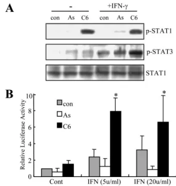
at 30 min. We found that microglia STAT-1 phosphorylation was increased when cells were seeded onto the C6 glioma-de-rived matrix and that the addition of IFN-␥ for 30 min had no further enhancing effect on this response (Fig. 4A). In contrast, the level of IFN-␥-mediated STAT-3 phosphorylation was enhanced in microglia seeded onto C6 glioma cell-derived mat-rices. Previous studies have demonstrated that IFN-␥ activates STAT-1 and -3, resulting in induction of genes containing an upstream GAS consensus sequence (28). BV2 cells were tran-siently transfected with GAS-luc reporter constructs. We found that IFN-␥ enhanced GAS-luc activity in BV2 cells seeded on C6 glioma cell-derived matrices but not astrocyte-derived matrices.
C6 Glioma Cell-derived Matrix Induces Microglia NF-B Activation—NF-B is a critical regulator of iNOS induction
(23, 28). BV2 cells were seeded on astrocyte or C6 matrices in the presence or absence of 5 or 20 units/ml IFN-␥ for 30 min, after which cells were harvested, and NF-B DNA binding activity was measured using gel mobility shift assays. We found that BV2 cells seeded on C6 glioma cell-derived matrices expressed NF-B DNA binding activity, and this was increased by IFN-␥ stimulation (Fig. 5A). Microglia NF-B transcription activity was then examined. BV2 cells were transfected with a plasmid containing a triple NF-B binding site and a luciferase reporter gene. We found that seeding onto the C6 glioma cell-derived matrix greatly increased NF-B transcription activity (Fig. 5A).
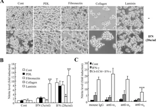
The Effect of Individual ECM Matrix Components on IFN- ␥-mediated NO Release by BV2 Cells—BV2 cells were seeded on either uncoated plastic culture dishes or on dishes coated with different ECM substrates, namely fibronectin, laminin, colla-gen, or PDL. Visual examination after 1 h of incubation showed that BV2 cells were well attached to uncoated and fibronectin-or PDL-coated dishes, weakly attached to laminin-coated dishes, and not attached to collagen-coated dishes (data not shown). After 24 h of culture, visual examination showed that BV2 cells on uncoated and fibronectin- and PDL-coated dishes FIGURE 3. IFN-␥-stimulated proinflammatory gene mRNA levels in
micro-glia cultured on astrocyte- or glioma cell-derived matrices. A and B, rat
primary microglia cells were seeded onto uncoated plastic culture dishes (con) or dishes coated with astrocyte-derived (As) or C6 glioma cell-derived matrices (C6). Cells were incubated with 50 units/ml IFN-␥ for 24 h, and total RNA was then isolated. iNOS and IL-6 mRNA expression was determined using specific primers in reverse transcription-PCR assays, with glyceralde-hyde-3-phosphate dehydrogenase mRNA levels used as a control (A) and iNOS protein levels in total cell lysates (30g) measured using immunoblot-ting (B). C and D, BV2 cells were seeded onto uncoated plastic culture dishes or dishes coated with astrocyte- or C6 glioma cell-derived matrices. Cells were stimulated with 5 or 20 units/ml IFN-␥ for 24 h, and total RNA was then iso-lated. iNOS, IL-6, IRF-1, TNF-␣, and cyclooxygenase-2 (COX-2) mRNA levels were measured using specific primers in reverse transcription-PCR assays (C). iNOS protein levels in total cell lysates (30g) were measured by immuno-blotting (D). Data are representative of three independent experiments.
FIGURE 4. STAT-1 and -3 phosphorylation and GAS-luciferase activity in
BV2 cells cultured on astrocyte- or C6 glioma cell-derived matrices in the presence or absence of IFN-␥. BV2 cells were cultured on uncoated plastic
culture dishes (con) or dishes coated with astrocyte-derived (As) or C6 glioma cell-derived matrices (C6). A, cells were stimulated with 20 units/ml IFN-␥ for 30 min. STAT-1 and -3 phosphorylation levels were measured and compared with unphosphorylated total STAT-1 protein levels in total cell lysates (30g) using immunoblotting. B, transcriptional activity of STAT proteins was deter-mined using a reporter assay system. BV2 cells were transfected with a GAS-luc reporter plasmid containing STAT binding sites and then incubated for 24 h with 5 or 20 units/ml IFN-␥, after which luciferase activity was deter-mined. The -fold induction of luciferase activity was determined by normal-izing transfection efficiency using cotransfected-galactosidase activity. Bars, mean⫾ S.E. from four independent experiments. *, the value was greater than that observed in uncoated culture dishes (con) following the addition of IFN-␥.
at Ewha Medical Library on March 22, 2017
http://www.jbc.org/
were well spread and that the addition of IFN-␥ resulted in typical elongated cells with processes (Fig. 6A). In contrast, BV2 cells on laminin-coated dishes were round and loosely attached, and the addition of IFN-␥ enhanced cell adhesion and spread-ing. BV2 cells in collagen-coated dishes were nonadherent and round (but were viable according to trypan blue staining (data not shown)), and the addition of IFN-␥ did not appear to mark-edly affect adhesion. Medium was collected after 24 h to test for NO production. We found that compared with BV2 cells on noncoated dishes, IFN-␥-stimulated microglia NO production was greater in cells on laminin-coated dishes and lesser in cells on collagen-coated dishes (Fig. 6B).
In order to address the role of specific integrin in microglial response to IFN-␥, we examined microglial NO in the presence of different integrin function-blocking antibodies. As shown in Fig. 6C, microglial NO release in response to IFN-␥ was reduced significantly by a well characterized integrin␣6 -block-ing antibody (GoH3) (42.9⫾ 15.6% of control IgG; p ⬍ 0.001) but not by␣1(mAb 1973Z) or␣2(mAb 1952Z) monoclonal antibodies.
IFN-␥ Down-regulates Activation of Cell Adhesion/Migration Molecules—It has been previously shown that microglial adhe-sion and morphology vary greatly, depending upon the type of ECM substrate (20). Therefore, we investigated how matrices of C6 glioma cells influence microglia cell adhesion/migration molecules. BV2 cells were seeded on dishes that were either uncoated (control) or coated with astrocyte or C6 glioma cell-derived matrices and then incubated for 24 h with/without 20
units/ml IFN-␥, after which they were examined under a micro-scope and harvested to assess cell adhesion molecule expression/ phosphorylation/activation. BV2 cells cultured on astrocyte- or C6 glioma cell-derived matrices were round and adhered strongly to the dishes (Fig. 7A). In the absence of IFN-␥, the morphology of BV2 cells cultured on C6 glioma cell-derived matrices was more aggregated compared with the well spread cells observed on astro-cyte-coated or uncoated control plates. The presence of IFN-␥ resulted in cells on the C6 glioma cell-derived matrix showing a more round and loosely bound phenotype compared with the well spread amoeboid-shaped cells in the control culture or in the astrocyte ECM culture (Fig. 7A). We then investigated cell adhe-sion molecules and found that IFN-␥ down-regulated activation of Rac and phosphorylation of FAK, cofilin, and Src but had no effect on the levels of these proteins (Fig. 7B). C6 matrices did not influ-ence the expression/activation of adhesion molecules either alone or in combination with IFN-␥.
DISCUSSION
Although microglia have been shown to play a role in immune defense against glioma cell proliferation (29, 30), it is unclear whether the presence of intratumoral microglia repre-sents a nonspecific reaction to tissue injury, whether microglia are manipulated by gliomas to support glioma growth, or whether microglia contribute to tumor cell death.
Using both the BV2 mouse microglial cell line and primary cultured rat microglial cells, the present study investigated the influence of glioma cell insoluble matrix substrates on both nonactivated (without IFN-␥) and activated (with IFN-␥) microglial responses. The study found that when seeded onto C6 glioma cell-derived matrices, microglia displayed enhanced IFN-␥-stimulated iNOS/NO induction, and this appeared to involve increased STAT-1 and -3 and NF-B activity. The study also found that the purified ECM molecules collagen and lami-nin differentially affected microglia IFN-␥-stimulated NO release. Laminin may be a potentially key ECM molecule to regulate microglia activation.
A previous study reported that fibronectin and vitronectin, but not laminin, promoted microglia activation by inducing major histocompatibility complex class I and also increased expression of the␣41and Mac-1 integrins (20). In the present study, laminin enhanced, whereas collagen suppressed, IFN- ␥-mediated NO generation. Our experiments demonstrated that microglia express the major laminin receptor subunit␣6, and this was increased by IFN-␥, whereas microglia displayed no expres-sion for the major collagen binding integrin subunits␣1and␣2 (data not shown). Furthermore, microglial NO release in response to IFN-␥ was specifically reduced by a well characterized integrin ␣6-blocking antibody (GoH3). This result shows that enhanced NO release by glioma ECM in response to IFN-␥ seems to be crit-ically regulated by␣6, a major laminin receptor of microglia.
It is possible that C6 glioma cells produce more laminin com-pared with normal astrocytes, which may cause increased microglia NO release. However, we found that neither STAT nor NF-B was activated in microglia cultured on laminin-coated plates (data not shown), yet these transcription factors were activated when microglia were cultured on C6 glioma cell-derived matrices. It may be that glioma cells produce and FIGURE 5. NF-B activity in microglia cultured on astrocyte- or C6 glioma
cell-derived matrices. BV2 cells were cultured on uncoated plastic culture
dishes (con) or dishes coated with astrocyte-derived (As) or C6 glioma cell-derived matrices (C6). A, cells were incubated with IFN-␥ (20 units/ml) for 30 min. NF-B DNA binding activity in 6-g nuclear extracts was measured using gel mobility shift assays. B, NF-B transcriptional activity was assayed in BV2 cells after transfection of cells with an NF-B-luc reporter plasmid containing three NF-B binding sites. Transfected cells were incubated with 20 units/ml IFN-␥, and luciferase activities were determined after 24 h. The -fold induction of luciferase activity was determined by normalizing transfection efficiency using cotransfected-galactosidase activity. Data are representative of four (A) or three (B) separate experiments. *, a significant difference from luciferase activity in cells of control culture (con).
at Ewha Medical Library on March 22, 2017
http://www.jbc.org/
release different amounts or differ-ent combinations of ECM sub-strates compared with normal astrocytes, and certain combina-tions of ECM molecules or unknown ECM components may be critically involved in microglia iNOS induction. Indeed, we have found that mRNA levels of fibronectin, laminin, collagen, and vitronectin are similar in both C6 glioma cells and normal astrocytes (data not shown). However, we have yet to compare these two cell types in terms of the extracellular levels of these ECM proteins, mainly because such assays are difficult due to the proteins having an insoluble nature and a large degree of modification (especially glycosylation). Thus, defining the exact nature, the pro-portion, and the biochemical modi-fication of each glioma cell ECM component and the roles of these components in microglial iNOS induction may not be possible in the near future. Nonetheless, we main-tain that our study has some value in establishing that gliomas them-selves may regulate microglial immune responses via components of their insoluble ECM.
The present study also found that IFN-␥ down-regulated the activa-tion of cell molecules associated with cell adhesion/migration, including FAK, cofilin, Src, and Rac, and that the activation of these mol-ecules was not specifically affected when microglia were seeded on C6 glioma cell-derived matrices. With virtually all cell types, ECM-recep-tor binding leads to activation of the focal adhesion complex, and FAK is well suited to integrate these diverse signaling activities (31, 32). Auto-phosphorylation of FAK initiates the formation of dynamic molecular complexes that contain numerous signaling proteins (e.g. Src, p85 reg-ulatory subunit of phosphatidylino-sitol 3-kinase, and mitogen-acti-vated protein kinases). The phosphatidylinositol 3-kinase and mitogen-activated protein kinase pathways are important in the regu-lation of microglial activation/NO production (23). The mechanisms FIGURE 6. IFN-␥-stimulated NO release in BV2 cells cultured on various purified ECM components and
the effect of microglial laminin receptor␣6. A and B, BV2 cells were cultured on uncoated plastic dishes or
dishes coated with PDL, fibronectin, collagen, or laminin, with or without 5 or 20 units IFN-␥. After 24 h, microglial cell phenotypes were assessed using phase-contrast light microscopy (A), and NO production was determined by measuring culture medium nitrite levels (B) (-fold increases are shown). Bars, mean⫾ S.E. from four independent experiments. *, the value was significantly greater than that observed following the addition of IFN-␥ to cells in uncoated dishes. **, the value in collagen matrices was lower than that under normal culture conditions following the addition of IFN-␥. C, BV2 cells were preincubated with normal mouse IgG (100 g/ml), anti-integrin␣1mAb (mAb 1973Z) (50g/ml), anti-integrin ␣2mAb (mAb 1950Z), or anti-integrin␣6(GoH3) (50
g/ml) for 60 min prior to plating to a control plastic culture dish (cont and IFN-␥) or C6 cell-derived matrices (C6-ECM⫹ IFN-␥). Cells were then stimulated with 5 units/ml IFN-␥ for 24 h, and NO production was deter-mined by measuring medium nitrite concentration. ***, the value was significantly reduced compared with IFN-␥-treated BV2 cells seeded on C6-ECM. Data are means ⫾ S.D. of triplicate determinations.
FIGURE 7. IFN-␥-mediated down-regulation of cell adhesion/migration protein activation and microglia
morphological changes in microglia cultured on C6 glioma cell-derived matrices. BV2 cells were seeded
onto control uncoated culture dishes (Cont) or onto dishes coated with C6 glioma cell-derived matrices (C6-ECM) or coated with astrocyte-derived matrices (Ast-(C6-ECM) and then incubated with or without 20 units/ml IFN-␥ for 24 h, after which whole cell lysates were prepared, and cell phenotype was observed. A, cell morphol-ogy was assessed using phase-contrast light microscopy. Data are representative of three independent exper-iments. B, equal masses of lysate protein were analyzed by immunoblotting using specific antibodies. For Rac1-GTP analysis, pull-down assays were performed in which cell lysates were incubated with recombinant GST-PAK1-(76 –150) bound to glutathione-coated Sepharose beads.
at Ewha Medical Library on March 22, 2017
http://www.jbc.org/
underlying IFN-␥-mediated down-regulation of cell-adhesion/ migration signaling remains to be elucidated. Furthermore, future studies should be followed to examine the role of adhe-sion signaling in iNOS/NO regulation in microglia.
The unusually high levels of nitrite detected in IFN-␥-treated microglia cells cultured on glioma cell-derived mat-rices probably reflect a nonspecific rather than specific reac-tion by microglial cells to the insoluble ECM components. Microglia responded similarly but to a lesser degree when cultured on top of glioma cell monolayers. This difference may be due to the matrix containing additional intracellular glioma cell-stimulatory factors that adhered to the matrix following glioma cell lysis during matrix preparation. If such insoluble intracellular factors were responsible for the addi-tional increase in NO production in IFN-␥-treated micro-glial cells, we might expect the addition of cellular extracts to increase nitrite production in IFN-␥-treated microglial cells. However, we found that incubation of BV2 cells with lysed whole cell extracts containing both soluble and insoluble cellular components prepared from detached C6 glioma cells caused only a mild increase in NO release and did not enhance IFN-␥-stimulated NO production (data not shown). The effect of the glioma cell-insoluble matrix on microglia iNOS/NO release was tumor cell-specific, since there was little or no such effect when microglia were grown in the presence of primary cultured rat astrocyte cells or astrocyte-insoluble matrix. Furthermore, astrocyte-insoluble matrices of other glioma cells, such as CRT-MG and U87MG, resulted in a similar stimulatory effect on iNOS/NO production in IFN-␥-stimulated microglia (data not shown). The effects of ECM from three nonglial tumor cell lines, two human lung cancer cell lines (A549 and H460), and human cervical cancer HeLa cells, on IFN-␥-mediated microglial NO generation were also examined. Interestingly, BV2 microglia cells seeded on matrices of three nonglial cancer cells did not show enhanced IFN-␥ response (data not shown). It is thus likely that glioma cells specifically secrete insoluble matrix com-ponents regulating microglial behavior. Despite these find-ings, the details surrounding glioma cell ECM components and their roles in microglia activation remain unknown. One obstacle in the study of ECM function is the difficulty in separating ECM components from other insoluble materials. A variety of insoluble substances with various modifications released by gliomas may mediate iNOS induction in micro-glia, and ECM expression may differ both between tumor types and even between tumors of the same type. Thus, the changes occurring in released or surface-insoluble C6 gli-oma cell molecules as a consequence of adhesive interaction with microglia probably play a key role in NO production by microglial cells. It is unlikely that the effect was due to lin-gering soluble factors, because the matrices were extensively washed before microglial cells were seeded.
The current findings support the view that microglia accu-mulation in gliomas reflects microglia participation in active immune defense through production of NO and that gliomas themselves may regulate this phenomena via their ECM com-position. Few studies have focused on the regulatory effects of tumor cell ECM molecules. We believe that the present study may assist in providing clues regarding the regulation of brain inflammation by tumor cell-derived insoluble components.
REFERENCES
1. Aloisi, F. (1999) Adv. Exp. Med. Biol. 468, 123–133
2. Jack, C., Ruffini, F., Bar-Or, A., and Antel, J. P. (2005) J. Neurosci. Res. 81, 363–373
3. Stoll, G., and Jander, S. (1999) Prog. Neurobiol. 58, 233–247 4. Morris, C. S., and Esiri, M. M. (1991) J. Neurol. Sci. 101, 47–58 5. Roggendorf, W., Strupp, S., and Paulus, W. (1996) Acta Neuropathol.
(Berl.) 92, 288 –293
6. Raines, E. W. (2000) Int. J. Exp. Pathol. 81, 173–182
7. Gladson, C. L. (1999) J. Neuropathol. Exp. Neurol. 58, 1029 –1040 8. Werb, Z., and Chin, J. R. (1998) Ann. N. Y. Acad. Sci. 857, 110 –118 9. Dustin, M. L., and Springer, T. A. (1991) Annu. Rev. Immunol. 9, 27– 66 10. Diamond, M. S., and Springer, T. A. (1994) Curr. Biol. 4, 506 –517 11. Huttenlocher, A., Sandborg, R. R., and Horwitz, A. F. (1995) Curr. Opin.
Cell Biol. 7,697–706
12. Lauffenburger, D. A., and Horwitz, A. F. (1996) Cell 84, 359 –369 13. Egan, R. A., and Vijayan, V. K. (1991) Brain Res. 568, 330 –334
14. Frisen, J., Haegerstrand, A., Risling, M., Fried, K., Johansson, C. B., Ham-marberg, H., Elde, R., Hokfelt, T., and Cullheim, S. (1995) Neuroscience 65, 293–304
15. Niquet, J., Gillian, A., Ben-Ari, Y., and Represa, A. (1996) Glia 16, 359 –367 16. Laywell, E. D., and Steindler, D. A. (1991) Ann. N. Y. Acad. Sci. 633, 122–141 17. Feuerstein, G. Z., Wang, X., and Barone, F. C. (1998)
Neuroimmunomodu-lation 5,143–159
18. Raivich, G., Bohatschek, M., Kloss, C. U., Werner, A., Jones, L. L., and Kreutzberg, G. W. (1999) Brain Res. Brain Res. Rev. 30, 77–105 19. Milner, R., and Campbell, I. L. (2002) J. Neurosci. 22, 1562–1572 20. Milner, R., and Campbell, I. L. (2003) J. Immunol. 170, 3850 –3858 21. Nakagawa, T., Kubota, T., Kabuto, M., Fujimoto, N., and Okada, Y. (1996)
J. Neurooncol. 28,13–24
22. Kloss, C. U., Bohatschek, M., Kreutzberg, G. W., and Raivich, G. (2001) Exp. Neurol. 168,32– 46
23. Kim, W. K., Hwang, S. Y., Oh, E. S., Piao, H. Z., Kim, K. W., and Han, I. O. (2004) J. Immunol. 172, 7015–7023
24. Sola, C., Casal, C., Tusell, J. M., and Serratosa, J. (2002) Eur. J. Neurosci. 16, 1275–1283
25. Mettouchi, A., Klein, S., Guo, W., Lopez-Lago, M., Lemichez, E., Westwick, J. K., and Giancotti, F. G. (2001) Mol. Cell 8, 115–127 26. Han, I. O., Kim, H. S., Kim, H. C., Joe, E. H., and Kim, W. K. (2003)
J. Neurosci. Res. 73,659 – 669
27. Hwang, S. Y., Jung, J. S., Lim, S. J., Kim, J. Y., Kim, T. H., Cho, K. H., and Han, I. O. (2004) Biochem. Biophys. Res. Commun. 318, 691– 697 28. Kim, H. S., Whang, S. Y., Woo, M. S., Park, J. S., Kim, W. K., and Han, I. O.
(2004) J. Neuroimmunol. 151, 85–93
29. Labeur, M. S., Roters, B., Pers, B., Mehling, A., Luger, T. A., Schwarz, T., and Grabbe, S. (1999) J. Immunol. 162, 168 –175
30. Rosales, A. A., and Roque, R. S. (1997) Brain Res. 748, 195–204 31. Burridge, K., and Chrzanowska-Wodnicka, M. (1996) Annu. Rev. Cell Dev.
Biol. 12,463–518
32. Guan, J. L. (1997) Matrix Biol. 16, 195–200
at Ewha Medical Library on March 22, 2017
http://www.jbc.org/
Inn-Oc Han
Yoon-Jung Kim, So-Young Hwang, Ji-Sun Hwang, Jung-Weon Lee, Eok-Soo Oh and
Inducible Nitric-oxide Synthase/Nitric Oxide Production in BV2 Microglial Cells
doi: 10.1074/jbc.M610219200 originally published online November 2, 2007 2008, 283:2526-2533.
J. Biol. Chem.
10.1074/jbc.M610219200 Access the most updated version of this article at doi:
Alerts:
When a correction for this article is posted •
When this article is cited •
to choose from all of JBC's e-mail alerts Click here
http://www.jbc.org/content/283/5/2526.full.html#ref-list-1
This article cites 32 references, 5 of which can be accessed free at
at Ewha Medical Library on March 22, 2017
http://www.jbc.org/