illing materials: evaluation by micro-computed tomography
3
연세대학교 치과대학 치과보존과학교실1), 부산대학교 치의학전문대학원 치과보존과학교실2),
국민건강보험공단일산병원 치과보존과3)
김 민 영1), 김 현 철2), 곽 상 원2), 윤 태 철3), 김 의 성1)*
Effect of Nd:YAG laser irradiation on
adherence of retrograde filling
materials: evaluation by
micro-computed tomography
Effect of Nd:YAG laser irradiation on adherence of retrograde filling materials: evaluation by micro-computed tomography
1)Microscope Center, Department of Conservative Dentistry and Oral Science Research Center, College of Dentistry, Yonsei University
2)Department of Conservative Dentistry, School of Dentistry, Pusan National University 3)Department of Conservative Dentistry, National Health Insurance Service Ilsan Hospital Minyoung Kim1), Hyeon-cheol Kim2), Sang Won Kwak2), Tai Cheol Yoon3), Euiseong Kim1)*
Background/Purpose: The purpose of this study was to evaluate the effect of Nd:YAG irradiation on adherence of retrograde filling materials (mineral trioxide aggregate [MTA] and Super-EBA) by micro-computed tomography (CT) measurement and to observe the dentinal surface after irradiation by scanning electron microscopy (SEM).
Materials and methods: Forty retrofilling models using extracted human teeth were divided into four groups according to the material and method used: ProRoot MTA (MTA group), Super-EBA (EBA group), MTA with Nd:YAG laser irradiation (LMTA group), and Super-EBA with Nd:YAG laser irradiation (LEBA group). All specimens were stored in 100% humidity for 24 hours until micro-CT was performed. The gap volume of the tooth/material interface was measured using the CTAn program. In six samples, the laser-irradiated dentin surface was observed using SEM.
Results: The mean percent difference in gap volume was not statistically significant between the Nd:YAG laser-irradiated groups and non-irradiated in both materials(P > 0.05). The gap volume in the MTA group was significantly lower than that in the EBA group (P < 0.05). Examination of the non-irradiated specimens by SEM showed patent dentinal tubules. In contrast, alterations in the texture of the dentin surface and obliteration of the dentinal tubules were evident in the Nd:YAG laser-irradiated specimens.
Conclusion: In this study, changes in the dentinal surface after Nd:YAG irradiation did not affect adherence between the apical filling material and the dentin wall.
Key words : apical seal; dentinal surface; laser device; microleakage; surgical endodontics; retrofill
ABSTRACT
Corresponding Author Euiseong Kim DDS, MSD, PhD
Professor, Department of Conservative Dentistry Yonsei University, Seoul, Korea 50 Yonsei-ro, Seodaemnu-gu, Seoul, 03722, Korea
Ⅰ. Introduction
Surgical endodontics is often indicated when nonsurgical endodontic treatment is unsuccessful and when its results are not predictable. According to Harty et al.1), the most important determinant of the success of apical surgery is the efficiency of the apical seal. Some irritants may remain after cleansing and shaping of the root canal system, and could gain access to the periapical tissues via the interface between the filling materials and dentin in the root canal wall. Therefore, the major objective of periradicular surgery with placement of an adequate root-end filling is to provide an apical seal that inhibits migration of irritants2).
Various types of materials have been proposed for sealing the root-end cavity, including amalgam, Cavit, gutta-percha, composite resins, zinc oxide-eugenol cements, alloys, glass ionomer, and gold foil3-6). Historically, amalgam has been the most widely used root-end filling material, but recent studies have shown less leakage with alternative filling materials3, 4). Intermediate restorative material and Super-EBA, a reinforced zinc oxide-eugenol cement, are alternatives to amalgam, and have been shown to provide a better seal3). Further, mineral trioxide aggregate (MTA) has shown promise as a root-end filling material5, 6).
The laser device is an important tool with a wide range of biomedical applications. Maillet et al7). have recommended use of the Nd:YAG laser to optimize apical surgery. Irradiation from this device promotes melting of the dentin surface,
which may result in obliteration of the dentinal tubules and decreased permeability7, 8). These effects could reduce apical microleakage, which is one of the major factors associated with unsuccessful periapical surgery.
In the past, leakage of root-end filling materials has been measured by penetration of dyes, isotopes, and microorganisms. All of these techniques have been shown to have shortcomings9). The extent of leakage may be quantified using a length scale, and these methods may be destructive or semi-quantitative, relying on visual assessment by examiners. On the other hand, assessment using micro-computed tomography (CT) is not destructive and allows three-dimensional visualization and repeated measurement. This method has been used to evaluate microleakage caused by polymerization shrinkage of restorative materials and to assess the quality of root canal filling10, 11).
The purpose of this in vitro study was to evaluate the effect of Nd:YAG laser irradiation on apical leakage after retrograde filling using MTA and Super-EBA cement and to observe the dentinal surface after irradiation by scanning electron microscopy
Ⅱ. Materials and Methods
Sample preparation
Forty six single-rooted human teeth, recently extracted for periodontal reasons, were collected
illing materials: evaluation by micro-computed tomography
for use in this study. Teeth with root caries, previous root canal filling, fracture, multiple canals, and/or excessive curvatures were excluded. All the teeth were cleared of attached soft tissue and stored in normal saline solution until use.
Clinical crowns were sectioned at the cement-enamel junction with a diamond bur in a high-speed handpiece with continuous water spray. Patency of the apical foramen was determined with a size 15 file. When the file tip appeared flush with the apical foramen, the length of the file was recorded and the working length was determined to be 1 mm short of the measured length. The root canal system was instrumented by the crown-down technique using ProTaper Universal nickel-titanium rotary instruments (Dentsply Maillefer, Ballaigues, Switzerland) to the size of F3. NaOCl 2.5% was used to irrigate the canals throughout the instrumentation process. After instrumentation, the smear layer was removed using 5 mL of 17% ethylenedia minetetraacetic acid for 1 minute and a final rinse with 5 mL of 2.5% NaOCl. After being cleaned and shaped, the canals were dried with paper points and obturated with gutta-percha and AH Plus sealer(Dentsply DeTrey, Konstanz, Ger many) with the continuous wave of condensation technique.
An apical resection at 90° to the long axis of the tooth was made at 3 mm from the root end using a high-speed diamond bur. Root-end cavities of 3 mm were prepared with an ultrasonic tip(KiS, Obtura Spartan, Fenton, MO, USA).
The teeth were randomly divided into four
groups comprising 10 teeth each. Root-end fillings were performed with ProRoot MTA (Dentsply Tulsa Dental, Tulsa, OK, USA) without laser treatment(MTA group) or Super-EBA(Bosworth Company, Skokie, IL, USA) without laser treatment(EBA group). In the LMTA and LEBA groups, the retrograde cavities were irradiated(150 mJ; 10 Hz; 1.5 W, 10 seconds) with Nd:YAG laser(B&B Systems, Samsung Electronics, Seoul, Korea) before retrograde filling with MTA or Super-EBA. All the procedures were undertaken by a single operator using dental microscopy(OPMI Pico, Carl Zeiss, Göttingen, Germany). The root-end filled specimens were stored in an incubator with a humidity of 100% and a temperature of 37°C.
Micro-CT scanning
Each specimen was scanned using micro-CT (Skyscan 1076, SkyScan, Kontich, Belgium; X-ray source voltage 100kV; beam current 100 A; filter Al 0.5mm thick; pixel size 9 m; exposure time 4700msec). All CT images were three-dimensionally reconstructed by NRecon software (Skyscan) and the gap volume of the tooth/material interface was measured using the CTAn(Skyscan) program.
Micro-CT analysis was performed by a single examiner who didn’t know the grouping to assess the gap volume of the tooth/material interface. The range of analysis for each specimen was up to 2 mm from the resected root-end surface, and the area of the filling material and interfacial gap
was set depending on the radiographic density: density range 80-255; filling material 0-23; interfacial gap(obtained by averaging). Based on these settings, the volume of filling material (VM)
and the volume of the interfacial gap(VG) were
measured, and finally the percentage of the interfacial gap volume to the total volume of retrograde cavity was calculated(VG/VG+VM).
Observation by SEM
Six specimens without root-end fillings were produced in the same way used for the four groups. The retrocavities in three specimens were irradiated by Nd:YAG laser and the remaining specimens which were not irradiated served as the control group. The six specimens were then longitudinally split for observation of the inner dental surface by scanning electron microscopy (SEM, S-3000N, Hitachi, Tokyo, Japan). The samples were dehydrated in an ascending series of ethanol concentrations. The completely dehydrated specimens were mounted separately
on aluminum stubs coated with gold/palladium, and then examined by SEM.
Statistical analysis
The two-sample t-test was performed using PASW Statistics version 18.0 software(SPSS Inc., Chicago, IL, USA) to test for statistically significant differences between the groups. A value of P < 0.05 was considered to indicate statistical significance.
Results
The gap volume of the teeth in each experimental group is presented in Table 1. The gap volume in the MTA group was significantly lower than that in the EBA group(P < 0.05). The laser-irradiated LMTA and LEBA groups were not significantly different from the non-irradiated groups(P > 0.05).
Micro-CT images reconstructed three-dimensionally showed that the EBA group had a
Figure 1. Three-dimensional reconstruction of micro-computed tomography images of retrograde filling material and surrounding gap (dots). (A) Mineral trioxide aggregate group. (B) EBA group
illing materials: evaluation by micro-computed tomography
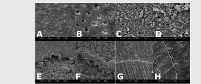
larger gap volume than the MTA group(Figure 1). Examination of the non-irradiated specimens by SEM showed patent dentinal tubules(Figures 2A and 2B). On the other hand, changes in the texture of the dentin surface and obliteration of dentinal tubules were evident in the Nd:YAG laser-irradiated specimens(Figures 2C and 2D). An irregular surface was formed by melting and recrystallization of the dentin particles, and the dentinal tubules were partially blocked. Super EBA/tooth interface(Figure2 G, H) showed
crack-like gap while mineral trioxide aggregate/tooth interface(Figue2 E, F) showed no gap.
Ⅲ. Discussion
The main objective of apical surgery is to provide a favorable apical seal that prevents movement of bacteria and diffusion of bacterial products from the root canal system into the
Table 1. Mean percent gap volume of the tooth/material interface in each study group
Notes: There were no significant differences between the laser and non-laser groups (p > 0.05).
a,bThe MTA group showed a significantly smaller percent gap volume (p < 0.05). Abbreviations: MTA, mineral trioxide aggregate; SD, standard
deviation
MTA 0.92 ± 2.06a
Super-EBA 6.60 ± 6.99b
Laser irradiation/MTA 1.36 ± 1.34a
Laser irradiation/Super-EBA 5.51 ± 5.34b
Group Mean ± SD
Figure 2. Scanning electron micrographs of the dentin surface after preparation of the cavity (A-D) and material/tooth interface after retrograde filling (E-H). (A, B) Dentin surface of the cavity without laser irradiation (A, 2000×, B; 5000×). (C, D) Cavity surface after laser irradiation (C, 2000×; D, 5000×). (E, F) Mineral trioxide aggregate/tooth interface (E, 300×; F, 1000×). (G, H) Super EBA/tooth interface (G, 300×; H, 1000×).
periapical tissues.
A large number of in vitro studies dealing with the marginal adaptation and sealing ability of retrograde materials has been published. The methodology of these leakage studies and their results, which are often contradictory, are now being questioned with regard to their clinical relevance12-14). Factors such as choice of storage
solution, pH, molecular weight of the dye used, and entrapped air pressure may have a critical effect on the outcome of these in vitro studies9, 12).
The most widely used method, i.e., penetration of dye or protein and linear measurement of the tracer, should be considered as a semiquantitative technique because it does not provide any information about the volume of penetrated tracer. Further, these methods are difficult to reproduce because the specimens are split or cross-sectioned before measurement15).
The model bacterial system is more clinically relevant16); however, the results of studies using
this method may be contradictory depending on the penetrating depth and bacterial species
used17-20).
In the present study, micro-CT was used to measure the gap volume of the tooth/material interface after retrograde filling. High-resolution micro-CT is an emerging technology with several promising applications in dentistry, and its use has increased markedly during the past two decades10, 21, 22). In the field of endodontic
research, micro-CT technology has been used to evaluate root canal anatomy and to assess root canal morphology after instrumentation21, 22).
Evaluation of root canal obturation has also been
possible by measuring the percentage volume of voids and gaps in obturated root canals using micro-CT10).
To our knowledge, this study is one of the first to use micro-CT to measure the volume of interfacial gaps in retrograde fillings. Although the gap volume was extremely small and the difference that occurred depended on the setting, measurement by micro-CT and analysis can provide more accurate information than a two-dimensional technique. It has been reported to be a stable, repeatable and nondestructive method for indirect evaluation of the sealing ability of retrofilling materials21).
In this study, when comparing the gap volume of the tooth/material interface using micro-CT, the mean gap volume in the MTA group was significantly lower than in the EBA group. This result might be due to the physical properties of the materials themselves. While MTA has an affinity for moisture, needs moisture to set and it expands slightly, Super-EBA is sensitive to moisture and the manipulation technique used. This sensitivity might be the cause of crack-like discrepancy. In particular, an inadequate powder-to-liquid ratio or mixing with bubbles causes shrinkage, resulting in long-term leakage. Therefore, Super-EBA needs to be used with caution23).
Irradiation with high-intensity lasers have been shown to reduce both bacterial levels and dentinal permeability24). An in vitro study using
penetration of methylene blue to evaluate the effect of laser irradiation on the sealing ability of retrograde filling materials demonstrated that
illing materials: evaluation by micro-computed tomography
specimens irradiated with laser had lower infiltration indices than controls25). Although still
controversial, this effect has been explained by morphologic and physical changes in the dentin surface after laser irradiation. During laser irradiation, energy is absorbed in calcified tissues (containing hydroxyapatite for example) causing thermochemical ablation and recrystallization. The altered dentinal surface shows glazing and melting of the smear layer and surface dentin, along with occlusion of the dental tubules26, 27).
However, in the present study, which used micro-CT for analysis, the mean percent gap volume in the Nd:YAG laser-irradiated group was not significantly different from that in the non-irradiated group. The results indicate that changes in the dentinal surface after Nd:YAG irradiation did not alter the pattern of contact between the apical filling material and the dentin wall. Nevertheless, observation by SEM confirmed that the smear layer and debris could be removed and the dentinal tubule could be obliterated after Nd:YAG laser irradiation.
There are two avenues by which leakage could occur at the apex of a root sealed with a retrograde filling. The first is by apical microleakage, i.e., leakage along the interface between the filling material and the canal wall, and the second is by flow of fluids and substances along open tubules at the resected root end, i.e., via permeable apical dentin28).
This study has shown that Nd:YAG irradiation can block the second pathway, but not the first one.
Micro-CT analysis is an indirect way of evaluating the sealing ability of retrograde fillings. However, the results of more clinically relevant research using a dye or bacterial penetration technique vary depending on the experimental factors or conditions used. From this point of view, because it is more stable, rapid to perform, and non-destructive, the micro-CT technique would be a useful supplementary means of evaluating the sealing ability of retrograde fillings. In particular, being able to repeat CT scanning would make it possible to evaluate changes in sealing ability over time. Future studies of the various retrofilling materials available and their sealing ability using such non-destructive techniques would help to increase the success rate of endodontic surgical interventions.
Within the limitation of this study, Nd:YAG laser irradiation did not have any effect on the decrease in tooth/material gap volume. However, SEM confirmed that Nd:YAG laser irradiation alters the texture of the dentin surface but does not change the adherence between the apical filling material and the dentin wall. Additionally, form the result of this study, MTA has better sealing ability as a root-end filling material than Super-EBA.
Ⅳ. Conflicts of interest
The authors deny any conflicts of interest related to this study.
Ⅴ. Acknowledgements
Minyoung Kim and Hyeon-Cheol Kim contributed equally to this work and share the first authorship. This research was supported by
Basic Science Research Program through the National Research Foundation of Korea(NRF) funded by the Ministry of Education (2015R1D1A1A09057552).
1. Harty FJ, Parkins BJ, Wengraf AM. The success rate of apicectomy. A retrospective study of 1,016 cases. Br Dent J 1970;129(9):407-413.
2. Gutmann JL, Harrison JW. Surgical endodontics. Boston: Blackwell Scientific Publications, 1991. 3. Dorn SO, Gartner AH. Retrograde filling materials: a
retrospective success-failure study of amalgam, EBA, and IRM. J Endod 1990;16(8):391-393.
4. Frank AL, Glick DH, Patterson SS, Weine FS. Long-term evaluation of surgically placed amalgam fillings. J Endod 1992;18(8):391-398.
5. Lee SJ, Monsef M, Torabinejad M. Sealing ability of a mineral trioxide aggregate for repair of lateral root perforations. J Endod 1993;19(11):541-544. 6. Torabinejad M, Watson TF, Pitt Ford TR. Sealing
ability of a mineral trioxide aggregate when used as a root end filling material. J Endod 1993;19(12):591-595.
7. Maillet WA, Torneck CD, Friedman S. Connective tissue response to root surfaces resected with Nd:YAG laser or burs. Oral Surg Oral Med Oral Pathol Oral Radiol Endod 1996;82(6):681-690. 8. Pozza DH, Fregapani PW, Xavier CB, et al. CO(2),
Er: YAG and Nd:YAG lasers in endodontic surgery.
J Appl Oral Sci 2009;17(6):596-599.
9. Oliver CM, Abbott PV. Entrapped air and its effects on dye penetration of voids. Endod Dent Traumatol 1991;7(3):135-138.
10 Hammad M, Qualtrough A, Silikas N. Evaluation of root canal obturation: a three-dimensional in vitro study. J Endod 2009;35(4):541-544.
11. Zeiger DN, Sun J, Schumacher GE, Lin-Gibson S. Evaluation of dental composite shrinkage and leakage in extracted teeth using X-ray microcomputed tomography. Dent Mater 2009;25(10):1213-1220.
12. Gondim E, Jr., Kim S, de Souza-Filho FJ. An investigation of microleakage from root-end fillings in ultrasonic retrograde cavities with or without finishing: a quantitative analysis. Oral Surg Oral Med Oral Pathol Oral Radiol Endod 2005;99(6):755-760.
13. Xavier CB, Weismann R, de Oliveira MG, et al. Root-end filling materials: apical microleakage and marginal adaptation. J Endod 2005;31(7):539-542. 14. Pichardo MR, George SW, Bergeron BE, et al.
Apical leakage of root-end placed SuperEBA, MTA, and Geristore restorations in human teeth
illing materials: evaluation by micro-computed tomography
previously stored in 10% formalin. J Endod 2006;32(10):956-959.
15. Wu MK, Wesselink PR. Endodontic leakage studies reconsidered. Part I. Methodology, application and relevance. Int Endod J 1993;26(1):37-43.
16. Yildirim T, Er K, Tasdemir T, et al. Effect of smear layer and root-end cavity thickness on apical sealing ability of MTA as a root-end filling material: a bacterial leakage study. Oral Surg Oral Med Oral Pathol Oral Radiol Endod 2010;109(1):e67-72.
17. Torabinejad M, Rastegar AF, Kettering JD, Pitt Ford TR. Bacterial leakage of mineral trioxide aggregate as a root-end filling material. J Endod 1995;21(3):109-112.
18. Scheerer SQ, Steiman HR, Cohen J. A comparative evaluation of three root-end filling materials: an in vitro leakage study using Prevotella nigrescens. J Endod 2001;27(1):40-42.
19. Mangin C, Yesilsoy C, Nissan R, Stevens R. The comparative sealing ability of hydroxyapatite cement, mineral trioxide aggregate, and super ethoxybenzoic acid as root-end filling materials. J Endod 2003;29(4):261-264.
20. Maltezos C, Glickman GN, Ezzo P, He J. Comparison of the sealing of Resilon, Pro Root MTA, and Super-EBA as root-end filling materials: a bacterial leakage study. J Endod 2006;32(4):324-327.
21. Jung M, Lommel D, Klimek J. The imaging of root canal obturation using micro-CT. Int Endod J
2005;38(9):617-626.
22. Barletta FB, Rahde Nde M, Limongi O, et al. In vitro comparative analysis of 2 mechanical techniques for removing gutta-percha during retreatment. J Can Dent Assoc 2007;73(1):65. 23. Song M, Shin SJ, Kim E. Outcomes of endodontic
micro-resurgery: a prospective clinical study. J Endod 2011;37(3):316-320.
24. Bergmans L, Moisiadis P, Teughels W, et al. Bactericidal effect of Nd:YAG laser irradiation on some endodontic pathogens ex vivo. Int Endod J 2006;39(7):547-557.
25. Oliveira RG, Gouw-Soares S, Baldochi SL, Eduardo CP. Scanning electron microscopy (SEM) and optical microscopy: effects of Er:YAG and Nd:YAG lasers on apical seals after apicoectomy and retrofill. Photomed Laser Surg 2004;22(6):533-536. 26. Lee BS, Lin CP, Lin FH, Lan WH. Ultrastructural
changes of human dentin after irradiation by Nd:YAG laser. Lasers Surg Med 2002;30(3):246-252.
27. Gurbuz T, Ozdemir Y, Kara N, et al. Evaluation of root canal dentin after Nd:YAG laser irradiation and treatment with five different irrigation solutions: a preliminary study. J Endod 2008;34(3):318-321.
28. Gilheany PA, Figdor D, Tyas MJ. Apical dentin permeability and microleakage associated with root end resection and retrograde filling. J Endod 1994;20(1):22-26.